382
Porciuncula CGG et al. Cleidocranial dysostosis: a report on two familial cases
Radiol Bras. 2013 Nov/Dez;46(6):382–384
Cleidocranial dysostosis: a report on two familial cases
*
Disostose cleidocraniana: relato de dois casos familiares
Carlos Guilherme Gaelzer Porciuncula1, Ricardo Ferreira de Lira2, Maria Lúcia Lima Soares3, Diego Lisboa Araújo2, Lucas Rocha Mota4, Larine Ferreira Lira5
Cleidocranial dysostosis is a rare genetic syndrome with an autosomal dominant inheritance pattern. The most common manifestations include clavicular aplasia or hypoplasia, open fontanelles and abnormal dentition. The present report describes two familial cases whose late diagnosis was made by means of clinical and radiographic findings. The treatment was radical, with complete surgical teeth extraction and making of total dental prosthesis.
Keywords: Cleidocranial dysplasia; Hypoplasia of clavicles; Supernumerary teeth.
Disostose cleidocraniana é uma síndrome genética rara com padrão de herança autossômica dominante. Suas mani-festações mais comuns são aplasia ou hipoplasia de clavículas, fontanelas abertas e dentição anômala. Este estudo relata dois casos clínicos familiares cujo diagnóstico foi feito tardiamente por meio de achados clínicos e radiográficos. Foi feito tratamento radical com remoção dos dentes e confecção de prótese total.
Unitermos: Disostose cleidocraniana; Hipoplasia de clavícula; Dentes extranumerários.
Abstract
Resumo
* Study developed at Hospital Universitário Professor Alberto Antunes – Universidade Federal de Alagoas (HUPAA-UFAL), Ma-ceió, AL, Brazil.
1. PhD, MD, Geneticist, Associate Professor of Medical and Clinical Genetics, School of Medicine, Universidade Federal de Alagoas (UFAL), Maceió, AL, Brazil.
2. Graduate Students of Medicine, Universidade Federal de Alagoas (UFAL), Maceió, AL, Brazil.
3. Neuroradiologist, Professor of Radiology and Imaging Diagnosis, School of Medicine, Universidade Federal de Alagoas (UFAL), Maceió, AL, Brazil.
4. Graduate Student of Biological Sciences, Universidade Federal de Alagoas (UFAL), Maceió, AL, Brazil.
5. Graduate Student of Dentistry, Universidade Federal de Alagoas (UFAL), Maceió, AL, Brazil.
Mailing Address: Ricardo Ferreira de Lira. Avenida Durval de Góes Monteiro, 4229, Condomínio Village Planalto, Quadra A, Casa 135, Tabuleiro dos Martins. Maceió, AL, Brazil, 57061-290. E-mail: [email protected].
Received March 8, 2013. Accepted after revision May 10, 2013.
Porciuncula CGG, Lira RF, Soares MLL, Araújo DL, Mota LR, Lira LF. Cleidocranial dysostosis: a report on two familial cases. Radiol Bras. 2013 Nov/Dez;46(6):382–384.
0100-3984 © Colégio Brasileiro de Radiologia e Diagnóstico por Imagem
CASE REPORT
development had occurred after exodontics in that region. At physical examination, the following findings were observed: low stat-ure, cranial bones bulging and failure in fontanelles closure, subtle exophthalmos, middle facial third hypoplasia, ogival pal-ate, occlusal disharmony, and multiple cari-ous lesions. Cephalometrics demonstrated cephalic perimeter corresponding to 60 cm with brachycephalus. Panoramic radiogra-phy demonstrated the presence of multiple unerupted teeth, with retained permanent teeth, innumerable supernumerary teeth and dentigerous cysts (Figure 1).
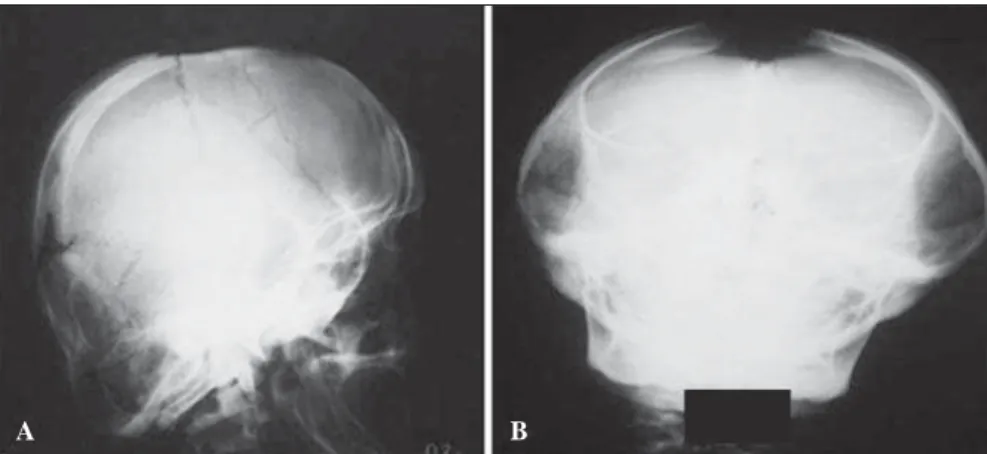
Skull radiography showed parietal and frontal bones bulging with depressed me-dian sagittal sulcus, corresponding to persis-tence of the anterior fontanelle (Figure 2). Clavicular hypoplasia that had been ob-served at physical examination, was also confirmed by means of radiography. The shoulders mobility was unusual and the patient was able to move them to the mid-line. Skeletal alterations were also identi-fied at pelvis radiography, revealing failure in the pubic symphysis ossification and deformity of the left hip, suggesting the presence of a sequel from metaphyseal dysplasia.
The patient reported, in his familial his-tory, many relatives with similar character-istics. The heredogram demonstrated the chondral and membranous bone tissues. It
may be related to delayed ossification of the skull, pelvis and extremities in cases of cleidocranial dysostosis(4). The diagnosis of such disorder is based on clinical and ra-diological findings, and the following triad is considered pathognomonic: presence of multiple supernumerary teeth; partial or total absence of the clavicles; and open sagittal suture and fontanelles. In the ab-sence of any of such findings one should consider other disorders for a possible dif-ferential diagnosis such as pycnodysostosis that is different from cleidocranial dysos-tosis for the presence of bones fragility, nanism and partial agenesis of hand and foot phalanges(5,6).
The present study is aimed at reporting two cases of cleidocranial dysostosis in a single family, as well as describing clinical and radiological findings which allow the diagnosis.
CASES REPORT
A 28-year-old, male patient with history of supernumerary teeth and dysmorphic signs was referred by the School of Odon-tology – Universidade Federal de Alagoas for consultation with the geneticist. At the anamnesis, the patient complained of a “lump” in the left maxillary region whose
INTRODUCTION
endo-383
Porciuncula CGG et al. Cleidocranial dysostosis: a report on two familial cases
Radiol Bras. 2013 Nov/Dez;46(6):382–384 dominant, monogenic autosomal etiology of the condition (Figure 3).
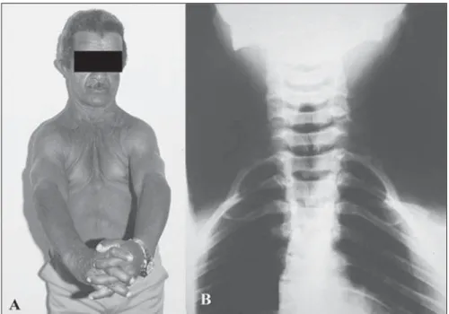
In the patient’s progenitor, the indi-vidual III-1, clinical evaluation revealed low stature, brachycephalus, cranial bones
bulging and frontal bone prominence, open sagittal suture at palpation, subtle exoph-thalmos, middle facial third hypoplasia, ogival palate, occlusal disharmony and anomalous dentition. Additionally, at
pal-pation, hypoplasia of both clavicles was observed, leading to anomalous shoulders mobility. Such a finding was confirmed by radiography of the clavicles (Figure 4).
Because of the generalized occlusal dis-harmony, absence of teeth, innumerable deep carious lesions and multiple dentiger-ous cysts, it was opted for complete surgi-cal teeth extraction and making of a total dental prosthesis.
Both patients present clinical manifes-tations that are different in intensity and va-riety, thus demonstrating the variable expressivity of cleidocranial dysostosis.
DISCUSSION
The clinical and radiological findings observed in the present cases correspond to the pathognomonic triad for the diagnosis of cleidocranial dysostosis – presence of multiple supernumerary teeth, partial or to-tal absence of clavicles and failure in sag-ittal suture and fontanelles closure –, as well as familial recurrence compatible with the dominant, monogenic autosomal etiol-ogy. Thus, the diagnoses of cleidocranial dysostosis was established, and the patients were referred for genetic counseling.
Based on the present report, one ob-serves the relevance of the dentist’s role in the diagnosis of the condition, since most of times this is the first professional con-sulted about the patient’s complaint. Addi-tionally, it should be highlighted that, dur-ing the anamnesis, it is necessary to inves-tigate the familial history, due to the high risk for recurrence.
According to Tanaka et al. and Gassen et al., it is clear that there is a necessity for a multidisciplinary approach in the assis-tance to patients with cleidocranial dysos-tosis. Specialists in odontology, psychol-ogy, speech therapy, geneticists, endocri-nologists and otorhinolaryngologists should be involved(6,7). According to Silva Júnior et al., the early diagnosis of such a condition is extremely important to mini-mize oral cavity alterations with a view on a functional adaptation and a better qual-ity of life for the individual(8).
REFERENCES
1. Lovell W. Lovell & Winter’s Pediatric ortho-paedics. 6th ed. Philadelphia: Lippincott Williams & Wilkins; 2006.
Figure 1. Panoramic radiography showing deciduous teeth permanence and unerupted permanent teeth, besides the presence of multiple supernumerary teeth and numerous dentigerous cysts. Maxillary hypo-plasia and hypoplastic zygomatic arch.
Figure 3. Heredogram of the family with members affected throughout several generations, demonstrat-ing dominant autosomal inheritance. The individual IV-5 corresponds to the patient, and the III-1, to his progenitor, both affected by the condition, as described in the present cases report.
Figure 2. Cranial radiography demonstrating macrocrania, brachycephalus with frontal and parietal bones bulging, and persistence of anterior fontanelle, besides Wormian bones and mastoid hypopneumatization.
384
Porciuncula CGG et al. Cleidocranial dysostosis: a report on two familial cases
Radiol Bras. 2013 Nov/Dez;46(6):382–384 2. Marussi VHR, Mariz FEN, Moraes AC, et al. Di-sostose cleidocraniana: relato de caso. Rev Ima-gem. 2008;30:79–82.
3. Serratine ACP, Rocha R. Displasia cleidocraniana – apresentação de um caso clínico. Arq Cat Méd. 2007;36: 109–12.
4. Machado CV, Pastor IMO, Rocha MCBS. Carac-terísticas clínicas e radiográficas da displasia clei-docraniana – relato de caso. RFO UPF. 2010;15: 302–6.
5. El-Gharbawy AH, Peeden JN Jr, Lachman RS, et al. Severe cleidocranial dysplasia and hypophos-phatasia in a child with microdeletion of the C-ter-minal region of RUNX2. Am J Med Genet A. 2010;152A:169–74.
6. Tanaka JL, Ono E, Filho EM, et al. Cleidocranial dysplasia: importance of radiographic images in diagnosis of the condition. J Oral Sci. 2006;48: 161–6.
7. Gassen HT, Marchiori M, Silva SO, et al. Relato de dois casos familiares de disostose Cleidocra-niana. Rev Fac Odont UFP. 2006;11:31–5.
8. Silva Júnior AN, Hernandez PAG, Vargas IA, et al. Displasia cleidocraniana: aspectos clínicos e radio-gráficos e relato de um caso clínico. Rev Ciênc Méd Biol. 2007;6:122–7.