Disposable
Versus
Reusable Laryngeal
Tube Suction for Ventilation in
Patients Undergoing Laparoscopic
Cholecystectomy
Afshin Amini, M.D., Farid Zand, M.D., and Masoud Maghbooli, M.D.
INTRODUCTION
The laryngeal tube suction II (LTS-II; VBM, Medizintechnik GmbH, Sulz, Germany) is a recent version of the laryngeal tube (LT) family of supraglottic airway devices originally in-tended for emergency airway management including out of hospital use 1,2 and unexpected difficult airway situations 3,4,
but which are currently also used during general anaesthe-sia 5-11. The original LT consists of an airway tube made of
silicone and two cuffs. The distal and proximal cuffs block the oesophageal inlet and the pharyngeal space above the larynx, respectively, while the holes in the shaft between the cuffs allow ventilation. The laryngeal tube suction (LTS) introduced in 2002, is the dual-lumen version of the LT. The LTS has an oesophageal drainage tube that isolates the respiratory and alimentary tracts and allows passage of a gastric tube into the oesophagus 12,13. In 2004 the LTS was modified into the
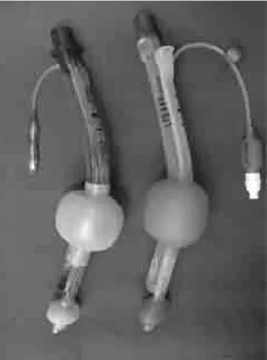
LTS-II 7,14. The major modifications include 1) a longer shaft 2)
a more pointed tip, and 3) an oval shaped distal cuff to better fit the oesophageal inlet. Two types of LTS-II were introduced to clinical use, disposable and reusable type (Figure 1). Dis-posable type (LTS-D) is made from latex free PVC material only for single use but reusable type (LTS-II) is made from latex free silicone material for frequent use (up to 50 times) after cleaning and sterilization by autoclave (according to the manufacturer’s instructions). Given increasing safety concerns about the inability to adequately clean the reusable airway vices and the potential for cross contamination by these de-vices 15, disposable equipment is being used increasingly.
The present investigation compared the performance of the two types of LTS-II (disposable and reusable type) in a rand-omized controlled study. Primary outcome was success rate in maintaining the airway with acceptable oxygenation and ventilation under conditions with elevated intra-abdominal pressure induced by capnoperitoneum. Secondary outcomes were airway seal pressure, insertion time, fiberoptic view and postoperative airway morbidity.
METHODS
After approval by institutional review board and obtaining writ-ten informed consent, 60 ASA I and II patients, aged 16 to 81yr, undergoing elective laparoscopic cholecystectomy in the supine position were recruited. Patients were excluded if they were morbidly obese (body mass index > 35 kg.m-2),
had upper respiratory tract symptoms in the previous 10 days, had history of chronic obstructive pulmonary disease, or an increased risk of aspiration (not fasting, diabetes mellitus, or oesophageal pathology). The patients were randomly allo-cated, based on computer-generated randomized numbers to receive either a LTS-D (n = 30) or LTS-II (n = 30) for airway management. The size ofboth devices was chosen according to the manufacturer’s recommendations. Both devices were recently introduced into practice in our hospital and only one anaesthesia resident, who had performed about 10 LTS-D and LTS-II insertions before the start of the study, placed the devices.
Standard monitoring devices were installed before induction of anaesthesia. After oxygenation, anaesthesia was induced with midazolam 0.04 mg.kg-1, fentanyl 2 µg.kg-1, and morphine
0.1 mg.kg-1 followed by propofol 1.5 to 2 mg.kg-1.
Neuromus-cular blockade was provided by atracurium 0.5 mg.kg-1. After
3 min of oxygenation with bag and face mask ventilation, one of the devices was placed. Tube size selection was based on the height of each patient (according to manufacturer’s in-structions). Size 3 was used for patients shorter than 155 cm, size 4 for those between 155 and 180 cm and size 5 for those taller than 180 cm. Before insertion, the cuffs were deflated and a water-soluble lubricant (K-Y jelly) was applied on the posterior surface of the proximal cuff of the devices.
The head was extended on the neck (sniffing position) and jaw trust maneuver was performed for all patients. The tip of the device was placed against the hard palate and the device was advanced until resistance was felt. The cuffs were inflat-ed using a cuff pressure gauge (VBM, Minflat-edizintechnik, Sulz, Germany) to a pressure of 60 cmH2O. No technical difference
exists in the insertion of disposable and reusable LTS-II ac-cording to the manufacturer’s recommendations. The insertion time (T1) was considered as the time from removal of the face mask until delivery of the first breath through the assigned airway device. The total time of successful insertion (T2) were recorded as the time from removal of the face mask till the device was secured after all necessary maneuvers to optimize expiratory tidal volume and stop leakage. If more than one attempt was needed for maintaining the airway, the times T1 and T2 were not used in the data analysis. An effective airway was defined as bilateral chest movement and auscultation, normal value of partial pressure of end-tidal carbon dioxide (PETCO2 35 to 45 mmHg) and normal capnography curve. In
the case of an ineffective airway and ventilation, interventions such as jaw lift, more neck extension and changing the depth of the device were performed.
Subsequently, patients were ventilated with a tidal volume of 7 mL.kg-1 at a respiratory rate of 14 per minute. If the
anaes-thetist was unable to establish an effective airway using the device after three attempts, an endotracheal tube was inser-ted for airway management. If ventilation failed during proce-dure, the gas was released from the abdominal cavity and the trachea was intubated using a laryngoscope.
Anaesthesia was maintained with a mixture of isoflurane in oxygen. The efficacy of the airway seal created by each device was determined by measuring the minimum airway pressure at which gas audibly leaked around it using a fresh gas flow of 6 L.min-1 with the pressure-limiting valve completely closed.
Airway pressure was not permitted to exceed 35 cmH2O.
The position of the device in relation to the glottic opening was assessed using a flexible fibre-optic bronchoscope. Views were graded from 1 to 4; grade 1 = vocal cords entirely visible, 2 = vocal cords or arytenoid cartilages partially visi-ble, 3 = epiglottis only visible and 4 = no laryngeal structures visible. LTS has an alimentary tract for passage of a gastric tube into the stomach. Laparoscopy procedure was done un-der CO2 insufflation into the peritoneum and increased intra
abdominal pressure was expected. The ability to access the gastrointestinal tract via the drain tube offers the possibility of increased protection against gastric distension, regurgitation and pulmonary aspiration. In all cases a 16 French gastric tube was inserted via the posterior channel with first attempt. The presence of gastric distension was then studied with di-rect vision of stomach in the monitor of laparoscopy. Also the surgeon was asked to report any signs of gastric distension noted during laparoscopy. Intra-abdominal gas pressure was not permitted to exceed 14 cmH2O. Slight head-up and
right-up position was used during laparoscopy to optimize the view of surgeon. Data including arterial oxygen saturation meas-ured by pulse oximeter, PETCO2, and peak airway pressure
were recorded at 1 and 5 minutes and then with 15 minutes
intervals. If the patients were intubated with endotracheal tube the data were used only for the number of insertion attempts, insertion time (T1), fixation and manipulation time (T2), airway sealing pressure and fiberoptic laryngeal view.
At the end of operation, residual neuromuscular block was reversed with neostigmine and atropine. The isoflurane was discontinued 5 to 10 minutes before completion of the surgi-cal procedure. The devices were routinely removed after the patient regained consciousness and opened his or her mouth to command. Upper airway trauma was assessed by observ-ing the device for the presence of blood. The patients were interviewed by a trained nurse, blinded to the used device. All patients were questioned about symptoms of airway trauma such as sore throat, hoarseness, pain on swallowing, jaw and neck pain 1 and 6 hours after the end of operation. All the patients were also followed up for symptoms of lung morbidity such as cough, fever and purulent sputum for 5 days.
Continuous data were compared with independent sample t-test while categorical data were analyzed with Chi-square and Fisher tests. Statistical analysis was performed using SPSS software version 15 for Windows (SPSS Ltd, Surrey, UK). P < 0.05 was considered as statistically significant.
RESULTS
All the recruited patients completed the study. There was no significant difference between groups with regard to patient characteristics or duration of surgery (Table I). The results of the study are summarized in table II. After successful insertion of the device in 96.6% of patients in both groups, it was possi-ble to maintain oxygenation and ventilation of all patients until the CO2 gas was insufflated into the peritoneum. Six patients
in LTS-D and 7 patients in LTS-II group needed head and neck manipulations such as jaw lift, neck extension and changing the depth of device to optimize the ventilation conditions. The insertion attempt in one patient of each group has failed after three attempts and tracheal intubation was performed. The range of oxygen saturation at 1st, 5th, 15th, 30th minute
after insertion was 98 to 100% in LTS-D and 97 to 100% in
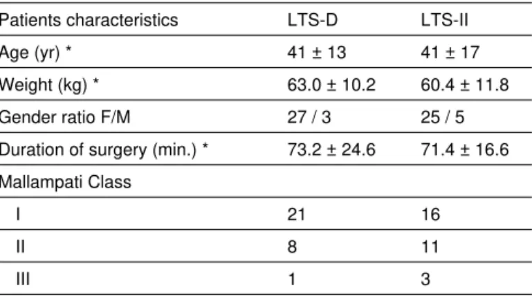
Table I – Patient Characteristics
Patients characteristics LTS-D LTS-II
Age (yr) * 41 ± 13 41 ± 17
Weight (kg) * 63.0 ± 10.2 60.4 ± 11.8
Gender ratio F/M 27 / 3 25 / 5
Duration of surgery (min.) * 73.2 ± 24.6 71.4 ± 16.6
Mallampati Class
I 21 16
II 8 11
III 1 3
* Data are presented as Mean + SD.
LTS-II group. PETCO2 was 28 to 44 mmHg in disposable, and
29 to 45 mmHg in reusable LTS-II group, at the 1st, 5th, 15th,
30th min after insertion of the devices.
In one patient with LTS-D and two patients with LTS-II after 15 to 30 minute of anaesthesia induction and 5 to 20 min-utes of peritoneal gas insufflation, adequate ventilation by these devices could not be obtained and PETCO2 was raised
(> 50mmHg) and interventions such as jaw lift, more neck
ex-tension and changing the depth of the devices were not effec-tive and endotracheal intubation was done.
Blood was noted on the cuff in six cases after removal of the LTS-D and in four cases of the LTS-II group. Postoperative airway morbidities are summarized in Table III.
DISCUSSION
Since the introduction of the supraglottic airway devices, few studies compared these different tools for airway man-agement in laparoscopic procedures 16-18. One study
con-cluded that Proseal Laryngeal Mask Airway (PLMA) is a reasonable alternative to conventional tracheal tube for gynecological laparoscopy with same advantages 17. Roth
and colleagues compared PLMA and LTS for ventilation in gynecological patients. They concluded both devices pro-vide a secure airway even under conditions of elevated intra-abdominal pressure 18. Our study demonstrates that
the LTS is a useful device for management of airway in laparoscopic cholecystectomy and the clinical performance of the disposable and the reusable LTS are similar with re-gard to oxygenation and ventilation.
We did not find any intraoperative or postoperative evidence of aspiration or lung morbidities. The first-time and overall in-sertion success rates of the both LTS groups was 90% and 96.66% respectively. The first time and overall insertion suc-cess rates for LTS-D group was 86.66% and 96.66% respec-tively and for LTS-II was 93.33% and 96.66% respecrespec-tively. In Kikuchi et al study the overall success rate of LTS-II inser-tion was 74% 19. This different result may be due to several
Table II – Details of Insertion, Airway Pressure, PETCO2, SpO2, and Fiberoptic Views for the LTS-D and LTS II
Disposable LTS-D (n=30)
Reusable LTS II (n=30)
P
Device insertion:
Successful attempts (one/two/three) # 26 / 3 / 0 28 / 1 / 0 –
Insertion time (s) * T1
T2
20.8 ± 11.6 73.3 ± 18.5
18.2 ± 4.8 65.5 ± 16.2
0.27 0.09
Ventilation:
Cases without leak # Mean SpO2 (%) *
24 99.1 ± 0.8
16 98.8 ± 0.8
0.02 0.16
PETCO2*
before capnoperitoneum (mmHg) after capnoperitoneum (mmHg)
34.2 ± 3.3 35.1 ± 3.2
33.5 ± 3.8 36.2 ± 4.3
0.40 0.28
Peak airway pressure *
before capnoperitoneum (cmH2O) after capnoperitoneum (cmH2O)
21.0 ± 6.2 24.2 ± 5.5
19.6 ± 6.0 22.7 ± 6.8
0.41 0.37
Successful gastric tube insertion # 30 30 –
Fiberoptic laryngeal view ## (1/2/3/4) 0 / 7 / 20 / 2 0 / 10 / 19 / 0 –
* Results expressed as Mean ± SD.
# Results are presented as absolute number of patients.
## 1. vocal cords entirely visible; 2. vocal cords or arytenoid cartilages partially visible; 3. epiglottis only visible; 4. no laryngeal structures visible
LTS-D – disposable laryngeal tube S; LTS II: laryngeal tube suction II.
Table III – Postoperative Airway Morbidities after Insertion of the LTS-D and LTS-II
Disposable LTS-D Reusable LTS II
Sore throat (+/–) 1st hour 6th hour
7 / 21 7 / 21
5 / 22 4 / 23
Hoarseness (+/–) 1st hour 6th hour
7 / 21 6 / 22
4 / 23 3 / 24
Dysphagia (+/–) 1st hour 6th hour
2 / 26 0 / 28
4 / 23 3 / 24
Jaw pain (+/–) 1st hour 6th hour
2 / 26 1 / 27
2 / 25 0 / 27
Neck pain (+/–) 1st hour 6th hour
6 / 22 4 / 24
6 / 21 5 / 22
Presence of blood (+/–) 6 / 22 4 / 23
Results are presented in absolute number of patients.
factors. First in our study insertion of all devices was done only by one anaesthetist but in Kikuchi’s study the insertion of devices was done by 35 anaesthetists with a wide range of experience in anaesthesia LTS placement. Second, we in-serted all tubes with gentle jaw thrust that can facilitate their insertion. Third, the LTS requires manipulations after insertion to obtain a clinically adequate airway 20 In our study
adjust-ment of the position of these airway tools was allowed but in the Kikuchi’s study this was not 19.
In the study of Mihai and colleagues insertion of LTS-II was successful on first attempt in 73% and with two attempts in 95% of patients 5. In this study neuromuscular relaxation was
not used, but in our study enough relaxation was achieved be-fore insertion of devices. The overall success rate of insertion of that study is comparable with ours. The mean insertion time was 40 seconds in Kikuchi’s study 19 and was 20s and 18s for
LTS-D and LTS-II groups respectively in our study. This differ-ence may be due to anaesthetists with different experidiffer-ence in Kikuchi’s study and our study and also maybe due to jaw-trust maneuver before insertion of devices.
The use of supraglottic airway devices under conditions of el-evated intra abdominal pressure requires an excellent airway seal to divide respiratory and alimentary tracts in a reliable manner due to the potential risk of regurgitation. There was significant difference in the quality of airway seal, represented by airway sealing pressure between two groups. Audible leak did not occur with LTS-D in 82% of cases and in LTS-II in 55% (p=0.02) in the airway pressure up to 35 cmH2O. In the cases
where audible leak occurred with airway pressures less than 35 cmH2O, the mean sealing pressure was 29 ± 5.09 cmH2O
and 27.15 ± 5.12 cmH2O for LTS-D and LTS-II groups
respec-tively (p = 0.50). A significant increase in peak airway pressure after induction of capnoperitoneum when compared to base-line values was found in both groups (p = 0.001 for LTS-D, p = 0.002 for LTS-II group). In the study of Roth et al they allowed the sealing pressure to rise up to 50 cmH2O for sealing
pres-sure meapres-surement and they calculate the difference between airway leak pressure and peak airway pressure and found that it may be considered as a rough estimate of the safety margin of these devices 18. We did not allow airway pressure rise to
more than 35 cmH2O and therefore no air leak occurred in the
majority of the cases, so we cannot comment for safety mar-gin of these devices. Fiberoptic assessment of the anatomical position of the LTS-D and LTS-II was performed through the airway tube. The vocal cords were visible (grade I to II) in 25% and 33.3% in the LTS-D and LTS-II groups respectively. Fiberoptic view of the vocal cords were considerably lower than reported in the literature 5,21. The cause of this difference
is unclear but LTS has two smaller medial orifices. Also we made no efforts to optimize this view and no manipulation for better view of glottis was done.
Failure of ventilation (PETCO2 > 50mmHg) occurred in one
patient in LTS-D and two in LTS-II groups after induction of capnoperitoneum. The reason for such failure is unclear. Anatomical differences of the airway may be responsible for this failure but no difference in airway examination existed between these patients and others who could be adequately
ventilated. The overall incidence of loss of airway after cap-noperitoneum in both groups was 5%, requiring interruption of surgery, deflation of the peritoneum, removal of LTS and endotracheal intubation. These episodes are intraoperative crises that must be regarded as adverse events.
The incidence of postoperative airway morbidities such as sore throat, dysphagia, hoarseness, jaw pain and neck pain although slightly higher after LTS-D insertion was not statisti-cally significant.
Some limitations of our study should be noted. We used pa-tients with normal airway during controlled positive pressure ventilation, therefore we cannot comment on results that could be obtained in spontaneously breathing patients and during difficult airway management. We did not allow the airway pres-sure rise more than 35 cmH2O so we cannot calculate
seal-ing pressure and could not determine the difference between sealing pressure and airway pressure that can estimate the safety margin of devices in conditions with increased intra ab-dominal pressure. Our intraoperative data collection was per-formed by a non-blinded observer, which is a possible source of bias. All postoperative throat complications, although asked by a blinded nurse, are subjective. All insertions were done by a relatively experienced anaesthetist and our findings may not apply to those with less experience.
In conclusion, our study suggests that the D and LTS-II are useful devices for airway management in laparoscopic procedures .Both disposable and reusable types are similar and can be used for this purpose in adult patients. However, further investigations that include large numbers of patients are necessary to resolve the issue of intraoperative failure of the airway after capnoperitoneum.
ACKNOWLEDGEMENTS
The LTS devices used in this study were provided by local dis-tributor of the manufacturer, VBM GmbH, Sulz, Germany. This was the only company involvement in this study. We thank for the operating room staff at Faghihi Hospital for their assistance and the Office for Development of Clinical Research of Nemazee Hospital, Shiraz, Iran for statistical analysis assistance.
REFERÊNCIAS – REFERENCES
1. Cook TM, Hommers C – New airways for resuscitation? Resuscita-tion, 2006;69:371-387.
2. Genzwuerker HV, Dhonau S, Ellinger K – Use of the laryngeal tube for out-of-hospital resuscitation. Resuscitation,2002;52:221-224. 3. Matioc AA, Olson J – Use of the laryngeal tube in two unexpected
diffi-cult airway situations: lingual tonsillar hyperplasia and morbid obesity. Can J Anaesth,2004;51:1018-1021.
4. Winterhalter M, Kirchhoff K, Groschel W et al. – The laryngeal tube for difficult airway management: a prospective investigation in pa-tients with pharyngeal and laryngeal tumours. Eur J Anaesthesiol 2005;22:678-682.
6. Amini A, Zand F, Sadeghi SE – A comparison of the disposable vs the reusable laryngeal tube in paralysed adult patients. Anaesthesia, 2007;62:1167-1170.
7. Asai T, Shingu K – The laryngeal tube. Br J Anaesth, 2005;95:729-736. 8. Asai T, Kawashima A, Hidaka I et al. – Laryngeal tube: its use for
controlled ventilation. Masui, 2001;50:1340-1341.
9. Ocker H, Wenzel V, Schmucker P et al. – A comparison of the la-ryngeal tube with the lala-ryngeal mask airway during routine surgical procedures. Anesth Analg,2002;95:1094-1097.
10. Gaitini LA, Vaida SJ, Somri M et al. – A randomized controlled trial comparing the ProSeal Laryngeal Mask Airway with the Laryngeal Tube Suction in mechanically ventilated patients. Anesthesiology, 2004;101:316-320.
11. Bein B, Carstensen S, Gleim M et al. – A comparison of the Proseal Laryngeal Mask Airway, the Laryngeal Tube S and the oesophageal tracheal combitube during routine surgical procedures. Eur J Anaes-thesiol,2005;22:341-346.
12. Dorges V, Ocker H, Wenzel V et al. – The Laryngeal Tube-S: a modi-fied simple airway device. Anesth Analg,2003;96:618-621.
13. Genzwurker H, Finteis T, Hinkelbein J et al. – Erste klinische Er-fahrungen mit dem neuen LTS. Ein Larynx-Tubus mit osophagealer Drainagemoglichkeit. Anaesthesist 2003 ;52:697-702.
14. Cook TM – The laryngeal tube sonda (LTS) and the LTS II. Acta An-aesthesiol Scand, 2006;50:521-522.
15. Blunt MC, Burchett KR – Variant Creutzfeldt-Jakob disease and dis-posable anaesthetic equipment-balancing the risks. Br J Anaesth, 2003;90:1-3.
16. Lu PP, Brimacombe J, Yang C et al. – ProSeal versus the classic laryngeal mask airway for positive pressure ventilation during laparo-scopic cholecystectomy . Br J Anaesth,2002;88:824-827.
17. Lim Y, Goel S, Brimacombe JR et al. – The ProSeal laryngeal airway is an effective alternative to laryngoscope-guided tracheal intubation for gynaecological laparoscopy. Anaesth Intensive Care,2007;35:52-56. 18. Roth H, Genzwuerker HV, Rothhaas A et al. – The ProSeal laryngeal
mask airway and the laryngeal tube suction for ventilation in gynaeco-logical patients undergoing laparoscopic surgery. Eur J Anaesthesiol, 2005;22:117-122.
19. Kikuchi T, Kamiya Y, Ohtsuka T et al. – Randomized prospective study comparing the laryngeal tube suction II with Proseal laryngeal mask airway in anesthetized and paralyzed patients. Anesthesiology2008;109:54-60. 20. Cook TM, Cranshaw J – Randomized crossover comparison of ProS-eal LaryngProS-eal Mask Airway with LaryngProS-eal Tube Sonda during anaes-thesia with controlled ventilation. Br J Anaesth,2005;95:261-266. 21. Zand F, Amini A, Sadeghi SE et al. – A comparison of the laryngeal
tube-S and Proseal laryngeal mask during outpatient surgical proce-dures.Eur J Anaesthesiol,2007;24:847-851.
RESUMEN
Amini A, Zand F, Maghbooli M – Tubo Laríngeo con succión desecha-ble Versus reutilizable para la ventilación de pacientes sometidos a la colecistectomía laparoscópica.
JUSTIFICATIVA Y OBJETIVOS: El tubo laríngeo con succión (LTS-II) es una versión reciente de los dispositivos supraglóticos reutiliza-bles que permiten el drenaje gástrico. En este estudio prospectivo y aleatorio, comparamos la inserción y la ventilación de LTS-II dese-chable (LTS-D) con la reutilizable (LTS-II) para la administración de las vías aéreas en condiciones asociadas con la presión abdominal elevada inducida por el neumoperitoneo.
MÉTODO: Sesenta pacientes ASA I y II sometidos a la colecistec-tomía laparoscópica electiva fueron aleatoriamente divididos para recibir el LTS-D (n = 30) o LTS-II (n = 30) para la administración de las vías aéreas. Después de la inducción de la anestesia general, los dispositivos se insertaron, y se verificó su correcta posición junto con la presión de salida de aire que también se midió. La facilidad de inserción, la calidad del sellado de las vías aéreas, la visualiza-ción fibrobroncoscópica, el riesgo de insuflavisualiza-ción gástrica, la inservisualiza-ción del tubo nasogástrico y la morbilidad faríngea postoperatoria fueron evaluadas.
RESULTADOS: Los índices de éxito del primer y segundo
in-tento se compararon en los de los grupos (86% vs. 93% y 96% vs. 96% en los grupos LTS-D y LTS-II, respectivamente); un paciente en cada grupo no pudo ser intubado después de ha-berlo intentado tres veces. Después de la insuflación, la ven-tilación de uno y de dos de los pacientes en los grupos LTS-D y LTS-II, respectivamente, falló y los pacientes necesitaron ser intubados con un tubo endotraqueal. El tiempo hasta que se su-ministró el primer volumen corriente a través del D y LTS-II fue de 20,8 ± 11,6 y 18,2 ± 4,8 segundos, respectivamente (p = 0,27), el tiempo de fijación y manipulación fue de 73,3 ± 18,5 y 65,5 ± 16,2 segundos, respectivamente (p = 0,096). El tubo na-sogástrico fue insertado en todos los pacientes. No se observaron diferencias significativas en los quejidos del postoperatorio.
CONCLUSIONES: Se pudieron obtener vías aéreas seguras en