Received from Programa de Pós-Graduação em Produtos Naturais e Sintéticos Bioativos, Universidade Federal da Paraíba, João Pessoa, PB, Brazil.
1. Laboratório de Tecnologia Farmacêutica Prof. Delby Fernandes de Medeiros, Universidade Federal da Paraíba, João Pessoa, PB, Brasil
2. PhD; Student in Pharmacology of Natural and Synthetic Bioactive Products, Universidade Federal da Paraíba. João Pessoa, PB, Brazil
3. PhD; Professor, Department of Pharmaceutical Sciences, Center for Health Sciences, Universidade Federal da Paraíba, João Pessoa, PB, Brazil
Submitted on October 27, 2011. Approved on January 23, 2012. Correspondence to:
Ana Carolina de Carvalho Correia, MD
Programa de Pós-Graduação em Produtos Naturais e Sintéticos Bioativos Universidade Federal da Paraíba
58051-970 – João Pessoa, PB, Brazil E-mail: [email protected]
REVIEW ARTICLE
Malignant Hyperthermia: Clinical and Molecular Aspects
Ana Carolina de Carvalho Correia
1,2, Polyana Cristina Barros Silva
1,2, Bagnólia Araújo da Silva
1,3Summary: Correia ACC, Silva PCB, Silva BA – Malignant Hyperthermia: Clinical and Molecular Aspects.
Content: Malignant hyperthermia (MH) is a potentially lethal pharmacogenetic disorder that affects genetically predisposed individuals. It mani-fests in susceptible individuals in response to exposure to Inhalant anesthetics, depolarizing muscle relaxants or extreme physical activity in hot environments. During exposure to these triggering agents, there is a rapid and sustained increase of myoplasmic calcium (Ca2+) concentration induced by hyperactivation of ryanodine receptor of skeletal muscle (RyR1), causing a profound change in Ca2+ homeostasis, featuring a hyper-metabolic state. RyR1, Ca2+ release channels of sarcoplasmic reticulum, is the primary locus for MH susceptibility. Several mutations in the gene encoding the protein RyR1 have been identified; however, other genes may be involved. Actually, the standard method for diagnosing MH sus-ceptibility is the muscle contracture test for exposure to halothane-caffeine (CHCT) and the only treatment is the use of dantrolene. However, with advances in molecular genetics, a full understanding of the disease etiology may be provided, favoring the development of an accurate diagnosis, less invasive, with DNA test, and also will provide the development of new therapeutic strategies for treatment of MH. Thus, this brief review aims to integrate molecular and clinical aspects of MH, gathering input for a better understanding of this channelopathy.
Keywords: Anesthetics, Inhalation; Calcium; Malignant Hyperthermia; Neuromuscular Blocking Agents; Ryanodine.
©2012 Elsevier Editora Ltda. All rights reserved.
INTRODUCTION
Malignant hyperthermia (MH) is a potentially fatal pharma-cogenetic disorder. During a crisis of MH, inhalational anes-thetics, muscle relaxants depolarizing (succinylcholine) or ex-treme physical activity in hot environments trigger a massive accumulation of calcium (Ca2+) in myoplasm, which leads to
an accelerated metabolism and skeletal muscle contractile activity. This hypermetabolic state generates heat and leads to hypoxemia, metabolic acidosis, rhabdomyolysis, and rapid increase in body temperature that can be fatal if not recog-nized and treated early 1,2.
This release of Ca2+ in myoplasm occurs due to a
mem-brane depolarization that induces conformational changes in L-type calcium channels (CAV-L) (or dihydropyridine
recep-tors [DHPRs]), which lead to Ca2+ release channels
activa-tion from sarcoplasmic reticulum (or ryanodine receptor sub-type-1 [RyR1] in skeletal muscle). This functional interaction
between DHPRs and RyRs, which transforms the electrical impulse into chemical substance, is commonly referred to as excitation-contraction coupling (E-C) 3,4.
Several mutations in the RyR1 gene have been already identified and implicated in a wide range of channelopathies, and this defect is primarily responsible for susceptibility to MH; however, other genes may be involved 2. This variation in
genes related to susceptibility to MH is the major cause of the syndrome’s different manifestations 5.
Thus, this paper aims to review the molecular and physi-ological bases of RyRs and outline the pathophysiphysi-ological and genetic factors involved in malignant hyperthermia, in order to provide a condensed and updated source of scientific infor-mation for healthcare professionals and incorporate molecular and clinical aspects for a better understanding of this chan-nelopathy.
RYANODINE RECEPTORS (RYRs)
Classification and location
Ryanodine receptors (RyRs) are high-conductance cation channels, which release Ca2+ from intracellular stores such as
the endo/sarcoplasmic reticulum (ER/SR) 6. RyRs are
ubiq-uitous in all cell types and are involved in a variety of cellular processes (E-C coupling, neurotransmission, secretion etc.) 4.
Molecular structure
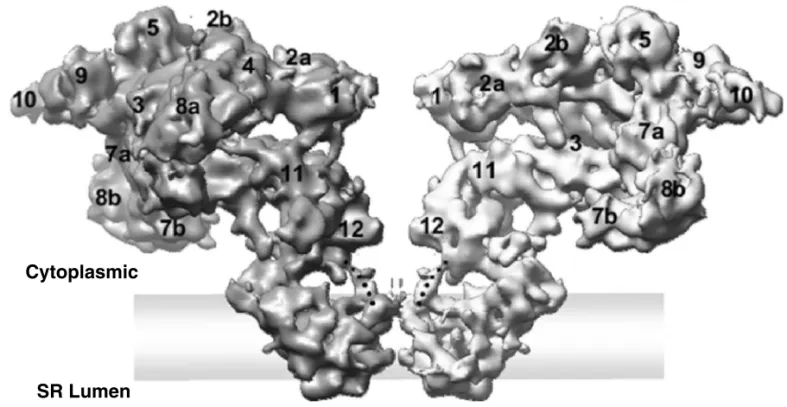
RyRs are homotetramers with a molecular mass of about 560 kDa, characterized by having a bell shape 8 (Figure 1).
It shows ~ 70% homologous amino acid sequence and the higher level of similarity is at the C-terminal region. In all isoforms, the C-terminal portion of the protein contains the transmembrane domains. According to systematic analysis, it is suggestedthat there are between 4 and 12 transmem-brane segments per RyR subunit 10 (Figure 2). There is also a
large N-terminal cytoplasmic domain containing binding sites for protein and other channel modulators (e.g., Ca2+
chan-nels) that control the RyRactivity state 15. Each RyR subunit
is closely associated with a 12 kDa protein, FKBP12, which modulates the opening parameters (probability of the channel being open and average time of opening) 16.
Activators and blockers
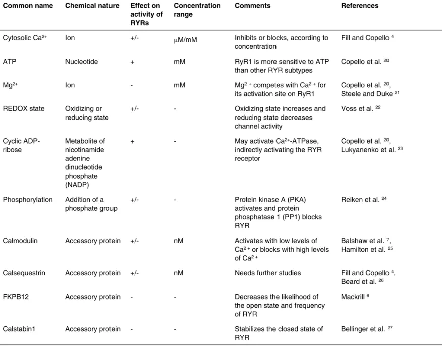
The various cellular processes, physiological agents, pharma-cological substances, and different associated proteins that regulate RyRs receptors are shown in Tables I and II.
Role of RyRs in excitation-contraction coupling (E-C)
There is clear evidence that RyRs interact with DHPRs near the T-tubule membrane. This functional interaction between DHPRs and RyRs is commonly referred to as E-C coupling,
which is the transformation of an electrical signal into a chemical signal, and these receptors play an important role in this process 4. The three genetically distinct isoforms of
RyR (RyR1, RyR2, and RyR3) show release of Ca2+ induced
by Ca2+ (ICRC), a process by which Ca2+ itself activates the
channel to release Ca2+ 28. DHFR is an L-type Ca2+ channel,
also known as CAV1, and the α-subunit of this channel is the
poforming unit that functions as a voltage sensor and re-sponds to changes in membrane potential. This α-subunit is the region in which there is binding of dihydropyridines. There are several isoforms of this channel classified according to their location. For example, subtype CaV1.1 is present in
skel-etal muscle and CaV1.2 in cardiac muscle 1.
In skeletal muscle, E-C coupling does not require the entry of extracellular Ca2+. The release of Ca2+ by RyR1
(the predominant isoform in skeletal muscle) is triggered by conformational change in the voltage sensor of DHFR in T-tubule depolarization. This Ca2+ release is referred to as
depolarization-induced Ca2+ release (DICR) 29. Structurally,
the DHFR-RyR1 complex organization is found in the ratio of 4:1, where RyR1 is physically coupled to four CaV1.1 4
(Figure 3A). In cardiac muscle, however, plasma membrane depolarization activates DHFR (CaV1.2) to allow the entry
of extracellular Ca2+ into cells. The entry of Ca2+, in turn,
triggers Ca2+ release by RyR2 (the predominant isoform in
heart) through CICR mechanism (Ca2+ release induced by
Ca2+ itself) 30. Structurally, the DHFR-RyR2 complex
orga-nization is very different from that found in skeletal muscle, with about one CaV1.2 for every 5-10 RyR2 not aligned in a
highly ordered manner 4 (Figure 3B).
Figure 1 Two Opposing Subunits of the RyR1 Tetramer are Displayed in a Side View. Figure adaptedfrom Serysheva et al. 9
Cytoplasmic
Table I – Exogenous Substances Regulating RyRs
Common name Chemical Nature Effect on activity of RYRs
Concentration range Pharmacological or clinical use
References
Ryanodine Alkaloid +/- nM-mM Inappropriate Fill and Copello 4
4-cloro-meta-cresol (4-CmC)
Chlorinated phenol + µM-mM Fungicide Mackrill 6,
Fessenden et al. 17
Caffeine Methylxanthine + mM Stimulant Mackrill 6
Dantrolene Hydantoin derivatives
- µM Treatment
of malignant hyperthermia, muscle spasticity
Mackrill 6,
Paul-Pletzer et al. 18
Procaine and tetracaine
Amino ester - µM-mM Local anesthetic Mackrill 6,
Brum et al. 19
Ruthenium red Polycationic dye - nM-µM - Mackrill 6
Table II – Physiological Agents Regulating RyRs
Common name Chemical nature Effect on
activity of RYRs
Concentration range
Comments References
Cytosolic Ca2+ Ion
+/-µM/mM Inhibits or blocks, according to concentration
Fill and Copello 4
ATP Nucleotide + mM RyR1 is more sensitive to ATP
than other RYR subtypes
Copello et al. 20
Mg2+ Ion - mM Mg2+ competes with Ca2+ for
its activation site on RyR1
Copello et al. 20, Steele and Duke 21 REDOX state Oxidizing or
reducing state
+/- - Oxidizing state increases and reducing state decreases channel activity
Voss et al. 22
Cyclic ADP-ribose
Metabolite of nicotinamide adenine dinucleotide phosphate (NADP)
+ - May activate Ca2+-ATPase,
indirectly activating the RYR receptor
Copello et al. 20, Lukyanenko et al. 23
Phosphorylation Addition of a phosphate group
+/- - Protein kinase A (PKA)
activates and protein phosphatase 1 (PP1) blocks RYR
Reiken et al. 24
Calmodulin Accessory protein +/- nM Activates with low levels of Ca2 + or blocks with high levels of Ca2 +
Balshaw et al. 7, Hamilton et al. 25
Calsequestrin Accessory protein +/- nM Needs further studies Fill and Copello 4, Beard et al. 26
FKPB12 Accessory protein - - Decreases the likelihood of
the open state and frequency of RYR
Mackrill 6
Calstabin1 Accessory protein - - Stabilizes the closed state of RYR
Correlated channelopathies
RyRs are encoded by three distinct genes located on hu-man chromosomes19q13.1 (RyR1), 1q42.1-1q43 (RyR2), and 15q14-q15 (RyR3) 31. Mutations in both RyR1 and RyR2
are correlated with disease 14. To date, over 100 mutations in
RyR1 have been identified and grouped into three regions of the protein: N-terminal, Central, and C-terminal 6. These
mu-tations have been implicated in a wide range of conditions, among them the susceptibility to malignant hyperthermia and various congenital myopathies, including central core disease, multiminicore myopathy with external ophthalmoplegia and, rarely, centronuclear myopathy. Although malignant hyper-thermia is predominantly inherited, the central core disease involves both autosomal dominant and recessive inheritance.
Multiminicore myopathy with external ophthalmoplegia is as-sociated with recessive inheritance and quantitative defects of RyR1 protein expression 32.
RyR2 mutations are associated with two forms of arrhyth-mia induced by stress, called catecholaminergic polymorphic ventricular tachycardia type 1, and a form of arrhythmogenic right ventricular dysplasia type 2. There are more than 80 mutations related to the RyR2 gene and they are clustered in three regions of the protein, similar to the distribution of changes in RyR1 6.
MALIGNANT HYPERTHERMIA
Concept
Malignant hyperthermia (MH), also known as malignant hy-perpyrexia, is a potentially lethal pharmacogenetic disorder which affects genetically predisposed individuals 2,33.
Figure 2 Models of Transmembrane regions of RyR1. (A) Model of Takeshima et al. 11; (B) Model of Zorzato et al. 12; (C) Model of Du et al. 13. Figure adapted from Hamilton 14.
Membrane
Membrane Membrane
Figure 3 Organization of DHPR-RyR Complex in Skeletal (A) and Cardiac (B) Muscles.
Figure adaptedfrom Fill et al. 4
A CYTOSOL
CYTOSOL
TETRAD
T-TUBULE
SR LUMEN
Etiology
There is clear evidence that individuals susceptible to MH have a skeletal muscle disorder associated with uncontrolled release of Ca2+ from sarcoplasmic reticulum 33. Two genes
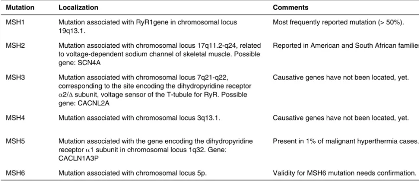
related to susceptibility to MH have been identified and at least four genes are in the process of positive identification 5
(Table III). Individuals susceptible to MH respond abnormally when exposed to inhalational anesthetics (halothane, enflu-rane, isofluenflu-rane, desfluenflu-rane, sevoflurane), depolarizing mus-cle relaxants (e.g. succinylcholine) or extreme physical activ-ity in hot environments 1. During exposure to these triggering
agents, there is a rapid and sustained growth of myoplasmic Ca2+ concentration due to RyR1 hyperactivation, which causes
a profound change in Ca2+ homeostasis and characterizes a
hypermetabolic state 27.
Epidemiology
MH was described in all ethnic groups and its susceptibility oc-curs equally in both sexes, although seizures are more com-mon in men. The incidence of anesthetic MH in adult patients is 1/50,000 and 1/15,000 in pediatric patients, although cases have been reported in extreme ages. Its true prevalence is difficult to define because some individuals present mild or no reactions and the variable penetrance of the inherited trait 34,35. The incomplete penetrance indicates that, although
the individual has the genetic mutation for MH susceptibility, it does not mean that this dysfunction will be expressed during the first or even after the exposure to a triggering agent 35.
Pathophysiology
Under normal conditions, the myoplasmatic levels of Ca2+
are controlled by RyR1, DHFR, and Ca2+-adenosine
triphos-phatase (Ca2+-ATPase) system 35. In MH crisis, there is intense
change in Ca2+ homeostasis in which RyR1 hyperactivation
causes an increase in cytoplasmic Ca2+ concentration, which
results in sustained activation of muscle contraction 18.
Some-times, the first symptom may be the presence of a masseter muscle spasm. This signal is considered by many authors as a sign of suspected syndrome5.
The process of muscle contraction and re-absorption of excess Ca2+ consume large amounts of ATP and
gener-ate excessive heat (hyperthermia), which is the hallmark of disease 18. Depletion of ATP stocks results in disruption of
the skeletal muscle membrane and there is leakage of cel-lular constituents, including potassium, creatine, phosphate, and myoglobin. Loss of potassium from muscle cells results in metabolic acidosis and cardiac arrhythmias 37. Decreased
concentration of ATP causes muscular rigidity, as the pres-ence of ATP is normally required to allow muscle relaxation, in addition to the combination of actin and myosin to allow muscle rigidity and inextensibility 18.
A potential increase in oxygen consumption through uncon-trolled glycolysis and aerobic metabolism leads to cell hypox-ia, progressive lactic acidosis, and excessive carbon dioxide generation 34. Thus, the most common initial signal of acute
malignant hyperthermia is an unexplained increase in values of capnography (EtCO2), a method that evaluates the
gradi-ent of CO2 present during expiration, in which excess expired
CO2 does not easily decreases with increased minute
ventila-Table III – Classification Summary of Genetic Mutations Associated with Susceptibility to Malignant Hyperthermia
Mutation Localization Comments
MSH1 Mutation associated with RyR1gene in chromosomal locus 19q13.1.
Most frequently reported mutation (> 50%).
MSH2 Mutation associated with chromosomal locus 17q11.2-q24, related to voltage-dependent sodium channel of skeletal muscle. Possible gene: SCN4A
Reported in American and South African families.
MSH3 Mutation associated with chromosomal locus 7q21-q22, corresponding to the site encoding the dihydropyridine receptor α2/∆ subunit, voltage sensor of the T-tubule for RyR. Possible gene: CACNL2A
Causative genes have not been located, yet.
MSH4 Mutation associated with chromosomal locus 3q13.1. Causative genes have not been located, yet.
MSH5 Mutation associated with the gene encoding the dihydropyridine receptor α1 subunit in chromosomal locus 1q32. Gene: CACLN1A3P
Present in 1% of malignant hyperthermia cases.
tion. This increased EtCO2 is associated with the presence of
tachycardia (due to sympathetic stimulation by acidosis) 5.
This hypermetabolic state generates heat and leads to hypoxemia, metabolic acidosis, rhabdomyolysis (breakdown and lysis of muscle cells) and a rapid increase in body temper-ature, which can be fatal if not recognized and treated early 2.
Signs and symptoms
The onset of acute malignant hyperthermia is characterized by one or more signals of systemic hypermetabolism during or immediately after administration of a triggering agent 34. The
first symptoms are tachycardia, hyperventilation, localized muscle stiffness, cyanosis, arrhythmias, excessive sweating and hyperthermia. The crisis of MH can manifest itself later on a recurring basis in up to 20% of cases, even after discontinu-ation of the triggering agent, and fever above 40°C, cyanosis, poor cutaneous perfusion, pressure instability, and general-ized muscle rigidity may occur 35.
Additional and potentially fatal complications include dis-seminated intravascular coagulation, congestive heart failure, intestinal ischemia, and limb compartment syndrome with a deep muscle edema 34.
Diagnosis
Clinical
MH diagnosis is based on clinical and laboratory findings. MH manifestation may be immediately after exposure to the trig-gering agents or even a few hours after its discontinuation. Without this prior exposure, it is usually impossible to identify a susceptible patient, which makes the clinical diagnosis very difficult 33,35,38.
Crises are classified according to clinical presentation and symptoms may vary from fulminant to abortive conditions, ac-cording to its intensity 35 (Table IV).
The most common initial symptoms are listed in Table V. Although nonspecific, these initial symptoms associated with exposure to triggering agents in the absence of other appar-ent cause will be sufficiappar-ent to establish a preliminary diagnosis of MH and immediately refer the patient to treatment. MH may evolve rapidly, presenting additional clinical and laboratory manifestations (Table V). Between 12 and 24 hours after the crisis onset, the peak plasma levels of creatine kinase (CPK) can be observed. Susceptibility confirmation will depend on caffeine-halothane contracture test (CHCT) outcome, indicat-ed only three months after the crisis’ onset 35.
Laboratory – Susceptibility to MH
Creatine kinase (CPK) at rest
Increased CPK is found in 50% of relatives of patients with an-esthetic malignant hyperthermia. The presence of increased CPK at rest, other than in strenuous exercise or muscle trau-ma, has relative value only in relatives of susceptible patients. Without additional explanation, high levels of CPK at rest raise the suspicion of myopathy. These changes are common and do not justify plasma CPK measurement in the general popu-lation 39.
Contraction test for exposure to caffeine-halothane (CHCT)
Even in classic cases, diagnosis confirmation is mandatory because it will be from the confirmed cases that the investiga-tion planning for relatives of those affected will be made. The standard test adopted for MH diagnosis is the contraction test of exposure to caffeine-halothane (CHCT) 35. Through
analy-sis of the contractile response to caffeine-halothane exposure, it is possible to discriminate patients as susceptible (MHS) when the answer to both caffeine and halothane is abnormal;
Table IV – Classification of Malignant Hyperthermia Crisis
Classical fulminant: potentially fatal, multiple metabolic and muscular manifestations. Fulminant
Moderate: metabolic and muscular manifestations without the severity of a classical fulminant. Abortive Mild: mild metabolic changes without muscle involvement.
Masseter muscle rigidity with evidence of muscle injury (e.g., increased serum creatine kinase and myoglobinuria).
Masseter muscle rigidity associated with metabolic changes (e.g., increased temperature, cardiac arrhythmias). Masseter muscle rigidity alone.
Masseter spasm
Sudden death or unexplained cardiac arrest during anesthesia.
Other: postoperative fever, rhabdomyolysis, renal failure, suspected family history
Atypical
negative (MHN) when the response to caffeine and halothane is normal; and equivocal (MHE) when the response to caffeine or halothane is abnormal. All patients diagnosed as MHE are treated as MHS due to their susceptibility. Clinical diagnosis is considered positive when there is a contracture ≥ 0.5 g for 3% halothane and ≥ 0.3 g at 2 mM of caffeine 40.
The procedure for muscle biopsy preparation varies de-pending on the laboratory. Some of them follow the United States’ protocol (97% sensitivity, but low specificity, with 22% false positives) while others follow the European protocol - also known as in vitro contracture test - which differs from the American protocol only by including the use of ryanodine or 4-chloro-m-cresol (99% sensitivity, 94% specificity) 5.
In Brazil, both the Muscle Biopsy Center of the Universi-dade Federal do Rio de Janeiro and the Study, Diagnosis and Malignant Hyperthermia Research Center (Cedhima) of the Escola Paulista de Medicina, Universidade Federal de São Paulo use the American protocol for the diagnosis of MH 41.
Genetic testing
From the first reported case of MH, it was suspected to be a family inherited disorder 40. With the demonstration that a
mutation in the gene encoding RyR1 in pig muscles was the basis of MH, a simple DNA test in humans to diagnose it in-creased the expectation. However, this expectation has not yet been achieved because there are many changes in skel-etal muscle that may be the cause for the different forms of the syndrome 42 (Table III).
The mutations associated with the six listed genes cor-respond to approximately 50% of families surveyed. In other families, the gene involved is still unknown 39. Furthermore,
despite the MHS1 mutation being the only direct genetic
cause for MHS, the additional presence of MHS3, MHS4, or MHS6 mutations may interact and increase the phenotype ex-pression in some individuals 40.
However, with time, an accuracy test based on DNA and applicable to most patients will be available and, once identi-fied the mutation in a case of MH, all family members may be tested for that specific mutation through a blood sample. A major international effort is underway to clarify the molecular genetic basis of MH 42.
Treatment
The internationally recommended protocol for treatment of malignant hyperthermia is based on discontinuation of expo-sure to the triggering agents, administration of specific drug (dantrolene), and support measures or measures aimed at preventing associated complications, such as:
1. Replacing the anesthesia circuit by another circuit un-contaminated by anesthetic agent;
2. Hyperventilating the patient with 100% oxygen; 3. External cooling and, if necessary, internal; 4. Correction of metabolic acidosis;
5. Reduction of hyperkalemia; 6. Correction of cardiac arrhythmias; 7. Maintenance of diuresis 33,35.
Dantrolene
Dantrolene was originally synthesized by Snyder et al. in 1967. It was found to have muscle relaxing properties after intravenous administration in animals. The studies
demon-Table V – Clinical Manifestations of Malignant Hyperthermia crisis
Clinical Laboratory
Early
Tachycardia
Progressive increase of exhaled CO2 Tachypnea
Localized muscle stiffness (including masseter rigidity) Cyanosis
Arrhythmias Hyperthermia Profuse sweating
Hypercapnia (respiratory acidosis) Metabolic acidosis Hyperlactacidemia Hyperkalemia Central venous desaturation
Late
Fever above 40°C Cyanosis Poor skin perfusion Pressure instability Generalized muscle rigidity
Myoglobinemia Increased serum creatine kinase
Increased serum creatinine Disseminated intravascular coagulation
strated that these relaxing properties are due to the depres-sion of excitation-contraction coupling (EC). It was initially used as a muscle relaxant in long-term treatment of skeletal muscle spasticity 43. Dantrolene has been used since 1975,
but currently its clinical use is restricted to malignant hyper-thermia 44,45.
Dantrolene blocks the RyRs, acts directly on RyR1 and RyR3 isoforms, reduces the channel activation by calmodu-lin, and reduces the channel sensitivity to Ca2+. RyR2 is not
blocked by dantrolene, which explains its lack of negative ino-tropic effect on the heart 7,10,46.
The molecular structure of dantrolene, a hydantoin de-rivative, is planar. It is highly lipophilic and, therefore, poorly water-soluble. This created problems for its introduction into clinical practice until the 1980s. Its widespread use had to wait for a suitable intravenous preparation 47. Currently, dantrolene
is available for intravenous use in vials containing 20 mg of lyophilized sodium dantrolene added to 3 g of mannitol to en-hance water solubility. The vial contents should be dissolved in 60 mL of water, which yields a final dantrolene concentra-tion of 0.33 mg.mL-1 at pH 9.5. The resulting alkaline solution
is highly irritating to peripheral veins and must be injected into a large vein or be rapidly infused 43.
Rapid preparation and administration of dantrolene are es-sential. Therapy begins with the administration of 2.5 mg.kg-1
and must be repeated every five minutes until the hyper-metabolic state normalization and disappearance of all MH symptoms 48. Continuous intravenous infusion of dantrolene at
10 mg.kg-1 should be given at least 24 hours after successful
initial therapy. Support therapy includes body cooling; admin-istration of sodium bicarbonate to treat acidosis; beta-blockers or lidocaine in case of cardiac arrhythmias persistency; and fu-rosemide and glucose-insulin infusion in case of hyperkalemia, hypercalcemia, and myoglobinuria. Thus, early diagnosis gen-erates a successful treatment in most patients 43.
Azumolene
Azumolene is 30 times more soluble in water than its ana-log, dantrolene. This is due to the replacement of the para-nitrophenyl group in dantrolene by the para-bromophenyl group. Compared to dantrolene, azumolene is equipotent for treatment and prevention of MH clinical manifestations dur-ing a crisis induced by halothane or succinylcholine. In vitro studies showed azumolene equipotential for relaxing porcine skeletal muscle and, in vivo, it was more potent for inhibit-ing the gastrocnemius muscle contractions. Therefore, this product may be useful for treating MH in the future. However, for economic reasons, it has not been introduced into clinical practiceyet 43.
PERSPECTIVES
The elucidation of the molecular genetic basis of MH has the perspective of making a pre-symptomatic diagnosis, without the need for biopsies, in addition to having a full understand-ing of the disease etiology. With the advancement in human genome mapping, there is a promising future for characteriz-ing new mutations related to this syndrome and unveilcharacteriz-ing the genetic heterogeneity of MH, as phenotypic variations may be caused by interactions of several genes, yet unknown, such as the RYR1 gene.
In recent years, a major breakthrough has occurred in un-derstanding the dynamics of Ca2+ release via RyR1 from the
REFERÊNCIAS/REFERENCES
1. Jurkat-Rott K, Lehmann-Horn F – Muscle channelopathies and critical points in functional and genetic studies. J Clin Invest, 2005;115:2000-2009.
2. Carpenter D, Robinson RL, Quinnell RJ et al. – Genetic variation in RYR1 and malignant hyperthermia phenotypes. Br J Anaesth, 2009;1-11.
3. Lueck JD, Goonasekera SA, Dirksen RT – Ryanodinopathies: Muscle Disorders Linked to Mutations in Ryanodine Receptors. Basic Appl Myol, 2004;14(5):345-358.
4. Fill M, Copello JA – Ryanodine receptor calcium release channels. Physiol Rev, 2002;82:893-922.
5. Gómez JRO – Anestesia en la hipertermia maligna. Rev Esp Aneste. siol Reanim, 2008;55:165-174.
6. Mackrill JJ – Ryanodine receptor calcium channels and their partners as drug targets. Biochem Pharmacol, 2010;79:1535-1543.
7. Balshaw DM, Yamaguchi N, Meissner G – Modulation intracellular Cal-cium-release channels by calmodulin. J Memb Biol, 2002;185:1-8. 8. Kovacs E, Xub L, Pasek, DA et al. – Regulation of ryanodine
recep-tors by sphingosylphosphorylcholine: Involvement of both calmodulin-dependent and -incalmodulin-dependent mechanisms. Biochem Biophys Res Commun, 2010;401:281-286.
9. Serysheva II, Ludtke SJ, Baker ML et al. – Subnanometer-resolution electron cryomicroscopy-based domain models for the cytoplas-mic region of skeletal muscle RyR channel. Proc Natl Acad Sci, 2008;105:9610-9615.
10. Meissner G – Molecular regulation of cardiac ryanodine receptor ion channel. Cell Calcium 2004;35:621-628.
11. Takeshima H, Nishimura S, Matsumoto T et al. – Primary structure and expression from complementary DNA of skeletal muscle ryanodi-ne receptor. Nature, 1989;339:439-445.
12. Zorzato F, Fujii J, Otsu K et al. – Molecular cloning of cDNA enco-ding human and rabbit forms of the Ca2+ release channel (ryanodi-ne receptor) of skeletal muscle sarcoplasmic reticulum. J Biol Chem, 1990;265(4):2244-2256.
13. Du GG, Avila G, Sharma P et al. – Role of the sequence surroun-ding predicted transmembrane helix M4 in membrane association and function of the Ca (2+) release channel of skeletal muscle sar-coplasmic reticulum (ryanodine receptor isoform 1). J Biol Chem, 2004;279(36):37566-37574.
14. Hamilton SL – Ryanodine receptors. Cell Calcium, 2005;38:253-260. 15. Marks AR – Ryanodine Receptors, FKBP12, and Heart Failure. Front
Biosci, 2002;7:970-977.
16. Samsò M, Wagenknecht T, Allen PD – Internal structure and visuali-zation of transmembrane domains of the RyR1 calcium release chan-nel by cryo-EM. Nature Struct Biol, 2005;12(6):539-544.
17. Fessenden JD, Perez CF, Goth S et al. – Identification of a Key De-terminant of Ryanodine Receptor Type 1 Required for Activation by 4-Chloro-m-cresol. J Biol Chem, 2003;278(31)28727-28735. 18. Paul-Pletzer K, Yamamoto T, Bhat MB et al. – Identification of a
dan-troleno-binding sequence on the skeletal muscle ryanodine receptor. J Biol Chem, 2002;277:34918-34923.
19. Brum G, Piriz N, DeArmas R et al. – Differential Effects of Voltage-Dependent Inactivation and Local Anesthetics on Kinetic Phases of Ca21 Release in Frog Skeletal Muscle. Biophys J, 2003;85:245-254. 20. Copello JA, Barg S, Sonnleitner A et al. – Differential activation by
Ca2+, ATP and caffeine of cardiac and skeletal muscle ryanodine re-ceptors after block by Mg2+. J Membr Biol, 2002;187:51-64.
21. Steele DS, Duke AM – Defective Mg2+ regulation of RyR1 as a causal factor in malignant hyperthermia. Arch Biochem Biophy, 2007;458:57-64.
22. Voss AA, Lango J, Ernst-Russell M et al. – Identification of hyperreac-tive cysteines within ryanodine receptor type 1 by mass spectrometry. J Biol Chem, 2004;279:34514-34520.
23. Lukyanenko V, Gyorke I, Wiesner TF et al. – Potentiation of Ca(2+) re-lease by cADP-ribose in the heart is mediated by enhanced SR Ca(2+) uptake into the sarcoplasmic reticulum. Circ Res, 2001;89:614-622.
24. Reiken S, Lacampagne A, Zhou H et al. – PKA phosphorylation ac-tivates the calcium release channel (ryanodine receptor) in skeletal muscle: defective regulation in heart failure. J Cell Biol, 2003;160:919-928.
25. Hamilton SL, Serysheva I, Strasburg GM – Calmodulin and Excitation-Contraction Coupling. News Physiol Sci, 2000;15:281-284.
26. Beard NA, Sakowska MM, Dulhunty AF et al. – Calsequestrin is an inhibitor of skeletal muscle ryanodine receptor calcium release chan-nels. Biophys J. 2002;82:310-320.
27. Bellinger AM, Mongillo M, Marks AR – Stressed out: the skel-etal muscle ryanodine receptor as a target of stress. J Clin Invest, 2008;118(2):445-453.
28. Endo M – Calcium-induced calcium release in skeletal muscle. Phys-iol Rev, 2009;89:1153-1176.
29. Murayama T, Kurebayashi N – Two ryanodine receptor isoforms in nonmammalian vertebrate skeletal muscle: Possible roles in excita-tionecontraction coupling and other processes. Progress in Biophys. and Mol. Biol, 2010;1-10.
30. Bers DM – Cardiac excitation–contraction coupling. Nature, 2002;415:198-205.
31. Islam MS – The ryanodine receptor calcium channel of beta-cells: molecular regulation and physiological significance. Diabetes, 2002;51:1299-1309.
32. Zhou H, Lillis S, Loy RE et al. – Multi-minicore disease and atypi-cal periodic paralysis associated with novel mutations in the skeletal muscle ryanodine receptor (RYR1) gene. Neuromuscular Disorders, 2010;20:166-173.
33. Rosenberg H, Davis M, James D et al. – Malignant Hiperthermia. Or-phanet encyclopedia, 2004;1-14.
34. Litman RS, Rosenberg H – Malignant Hyperthermia – Update on Sus-ceptibility Testing. Am Med Assoc, 2005;293(23):2918- 2924. 35. Amaral JLG, Carvalho RB, Cunha LBP et al. – Hipertermia Maligna.
In: Associação Médica Brasileira e Conselho Federal de Medicina (orgs). Projeto Diretrizes. São Paulo: AMB/CFM, 2009. Disponível em: <http://www.projetodiretrizes.org.br/projeto_diretrizes/058.pdf>. 36. Parness J, Bandschapp O, Girard T – The myotonias and
susceptibil-ity to malignant hyperthermia. Anesth Analg, 2009;109(4):1054-1064. 37. Ali SZ, Taguchi A, Rosenberg H – Malignant hyperthermia. Best Pract
Res Clin Anaesthesiol, 2003;17(4):519-533.
38. Hopkins PM – Malignant hyperthermia: advances in clinical manage-ment and diagnosis. Br J Anaesth, 2000;85(1):118-128.
39. Silva HCA, Bahia VS, Oliveira RAA et al. – Susceptibilidade à hiper-termia maligna em três pacientes com síndrome maligna por neuro-lépticos. Arq Neuropsiquiatr, 2000;58(3-A):713-719.
40. Hernandez JF, Secrest JA, Hill L et al. – Scientific advances in the genetic understanding and diagnosis of malignant hyperthermia. J Pe-riAnesth Nur, 2009;24(1):19-34.
41. Hors CP, Garicochea B – Bases genéticas da hipertermia maligna. Rev Bras Anestesiol, 1999;49(4):277-281.
42. Rosenberg H, Antognini JF, Muldoon S – Testing for malignant hyper-thermia. Anesthesiol, 2002;96:232-237.
43. Krause T, Gerbershagen MU, Fiege M et al. – Dantroleno: a review of its pharmacology, therapeutic use, and new developments. Anaesth, 2004;59:364-373.
44. Lin CM, Neeru S, Doufas AG et al. – Dantroleno reduces the threshold and gain for shivering. Anesth Analg, 2004;98(5):1318-24.
45. Hadad E, Cohen-Sivan Y, Heled Y et al. – Clinical review: treat-ment of heat stroke: should dantroleno be considered? Crit Care, 2005;9(1):86-91.
46. Muehlschlegel S, Sims JR – Dantroleno: mechanisms of neuroprotec-tion and possible clinical applicaneuroprotec-tions in the neurointensive care unit. Neurocrit Care, 2009;10(1):103-115.
47 Thorell WE, Leibrock LG, Agrawal SK – Role of RyRs and IP3 recep-tors after traumatic injury to spinal cord white matter. J Neurotrauma, 2002;19(3):335-342.