Received from Dokuz Eylül Universitesi Araştırma Uygulama Hastanesi Ameliyathaneleri,
İnciraltı, İzmir, Turkey.
1. MD; Associate Professor Doctor; Dokuz Eylül University, School of Medicine, Department of Anesthesiology and Reanimation (Formerly Bulent Ecevit University, School of Medicine, Department of Anesthesiology and Reanimation)
2. MD; Assistant Professor Doctor; Mustafa Kemal University, School of Medicine, Department of Anesthesiology and Reanimation (Formerly Bulent Ecevit University, School of Medicine, Department of Anesthesiology and Reanimation)
3. MD; Associate Professor Doctor; Bulent Ecevit University, School of Medicine, Department of Physiology
4. MD; Associate Professor Doctor; Bulent Ecevit University, School of Medicine, Department of Pathology
5. MD; Professor Doctor; Bulent Ecevit University, School of Medicine, Department of Physiology
6. MD; Professor Doctor; Bulent Ecevit University, School of Medicine, Department of Pathology
7. MD, Assistant Professor Doctor; Dokuz Eylül University, School of Medicine, Department of Anesthesiology and Reanimation (Formerly Bulent Ecevit University, School of Medicine, Department of Anesthesiology and Reanimation)
8. MD, Professor Doctor; Bulent Ecevit University, School of Medicine, Department of Anesthesiology and Reanimation
Submitted on June 13, 2012. Approved on July 30, 2012. Correspondence to: Volkan Hancı MD
Dokuz Eylül Universitesi Araştırma Uygulama Hastanesi Ameliyathaneleri, İnciraltı, İzmir Phone: +90.530.643.32.40
E-mail: [email protected]
SCIENTIFIC ARTICLE
Comparison of the Effects of Bupivacaine, Lidocaine, and
Tramadol Infiltration on Wound Healing in Rats
Volkan Hancı
1, Sedat Hakimo
ğ
lu
2, Haktan Özaçmak
3, Sibel Bekta
ş
4, Hale Sayan Özaçmak
5,
Ş
ükrü O
ğ
uz Özdamar
6, Serhan Yurtlu
7, I
ş
ıl Özkoçak Turan
8Summary: Hancı V, Hakimoğlu S, Özaçmak H, Bektaş S, Özaçmak HS, Özdamar ŞO, Yurtlu S, Turan IÖ – Comparison of the Effects of Bupiva-caine, LidoBupiva-caine, and Tramadol Infiltration on Wound Healing in Rats.
Background and objectives: The aim of this study was to investigate the effects of saline solution, bupivacaine, lidocaine and tramadol infiltration on wound healing in rats.
Method: Thirty-two male Wistar Albino rats were randomly separated into four groups, receiving 3 mL saline solution in control group (Group C, n = 8), 3 mL of 2% lidocaine in lidocaine group (Group L, n = 8), 3 mL of 0.5% bupivacaine in bupivacaine group (Group B, n = 8), and 3 mL of 5% tramadol in tramadol group (Group T, n = 8). Breaking-strength measurements, collagen bundle counting, and histopathologic evaluation were evaluated in the tissue samples taken from the rats.
Results: Comparing the control group with the groups where bupivacaine and lidocaine were used for wound infiltration, collagen production was lower, breaking-strength measurements showed reduced resistance while significantly high edema, vascularity, inflammation scores were found (p < 0.0125). Between the control and the tramadol group there were no significant differences in collagen production, breaking-strength measure-ments, and edema, vascularity, inflammation scores (p > 0.0125).
Conclusion: In our study, we found bupivacaine and lidocaine reduced the collagen production, wound breaking strength, and caused signifi-cantly high scores for edema, vascularity, and inflammation when compared to the control group. There was no significant difference between the control and the tramadol group. Results of this experimental preliminary study on rats support the idea that tramadol can be used for wound infiltration anesthesia without adverse effect on the surgical healing process. These results need to be verified in humans.
Keywords: Anesthesia, Local; Tramadol; Bupivacaine; Lidocaine; Wound Healing.
©2012 Elsevier Editora Ltda. All rights reserved.
INTRODUCTION
Infiltrating the wound with local anesthetics is increasingly used as a post-operative analgesia method due to its ease of
application, simplicity and few side effects 1-4. Surgical wound
infiltration, especially after minor to intermediate surgeries, re-duces post-operative opioid consumption and related
compli-cations, hospital stay time and costs 4.
Surgical wound infiltration has been proven to be an effec-tive analgesic and is widely used for post-operaeffec-tive pain re-lief after abdominal hysterectomy, cesarean section, inguinal hernia repair, lumbar disc hernia, prostatectomy and similar surgeries 5-8.
When infiltration analgesia is installedbefore the surgical
incision, it preemptively increases analgesic efficiency during and after the operation; additionally, it protects against chronic pain 3,5.
Local anesthetic agents commonly used for surgical wound infiltration include lidocaine, prilocaine, bupivacaine,
ropiva-caine and levobupivaropiva-caine 1,3-12. Tramadol is a synthetic
ana-logue of codeine, which acts through both opioid and non-opioid
mechanisms of action 1,13. Tramadol has shown similar effects
to local anesthetics on peripheral nerves 14-20. Tramadol may be
used as a local anesthetic agent for minor surgeries; similarly, it
may be used as an adjuvant to local anesthetics21. When added
as an adjuvant to local anesthetic agents, it has a similar effect to clonidine and can modify the effects of local anesthetics directly or indirectly by affecting sodium channels and, thus, contributing
to more effective analgesia 22-27.
post-oper-ative morbidity. For this reason, it is important to know not just the effects of the wound infiltration agents on postoperative pain but their effect in detail on wound healing and whether
they are a cause of morbidity in clinical use 3,28-31. Previous
research using experimental models and fibroblast tissue cul-tures from surgical wounds had looked at the effect of local anesthetic agents such as bupivacaine, prilocaine and
lido-caine on wound healing 3,28-31. There are no known studies on
the effects of tramadol, which may be used for wound
infiltra-tion and healing 14-21.
Our hypothesis was to investigate whether tramadol ap-plied subcutaneously on rats as a surgical wound infiltration anesthetic had any effects on wound healing. To test this hy-pothesis, the subcutaneous tissue of rats was injected with saline, tramadol, lidocaine and bupivacaine. The effects of these medications on wound healing were investigated by comparing wound stress test results and histopathologic col-lagen counts.
MATERIAL AND METHODS
The study was approved by the Animal Ethics Committee of Zonguldak Bülent Ecevit University Medical School. All ani-mals were treated in compliance with the recommendations of the university’s animal care committee and the principles of laboratory animal care (NIH publication No. 85-23, revised 1985). The rats were housed in a temperature-controlled room (24 ± 1°C) on a 12-hour-light – 12-hour-dark cycle and were fed with standard rat chow and water for 12 hours prior to the experimental protocol.
Thirty-two male Wistar Albino rats weighing between 250-300 grams were randomly separated into four groups of eight animals each. Surgical procedures were done under general
anesthesia, induced by intraperitoneal injection of 75 mg.kg-1
ketamine. The hair on the back of the animal was shaved af-ter the loss of cornea reflex and extremity drawing response were diminished. The area of the incision was cleaned with povidione iodine and was wiped dry with sterile gauges after two minutes.
The areas of the incisions were subcutaneously infiltrated with 3 mL doses of the study drug. The rats in the groups were infiltrated with normal saline in control group (Group C) (n = 8), 2% lidocaine in lidocaine group (Group L) (n = 8), 0.5% bupivacaine in bupivacaine group (Group B) (n = 8), and 5% tramadol in tramadol group (Group T) (n = 8).
After two minutes from study drug infiltration, a 3-cm surgi-cal incision including cutaneous and subcutaneous connec-tive tissue was done with a scalpel under sterile conditions and the tissues were joined with a 4.0 prolene suture. No anti-biotics were applied during or after the procedure. The wound was cared for once a day and the animals were euthanized at the end of the 8th day. A band of 6x2 cm tissue samples were taken from the incision line.
Breaking-Strength Measurements
For the mechanical tension tests, tissue samples strip shaped 5x5 mm were taken just from the middle of incision line. In these tests of scar breaking forces, power transducer (FDT 10-A, May IOBS 99; Commat Co., Ankara, Turkey) and data recording system (MP 30 B-CE; BIopac System, Inc., Santa Barbara, CA, USA) were used. Tissues were stretched at the two edges of the tensiometer. Forces leading to rupture of ob-tained scar are divided into sample size on each sample and
standardized as gram.cm-2 3,28.
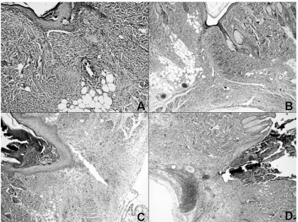
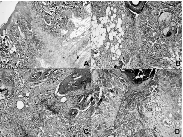
Histopathological evaluation
All samples were fixed in 10% formalin, embedded in paraffin, cut into 5 µm sections, and stained with hematoxylin-eosin (H&E). These sections were then examined under a light mi-croscope for histological changes by a blinded pathologist. Slides were scored for the presence of collagenization, vascu-larity, edema, and degree of acute and chronic inflammatory cells (0= none, 1= mild, 2= moderate and 3= severe). Mas-son’s trichrome was also applied for histochemical identifica-tion of collagenizaidentifica-tion. H&E and Masson’s trichrome stained
slides were reviewed by the same pathologist 3,28.
Morphometric analysis
Morphometric analysis was performed on Masson’s trichrome stained histological sections. The number of collagen bundles was measured with Leica, QWINPlus v.3.1.0 software using a Leica (DMLB-100S) microscope. Each slide was measured on one high-power field at x400 magnification including wound healing area; the mean number of collagen bundles of each
group was then calculated3,29.
Statistical analysis
The statistical analysis was performed using Statistical Pack-age for the Social Sciences (SPSS) version 16.0 for Windows (SPSS, Chicago, IL). For the scores and non-normally dis-tributed variables, comparison between groups was done by the Mann-Whitney U and the Kruskal Wallis test. The results were expressed as median (minimum-maximum). A p value < 0.05 after Bonferroni correction (p < 0.0125) was considered significant.
RESULTS
Three different methods were used to determine wound healing.
Breaking-strength findings
a significant difference between the control and Group L (p = 0.001), and Group B (p = 0.004). There was no significant difference between the control and the Group T (p = 0.029). The breaking-strength measurements between Group B and Group L (p = 0.336), Group B and Group T (p = 0.152), Group L and Group T (p = 0.021) were all similar (Table I).
Table I – The Breaking-Strength Measurements.
Group Force (gram.cm-2)
Group C 201.02 (145.12-230.53)
Group L 88.35 (37.46-165.51) *
Group B 124.88 (48.01-159.26) *
Group T 141.75 (105.35-209.52)
Median (min – max).
*: p < 0.0125 Compared with Group C, Mann Whitney U.
Morphometric findings
The second method was a morphometric analysis and colla-gen bundle counting. When all of the study groups were eval-uated according to the number of collagen bundles, we ob-served a significant difference between the control and Group L (p < 0.001), and Group B (p = 0.001). There was no signifi-cant difference between the control and Group T (p = 0.014).
Collagen bundle counts were significantly higher in Group T than in Group L (p = 0.001) and Group B (p = 0.004). There was no significant difference in the amount of collagen fibers between Group B and Group L (p = 0.338) (Table II).
Table II – Collagen Bundle Counts.
Group Collagen bundle counts
Group C 624 (494-710)
Group L 265 (221-492) * †
Group B 285 (261-510) * †
Group T 518,50 (420-591)
Median (min – max).
*: p < 0.0125 Compared with Group C, Mann Whitney U; †: p < 0.0125 Compa-red with Grup T, Mann Whitney U.
Histopathological findings
The third indicator of wound healing was determined by his-topathologic evaluation. When the working groups were com-pared in relation to edema, vascularity, inflammatory reaction and collagenization, there were significant differences be-tween the control group and Groups L and B. There was no significant difference between the control group and Group T. No significant differences were found between Groups B and L, Groups B and T and Groups L and T in terms of the histopathologic indicators of edema, vascularity, inflammatory reaction and collagenization (Table III).
Table III – Histopathologic Evaluation Scores.
Group Edema Vascularity
Inflamma-tory reaction
Collage-nization
Group C 1 (0-1) 0 (0-1) 1 (0-2) 3 (2-3)
Group L 2 (2-2)* 1 (1-3)* 2,5 (1-3)* 1 (1-2)*
Group B 2 (1-2)* 2 (1-3)* 3 (2-3)* 1 (1-2)*
Group T 1 (1-2) 1 (0-2) 2 (1-2) 2 (1-2)
Median (min – max).
*: p < 0.0125 Compared with Group C, Mann Whitney U.
DISCUSSION
Comparing the control group with the groups where bupi-vacaine and lidocaine were used for wound infiltration, col-lagen production was lower, breaking-strength measurements showed reduced resistance and significantly high edema, vascularity and inflammation scores were found. Between the control group and the tramadol group, there were no sig-nificant differences in collagen production, breaking-strength measurements, as well as edema, vascularity and inflamma-tion scores.
Surgical wound infiltration has been proven to be an effec-tive analgesic and is widely used for postoperaeffec-tive pain re-lief after abdominal hysterectomy, cesarean section, inguinal hernia repair, lumbar disc hernia, prostatectomy and similar
surgeries 1,5-8. Frequently used local anesthetic agents are
lidocaine, bupivacaine, ropivacaine and levobupivacaine 1,3-12.
Research on the effect of these and other local anesthetic agents used for wound infiltration on wound healing is limited
and results are controversial 3,28-31.
In a histopathologic study that included wound strength tests, rabbits were given 0.5% lidocaine, 2% lidocaine and 0.5% bupivacaine along a midline ventral abdominal wound. Comparing the control group and the test groups, there were no significant differences found in terms of wound tensile scores on any test. The same study emphasized that a com-parison of saline and local anesthetic infiltrated tissues found no significant difference in histopathologic results. They con-cluded that wound infiltration with lidocaine and bupivacaine had no effect on wound healing in midline abdominal incisions
in rabbits 28. Waite et al. 29 evaluated the effect of lidocaine
and bupivacaine on wound healing in mice and found that, although these anesthetics influenced local inflammation and
proteolytic factors, they had no effect on wound healing 29.
Other research found that lidocaine and bupivacaine inhib-ited collagen synthesis in fibroblast tissue cultures, and had
cytotoxic effects on different cell lines 30-35.
A study on guinea pig wound healing when given 1% lido-caine evaluated breaking strength, number of collagen fibers by morphometry, and histologic examination of collageniza-tion, edema, vascularity, and presence of acute and chronic in-flammatory cells. Comparisons with the control group showed that, though there was no significant difference in breaking strength measurements, the lidocaine group had significant vascularity and morphometric differences, as well as a lower
amount of collagen 30. While the application of lidocaine by
remained the same 30. Another study on the effects of local
anesthetic on human fibroblast found that lidocaine, bupiva-caine and ropivabupiva-caine produced dose-dependent cytotoxic
ef-fects on human fibroblast 31. Research on lidocaine in wound
infiltration of rats found collagenization and effects on mast
cell numbers in the wound 36. Aside from local anesthesia
affecting collagen fiber numbers and capillary veins, it may cause varying degrees of inflammation and edema along the
wound edges which can affect wound healing 3,37,38.
Past studies have found that the concentration of local an-esthetic affects wound healing while high concentrations of
local anesthetic delay healing 30,39,40. While doses inferior to
100 mcg.mL-1 of lidocaine had no effect on healing, in a study
of corneal epithelial cells, doses above 250 mcg.mL-1 delayed
epithelial healing in a dose-dependent fashion 41.
Tramadol may be used for peripheral nerve block and in
wound infiltration due to its anesthetic effects 14-20. No study
has been found evaluating the effects of tramadol on wound healing, which it is the aim of this study. In our search through the literature, we were not able to find any study that reports the histopathological and physical effects of tramadol on wound healing. We believe our study is the first to concentrate on this subject. Our aim is to evaluate the histopathological and band physical effects of tramadol on the healing of surgi-cal wounds when used for infiltration anesthesia.
While our study found results similar to previous studies on
the effects of bupivacaine and lidocaine on wound healing
30-41, no significant difference was found between tramadol and
the control group. The antibacterial properties of local anes-thetics and other agents used in wound infiltration are impor-tant. Previous research emphasized the antibacterial
proper-ties of bupivacaine 42-44. Controversy surrounds lidocaine
antibacterial properties though there are studies in the
litera-ture emphasizing its antibacterial properties 45. However,
bac-terial strains are not inhibited up to 2 hours after 1% lidocaine administration and when biopsy cultures are required within
two hours, lidocaine should be used 46. Previous research has
evaluated the antibacterial properties of tramadol 47.
Trama-dol exhibits dose and time dependent bactericidal activity for E. coli and S. epidermidis, and antibacterial against S. aureus
and P. aeruginosa strains. Researchers have emphasized
that tramadol may be helpful in reducing bacterial infection risk after local and regional anesthesia due to its antibacterial
properties 47. Our study found no trace of macroscopic
infec-tion at the wound site in any subject. This is in accordance with previous studies which emphasized the interaction of lo-cal anesthetic and tramadol’s antibacterial properties.
Local anesthetics are known for myotoxic effects and
when used in infiltration may cause myotoxicity 3,48.
Bupiva-caine carries that risk when used as a continuous peripheral
nerve block related to the duration of exposure 48,49. Lidocaine
also has myotoxic properties 50. However, tramadol, without
such effects, has been administered intramuscularly for many years 51.
REFERENCES
1. Ozyilmaz K, Ayoglu H, Okyay RD et al. – Postoperative Analgesic Effects of Wound Infiltration With Tramadol and Levobupivacaine in Lumbar Disk Surgeries. J Neurosurg Anesthesiol, 2012 Jul 2. [Epub ahead of print].
2. Beaussier M, Bouaziz H, Aubrun F et al.; les membres du comité dou-leur - ALR de la Sfar – Wound infiltration with local anesthetics for postoperative analgesia. Results of a national survey about its practi-ce in Franpracti-ce. Ann Fr Anesth Reanim, 2012;31:120-125.
3. Dere K, Sen H, Teksoz E et al. – The comparison of the effects of di-fferent doses of levobupivacaine infiltration on wound healing. J Invest Surg, 2009;22:112-116.
4. Johansson B Glise H, Hallerback B, Dalman P, Kristoffersson A – Pre-operative local infiltration with ropivacaine for postPre-operative pain relief after cholecystectomy. Anesth Analg, 1994;78:210-214.
5. Vigneau A, Salengro A, Berger J et al. – A double blind randomized trial of wound infiltration with ropivacaine after breast cancer surgery with axillary nodes dissection. BMC Anesthesiol, 2011;11:23. 6. Bari MS, Haque N, Talukder SA et al. – Postoperative pain relief
follo-wing inguinal hernia repair in children by wound infiltration with levo-bupivacaine. Mymensingh Med J, 2011;20:586-590.
7. Bilgin TE, Bozlu M, Atici S, Cayan S, Tasdelen B – Wound Infiltration with Bupivacaine and Intramuscular Diclofenac Reduces Postoperati-ve Tramadol Consumption in Patients Undergoing Radical Retropubic Prostatectomy: A Prospective, Double-blind, Placebo-controlled, Ran-domized Study. Urology, 2011;78:1281-1285.
8. Hernandez Palazon J, Tortosa Serrano JA, Burguillos Lopez S, Mo-lero MoMo-lero E – Infiltration of surgical wound with local anesthetic for postoperative analgesia in patients operated for lumbar disc hernia-tion: comparative study of ropivacaine and bupivacaine. Rev Esp Anestesiol Reanim, 2001;48:17-20.
9. Cnar SO, Kum U, Cevizci N, Kayaoglu S, Oba S – Effects of levobu-pivacaine infiltration on postoperative analgesia and stress respon-se in children following inguinal hernia repair. Eur J Anaesthesiol, 2009;26:430-434.
10. Sorbello M, Paratore A, Morello G et al. – Wound levobupivacaine continuous infusion for postoperative analgesia in living kidney do-nors: case-control study. Transplant Proc, 2009;41:1128-1131. 11. Kocabas S, Yedicocuklu D, Yuksel E, Uysallar E, Askar F – Infiltration
of the sternotomy wound and the mediastinal tube sites with 0.25% levobupivacaine as adjunctive treatment for postoperative pain after cardiac surgery. Eur J Anaesthesiol, 2008;25:842-849.
12. Memis D, Hekimoglu S, Kaya G, Atakan HI, Kaplan M – Efficacy of levobupivacaine wound infiltration with and without intravenous lor-noxicam for post-varicocoele analgesia: a randomized, double-blind study. Clin Drug Investig, 2008;28(6):353-359.
13. Lewis KS, Han NH – Tramadol: a new centrally acting analgesic. Am J Health Syst Pharm, 1997; 54: 643-52.
14. Khajavi MR, Aghili SB, Moharari RS et al. – Subcutaneous tramadol infiltration at the wound site versus intravenous administration after pyelolithotomy. Ann Pharmacother, 2009;43:430-435.
15. Kargi E, Babuccu O, Altunkaya H, Hosnuter M, Ozer Y, Babuccu B, Payasli C – Tramadol as a local anaesthetic in tendon repair surgery of the hand. J Int Med Res, 2008;36:971-978.
16. Kaki AM, Al Marakbi W – Post-herniorrhapy infiltration of tramadol versus bupivacaine for postoperative pain relief: a randomized study. Ann Saudi Med, 2008;28:165-168.
17. Demiraran Y, Ilce Z, Kocaman B, Bozkurt P – Does tramadol wound infiltration offer an advantage over bupivacaine for postoperati-ve analgesia in children following herniotomy? Paediatr Anaesth, 2006;16:1047-1050.
18. Altunkaya H, Ozer Y, Kargi E et al. – The postoperative analgesic effect of tramadol when used as subcutaneous local anesthetic. Anes-th Analg, 2004;99:1461-1464.
19. Altunkaya H, Ozer Y, Kargi E, Babuccu O – Comparison of local ana-esthetic effects of tramadol with prilocaine for minor surgical procedu-res. Br J Anaesth, 2003;90:320-322.
20. Desmeules JA, Piguet V, Collart L, Dayer P – Contribution of mono-aminergic modulation to the analgesic effect of tramadol. Br J Clin Pharmacol, 1996;41:7-12.
21. Yurtlu S, Hanci V, Kargi E et al. – The analgesic effect of dexketo-profen when added to lidocaine for intravenous regional anaesthesia: a prospective, randomized, placebo-controlled study. J Int Med Res, 2011;39:1923-1931.
22. Memis D, Turan A, Karamanlioglu B, Tükenmez B, Pamukçu Z – The effect of tramadol or clonidine added to intraperitoneal bupivacaine on postoperative pain in total abdominal hysterectomy. J Opioid Manag, 2005;1:77-82.
23. Tsai YC, Chang PJ, Jou IM – Direct tramadol application on sciatic nerve inhibits spinal somatosensory evoked potentials in rats. Anesth Analg, 2001; 92:1547-1551.
24. Acalovschi I, Cristea T, Margarit S, Gavrus R – Tramadol added to lidocaine for intravenous regional anesthesia. Anesth Analg, 2001;92:209-214.
25. Robaux S, Blunt C, Viel E et al. – Tramadol added bupivicaine, mor-phine and tramadol in rats. Agri, 2004;16:53-58.
26. Jou IM, Chu KS, Chen HH, Chang PJ, Tsai YC – The effect of intrathe-cal tramadol on spinal somatosensory-evoked potentials and motor-evoked responses in rats. Anesth Analg, 2003;96:783-788.
27. Guven M, Mert T, Gunay I – Effects of tramadol on nerve action po-tentials in rats: comparisons with benzocaine and lidocaine. Int J Neu-rosci, 2005;115:339-349.
28. Vasseur PB, Paul HA, Dybdal N, Crumley L – Effects of local anes-thetics on healing of abdominal wounds in rabbits. Am J Vet Res, 1984;45:2385-2388.
29. Waite A, Gilliver SC, Masterson GR, Hardman MJ, Ashcroft GS – Cli-nically relevant doses of lidocaine and bupivacaine do not impair cuta-neous wound healing in mice. Br J Anaesth, 2010;104:768-773. 30. Drucker M, Cardenas E, Arizti P, Valenzuela A, Gamboa A–
Experi-mental studies on the effect of lidocaine on wound healing. World J Surg, 1998;22:394-397; discussion 397-398.
31. Fedder C, Beck-Schimmer B, Aguirre J et al. – In vitro exposure of human fibroblasts to local anaesthetics impairs cell growth. Clin Exp Immunol, 2010;162:280-288.
32. Desai SP, Kojima K, Vacanti CA, Kodama S – Lidocaine inhibits NIH-3T3 cell multiplication by increasing the expression of cyclin-depen-dent kinase inhibitor 1A (p21). Anesth Analg, 2008;107:1592-1597. 33. Scherb MB, Han SH, Courneya JP, Guyton GP, Schon LC – Effect of
bupivacaine on cultured tenocytes. Orthopedics, 2009;32:26. 34. Harris KL, Bainbridge NJ, Jordan NR, Sharpe JR – The effect of
to-pical analgesics on ex vivo skin growth and human keratinocyte and fibroblast behavior. Wound Repair Regen, 2009;17:340-346. 35. Sturrock JE, Nunn JF – Cytotoxic effects of procaine, lignocaine and
bupivacaine. Br J Anaesth, 1979;51:273-281.
36. Rodrigues FV, Hochman B, Wood VT, Simões MJ, Juliano Y, Ferreira LM – Effects of lidocaine with epinephrine or with buffer on wound healing in rat skin. Wound Repair Regen, 2011;19:223-228. 37. Field FK, Kerstein MD – Overview of wound healing in a moist
envi-ronment (review). Am J Surg, 1994;167:2S-6S.
38. Luostarinen V, Evers H, Lytikainen MT et al. – Antitrombotic effects of lidocaine and related compounds on laser induced microvascular injury. Acta Anesth Scand, 1981;9:25-28.
39. Morris T, Appleby R – Retardation of wound healing by procaine. Br J Surg, 1980;67:391-395.
41. Bisla K, Tanelian DL – Concentration-dependent effects of lidocai-ne on corlidocai-neal epithelial wound healing. Invest Ophthalmol Vis Sci, 1992;33:3029-3033.
42. Rosenberg PH, Renkoven OV – Antimicrobial activity of bupivacaine and morphine. Anesthesiology, 1985;62:178-179.
43. Sakuragi T, Ishino H, Dan K – Bactericidal activity of 0.5 % bupivacai-ne with presentatives on microorganisms in the human skin flora. Reg Anesth, 1997;22:178-184.
44. Hodson M, Gajraj R, Scott NB – A comparison of the antibacterial acti-vity of levobupivacaine vs. bupivacaine: an in vitro study with bacteria implicated in epidural infection. Anesthesia, 1999;54:683-702. 45. Sedef Gocmen J, Buyukkocak U, Caglayan O, Aksoy A – In vitro
antibacterial effects of topical local anesthetics. J Dermatolog Treat, 2008;19:351-353.
46. Berg JO, Mössner BK, Skov MN, Lauridsen J, Gottrup F, Kolmos HJ – Antibacterial properties of EMLA and lidocaine in wound tissue biop-sies for culturing. Wound Repair Regen, 2006;14:581-585.
47. Tamanai-Shacoori Z, Shacoori V, Jolivet-Gougeon A et al. – The an-tibacterial activity of tramadol against bacteria associated with infec-tious complications after local or regional anesthesia. Anesth Analg, 2007;105:524-527.
48. Zink W, Seif C, Bohl JR et al. – The acute myotoxic effects of bupiva-caine and ropivabupiva-caine after continuous peripheral nerve blockades. Anesth Analg, 2003;97:1173-1179.
49. Nouette-Gaulain K, Bringuier S, Canal-Raffin M et al. – Time course of mitochondrial metabolism alterations to repeated injections of bupi-vacaine in rat muscle. Can J Anaesth, 2010;57836-57842.
50. Foster AH, Carlson BM – Myotoxicity of local anesthetics and rege-neration of the damaged muscle fibers. Anesth Analg, 1980;59:727-736.