ABSTRACT
Inluence of concentration, time and method of
application of citric acid and sodium citrate in
root conditioning
Rodrigo CAVASSIM1, Fábio Renato Manzolli LEITE2, Daniela Leal ZANDIM3, Andrea Abi Rached DANTAS4, Ricardo
Samih Georges Abi RACHED5, José Eduardo Cezar SAMPAIO6
1- DDS, MSc, PhD student, Department of Diagnostic and Surgery, School of Dentistry of Araraquara, UNESP - Univ. Estadual Paulista, Araraquara, SP, Brazil. 2- DDS, MSc, PhD, Adjunct Professor, Department of Semiology and Clinics, Dental School, Federal University of Pelotas - UFPel, Pelotas, RS, Brazil. 3- DDS, MSc, PhD, Department of Diagnostic and Surgery, School of Dentistry of Araraquara, UNESP - Univ. Estadual Paulista, Araraquara, SP, Brazil. 4- DDS, MSc, PhD, Assistant Professor, UNIARA Dental School, Araraquara, SP, Brazil.
5- DDS, PhD, Full Professor, Department of Diagnostic and Surgery, School of Dentistry of Araraquara, UNESP - Univ. Estadual Paulista, Araraquara, SP, Brazil. 6- DDS, PhD, Adjunct Professor, Department of Diagnostic and Surgery, School of Dentistry of Araraquara, UNESP - Univ. Estadual Paulista, Araraquara, SP, Brazil.
Corresponding address: Prof. Dr. José Eduardo Cezar Sampaio - Faculdade de Odontologia de Araraquara - UNESP - Departamento de Diagnóstico e Cirurgia Rua Humaitá, 1680 Araraquara - 14801-903 - SP - Brasil - Phone: +55 16 3301-6374 - Fax: +55 16 3301-6369 - e-mail: [email protected]
Received: October 28, 2010 - Modiication: August 5, 2011 - Accepted: September 5, 2011
O
bjective: The aim of this study was to establish the parameters of concentration, time and mode of application of citric acid and sodium citrate in relation to root conditioning. Material and Methods: A total of 495 samples were obtained and equally distributed among 11 groups (5 for testing different concentrations of citric acid, 5 for testing different concentrations of sodium citrate and 1 control group). After laboratorial processing, the samples were analyzed under scanning electron microscopy. A previously calibrated and blind examiner evaluated micrographs of the samples. Non-parametric statistical analysis was performed to analyze the data obtained. Results: Brushing 25% citric acid for 3 min,promoted greater exposure of collagen ibers in comparison with the brushing of 1% citric
acid for 1 minute and its topical application at 1% for 3 min. Sodium citrate exposed
collagen ibers in a few number of samples. Conclusion: Despite the lack of statistical signiicance, better results for collagen exposure were obtained with brushing application of
25% citric acid for 3 min than with other application parameter. Sodium citrate produced a few number of samples with collagen exposure, so it is not indicated for root conditioning.
Key words: Periodontics. Dental scaling. Smear layer. Collagen.
INTRODUCTION
Cementum is a mineralized tissue with primary function to insert the ligament fibers on the root surface and releasing oclusal forces to the surrounding alveolar bone. There is considerable interest in observing the changes that occur on the cementum surface inside periodontal pockets as a result of periodontal disease14,21.
Periodontitis-affected root surfaces are hypermineralized and contaminated with endotoxins and other biologically active substances1,20,25. It has been
suggested that endotoxin present in the cement could impair periodontal healing and should be removed to promote a more biologically
acceptable surface than the one obtained only after scaling and root planning7.
The most important event in the reattachment of the connective tissue is related to the adhesion of blood elements to the collagen present at the root surface, which retards the apical migration of the sulcular epithelium into the pocket13,26. ephitelial downgrowth is exacerbated
by the strong adhesion of bacterial products and endotoxins, especially originated from Gram negative bacteria. These bacterial compounds
have afinity to mineral structures such as the
cell wall lipopolysaccharides remain active even after bacterial death, not being removed by scaling and root planning1,14. These bacteria product
are found bound to the smear layer produced by the action of curettes, ultrasonic and rotary instruments1,3.
In an attempt to remove smear layer and demineralize the contaminated root surface, different approaches has been studied as the use of chemical agents and laser12,22,24,28.
Special attention has been focused on the use of chemical agents as a mean to obtain adequate preparation of the root to development of new connective attachment. Certain acids, especially citric acid has been used to clean the root surface by demineralization and, recently, some authors showed that demineralization of exposed dentin
can signiicantly increase the reattachment of
connective tissue to the root surface9,10.
Previous studies on tissue regeneration have
used tetracycline hydrochloride HCl to clean
the root surface because its bactericidal and demineralizing effect9,25. The parameters for root conditioning with tetracycline HCl were established
by Ishi, et al.12 (2008). However, the search for
a less acid substance to avoid tissue necrosis and probably provide better conditions for clot adhesion to the root surface, showed promising results employing ethylenediaminetetraacetic acid (eDTA)15,29. However the outcomes were not
so favorable in clinical trials. The use of eDTA gel as a root surface conditioning agent negatively affected the outcome of root coverage3. eDTA
might have inhibited blood element adsorption and adhesion to the dentin surface because of a possible incomplete removal of the gel from the root surface. In addition, eDTA is a calcium chelator; therefore, its residues may have inhibited or retarded coagulation events17. Citric acid is a
substance such capable of removing smear layer and opening dentinal tubules18,28. However, citric acid’s low pH may induce cytotoxic effects when
in contact with connective tissue16.
In an attempt to ind a substance that could
efficiently remove smear layer and expose
collagen ibers, Leite, et al.18 (2010) tested the sodium citrate in the clot stabilization. However,
this substance has not been tested before and the application parameters are not established yet.
In order to establish the application parameters for sodium citrate and ascertain that this substance is capable of smear layer removal, we suggested conducting this study. In addition, as citric acid presented better results than eDTA for clot stabilization18 and as citric acid combined with
platelet-derived growth factor-BB showed better results than eDTA and tetracycline hydrochloride on attachment of periodontal ligament cells on
root surfaces2, we proposed to test if a lower
concentration of citric acid applied by a shorter time is capable of removing smear layer and
exposing collagen iber. The aim of this study was
to establish concentrations, times and modes of ideal applications of citric acid and sodium citrate in removing smear layer and exposure of collagen
ibers.
MATERIAL AND METhODS
A total of 124 periodontally involved human teeth were obtained at the Oral and Maxillofacial Surgery and Periodontics clinics at the Araraquara School of Dentistry, UNeSP – Univ. estadual Paulista, Brazil. This study was approved by the
institutional Human Ethics Committee.
Sample preparation
The samples were prepared according to previous published data12 and briely described
as follows. For sample preparation the buccal and lingual root surfaces of each tooth were used. Two parallel grooves with approximately 0.8 mm deep were made using a high speed cylindrical bur (#3099 - Ø 1.6 mm) (KG Sorensen, Medical Burs, Cotia, SP, Brazil) under copious irrigation. One groove was made at the cementoenamel junction and another one approximately 3 mm
distant from the irst, in the apical direction. The
same bur was used to remove the surface layer of the root between the two grooves.
In order to create a smear layer, 50 apical to cervical strokes were performed using a sharp
#5-6 Gracey curette (Hu-Friedy, Hu-Friedy,
Chicago, IL, USA). A total of 495 samples measuring about 2x3 mm were obtained cutting
teeth with a lexible double faced diamond disc
(#7020 – Ø 0.22 mm – thickness: 0.15 mm) (KG Sorensen, Medical Burs, Cotia, SP, Brazil) at low speed. The samples were equally distributed among the groups.
The citric acid was applied in ive concentrations 0.5, 1, 2, 15 and 25% (ive groups n=45 samples
each). The sodium citrate was also divided into
ive groups with the following concentrations 3,
10, 20, 30 and 40% (n=45 each). The control group was represented by conditioning with saline solution (n=45).
Five different concentrations were tested in order to determine if lower concentrations, and consequently less aggressive substances, could be effective in removing smear layer and exposing
collagen ibers. In the same way, different modes
and times of application were evaluated in an attempt to reduce the contact of the substances with the periodontal cells.
according to the mode and time of application solutions. Three modes of application were used (n=15 each): 1) simple positioning of a small cotton pellet embedded in solution (passive application); 2) brushing application with a soft brush (Disposable Brush Tips Ø2, 3M eSPe, Seefeld, Germany); and 3) burnishing application (friction) with a small cotton pellet. each of these three subgroups was further divided into the three application periods of 1, 2, or 3 min (n=5 each).
Samples were dehydrated in an increasingly graded series of ethanol: 30, 50, 70, 80, 95 and 100%. Then, the samples were dried overnight in a dehydration jar (Corning, Corning Life Sciences, São Paulo, SP, Brazil), mounted on metallic stubs (Senai, São Paulo, SP, Brazil) and sputter-coated with a thin 25 nm layer of 99.99% pure gold (Balt-Tec SCD-050, Balt-Tec, Gnathole Farm,
Kettleshulme, High Peak, Cheshire, UK).
SEM evaluation
Two micrographs were obtained from the center area of each sample with 1,500x and
3,500x magniications, using a scanning electron
microscope operated at an accelerating voltage of 20 kV (Jeol T330 A; Jeol Ltd., Peabody, MA, USA). The micrographs were evaluated according to a
root surface modiication index adapted for this
study. A previously calibrated (kappa score=0.93) and experienced examiner18 evaluated three times
each image, with an interval of 15 days between each evaluation. The score attributed to each sample was the most prevalent score in the three evaluations. The adapted index used for this study
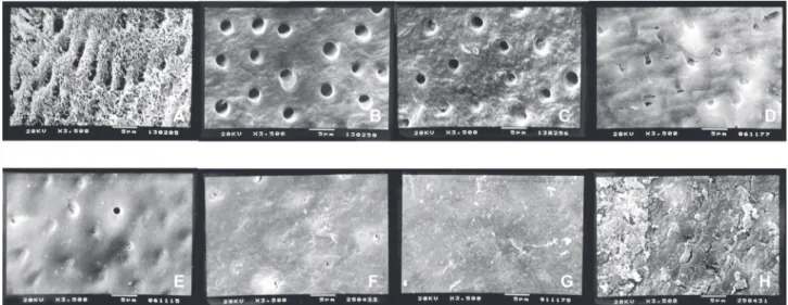
consisted of eight scores as shown in Figure 1.
Statistical analysis
The non-parametric analysis of variance (Kruskal-Wallis test) was applied to independently evaluate the effect of the three dependent variables: solution concentration, mode of application and period. The test was performed separated for both the substances evaluated.
The level of signiicance adopted was 5%. If p≤0.05, Dunn’s Multiple Comparison post hoc
test was applied to detect statistically signiicant
differences among groups. Statistical analysis was made with a computer software (GraphPad Prism 5.00; GraphPad Software Inc., San Diego, CA, USA).
RESULTS
Citric acid
Citric acid was evaluated at concentrations of 0.5, 1, 2, 15 and 25%. All concentrations were used for 1, 2 and 3 min under the passive, brushing or burnishing forms.
Statistically significant differences were found between control group and the other concentrations used (Table 1). even without
statistical signiicance, the concentration of 25%
and 15% showed less variation in score values (Figure 2), the same way that the concentration of 25% presented more samples with score 1 (Table 1). Score 1 represents complete smear
layer removal with dentin collagen ibers exposure
and complete opened dentin tubules, without
Figure 1- Root Modiication Index. A) Score 1. Complete smear layer removal with dentin collagen iber exposure.
Completely open dentinal tubules, without smear layer on root surface. B) Score 2. Complete smear layer removal. No
collagen iber exposure. Completely open dentin tubules. C) Score 3. Traces of smear layer remaining in the openings
smear layer on root surface. This is the aim of the chemical root conditioning (Figure 1).
The comparison of the different periods of application of citric acid, showed no differences among the groups (Figure 3). Furthermore, Table
1 shows a slight tendency of the 3-min application
to be more effective in exposing collagen ibers
(score 1).
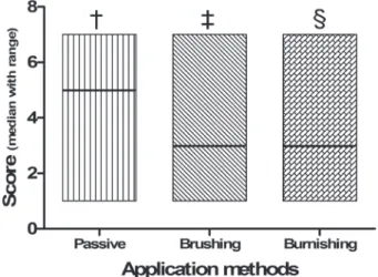
The same way, the evaluation of the different modes of application showed no significant
Citric acid concentration
Control 0.5% 1% 2% 15% 25%
n 45 45 45 45 45 45
Median/Mean (SD) 6.00/6.00 (0.78)
5.00/5.00 (1.20)
5.00/4.80 (1.40)
5.00/4.60 (0.75)
5.00/4.50 (1.30)
5.00/3.80 (1.80)
p Value A B B B B B
Number of samples with score 1 (%)
0 0 3 (6.67) 0 4 (8.89) 12 (26.67)
Periods of application
1 minute 2 minutes 3 minutes
n 90 90 90
Median/Mean (SD) 5.00/5.00 (1.60) 5.00/4.80 (1.00) 5.00/4.60 (1.60)
p Value NS NS NS
Number of samples with score 1 (%)
7 (7.78) 4 (4.44) 8 (8.89)
Modes of application
Passive Brushing Burnishing
n 90 90 90
Median/Mean (SD) 5.00/4.90 (1.50) 5.00/4.50 (1.70) 5.00/5.00 (0.79)
p Value NS NS NS
Number of samples with score 1 (%)
4 (4.44) 14 (15.55) 1 (1.11)
Kruskal-Wallis test and Dunn´s Multiple Comparison post hoc test. p=0.05
* Within the same category, scores with the same letter are not statistically different. SD= standard deviation
Table 1- Sample distribution for the citric acid concentrations, periods and modes of application
Figure 2- Median with range of the scores obtained for
the ive concentrations of citric acid and the control group.
Concentrations with the same symbol are not statistically different
difference between groups (Figure 4). However,
application by brushing seems to have favored the production of samples with more exposure of
collagen ibers as seen in Table 1.
Sodium citrate
The effect of sodium citrate in smear
layer removal was evaluated in ive different
concentrations and in a control group. Among the concentrations evaluated, the 3% showed less effective results than the others and
signiicant statistical difference was observed. The
concentrations of 10, 20, 30 and 40% showed no
statistically signiicant differences among them
(Figure 5). Table 2 shows the median values and standard deviation (SD) for the concentrations, periods of applications and modes of application.
The analysis of the application periods showed significant differences among the groups.
even though, the results for 2 and 3 min were statistically the same, the 3-min application seems to have been slightly better than others (Figure 6).
Regarding the mode of application, more favorable results were obtained with application by brushing or by burnishing than the passive
application (Figure 7). A statistically signiicant
difference was observed among the modes of application and a slight tendency of best results can be seen with the application by burnishing (Table 2).
Figure 4- Median with range of the scores obtained for the three application methods of the citric acid. Application methods with the same symbol are not statistically different
Figure 5- Median with range of the scores obtained for
the ive concentrations of sodium citrate and the control
group. Concentrations with the same symbol are not statistically different
Figure 7- Median with range of the scores obtained
for the three application methods of the sodium citrate. Application methods with the same symbol are not statistically different
DISCUSSION
With the methodology used in this study it is possible to evaluate the morphological aspect of the conditioned root surface. The micrographs
taken at 1,500 and 3,500x has excellent deinition
and quality for observation of the root surface. This methodology has been successfully used for this kind of evaluation12,17,18,24.
The SeM micrographs should represent the evaluated sample, but it is not possible to take a micrograph of the entire sample. To solve this
dificulty, the visualization was directed to the
center of the sample and all the micrographs were taken from the center of the sample.
Larger samples take longer to be sputter-coated and are difficult to be obtained. The samples used in this study were about 2x3 mm. These dimensions were obtained after sample reduction from the coronal root third and comprise approximately the measure between the cementoenamel junction and the furcation entrance in molars.
Despite the limitations presented, the methodology in this study has been extensively used before and is accepted as a method to
evaluate root conditioning and blood cell adhesion with in vitro studies12,17,18,22,24.
The use of different concentrations, application modes and times was efficient in determine and standardize the parameters of application of a chemical substances and allows the use of the obtained parameters in other studies such as studies to evaluate blood cell adhesion and periodontal ligament cell attachment to the root surface2,18.
The rationale for this study is that the evident contamination of the cementum and dentin with bacterial toxins could impair periodontal healing1. These products may be responsible for
a marked progressive and irreversible destruction of the periodontal structures of support. Scaling and root planning can be accomplished by manual instruments, rotary instruments, sonic and ultrasonic instruments. Such methods often do not appear to be totally effective in removing mineralized debris of the root surface. Furthermore, root instrumentation causes grooves and results in a root surface covered with smear layer, which contains remnants of dental calculus, contaminated cementum, bacterial endotoxin and subgingival plaque5,7,19.
Sodium citrate concentration
Control 3% 10% 20% 30% 40%
n 45 45 45 45 45 45
Median/Mean (SD) 6.00/6.00 (0.78)
5.00/5.00 (1.70)
3.00/3.80 (1.10)
3.00/3.20 (1.50)
3.00/3.20 (1.40)
3.00/3.60 (0.96)
p Value A B C C C C
Number of samples with score 1 (%)
0 0 0 3 (6.67) 4 (8.89) 0
Periods of application
1 minute 2 minutes 3 minutes
n 90 90 90
Median/Mean (SD) 5.00/4.70 (1.40) 5.00/4.20 (1.70) 3.00/3.40 (1.40)
p Value A A B
Number of samples with score 1 (%)
0 3 (3.33) 4 (4.45)
Modes of application
Passive Brushing Burnishing
n 90 90 90
Median/Mean (SD) 5.00/4.80 (1.60) 3.00/4.10 (1.50) 3.00/3.40 (1.60)
p Value A B C
Number of samples with score 1 (%)
1 (1.11) 2 (2.22) 4 (4.45)
Kruskal-Wallis test and Dunn´s Multiple Comparison post hoc test. p=0.05
* Within the same category, scores with the same letter are not statistically different. SD= standard deviation
Over the past decade a number of alternatives have been presented as compensation for the limitations inherent to the mechanical therapy.
In vitro11 and in vivo studies9,10 have emphasized
the conditioning of the root surface using different methods as an adjuvant treatment to scaling and root planning in regenerative procedures.
Several chemical agents have been proposed including citric acid10,23,27-29, eDTA4-8,17,23,24,
tetracycline hydrochloride12,13,15 among others.
An important factor that should be considered and is often neglected is the mode of application, time, composition and concentration of the
conditioning agent in addition to the pH of the substance used. Root surfaces exposed to bioilm
are hypermineralized20,25 and therefore more dificult to be both mechanically and chemically decontaminated, and thus, low pH substances
were proposed to aid smear layer removal.
With respect to pH, it was possible to
demonstrate that citric acid which possesses a
low pH, causes cell death when in contact with
periodontal ligament (PDL) cells16 and thus could
delay cell proliferation and repair of the area. Because of this necrotizing effect, the use of 24%
EDTA at neutral pH was proposed as an alternative for low pH substances6. This way, studies were
carried out to compare different formulations, concentrations, modes and length/duration of the application. Most studies have shown that eDTA is a substance that provides smear layer removal
and exposure of collagen ibers4,5,7,8,24. However,
researches on clot adherence to root surface bioengineered with eDTA obtained worse results when compared to citric acid and tetracycline hydrochloride18. This may be because eDTA is
an anticoagulant or even because it removes the calcium ions from the surface of root dentin that are important in blood clotting cascade18.
Considering these results, a new substance was proposed in the present study - sodium citrate - which has the same principle of eDTA, but is more biocompatible and is also currently used in blood collection bags. It was also evaluated the citric acid different concentrations in order to ascertain the optimal parameters for its use. Actually, the
problem of this substance is its low pH and its high
power to demineralization. This way, in the present study lower concentrations with consequently
higher pH were evaluated.
This high demineralization power can be observed after analysis of the SeM micrographs, in which we found that almost all groups of citric acid concentrations of 15, 20, 25 and 30% can cause chemical dissolution of tooth surface (score 5) with collagen destruction. These results disagrees with the majority of studies published10,15,27,28, not being
compatible with the principle of root conditioning,
which requires the exposure of dentin collagen
ibers for adhesion of the clot on the tooth surface.
Thus, these results suggest that the concentration should be decreased so that the smear layer could be effectively removed but without destroying the
collagen ibers network. This way, the citric acid
concentration was reduced to 0.5, 1.0 and 2.0% to try to solve the problem, but no difference was observed. Citric acid even at lower concentrations produced excessive demineralization not having a standard of performance (Table 1).
Despite being more biocompatible, sodium
citrate was not able to expose collagen ibers of
the root surface (Table 1) even in the group with best results, which was vigorous application for 3 min (Graphs 5 and 6).
CONCLUSION
Within the limitations of the methodology, it can be concluded that, despite the lack of statistical
signiicance, the best results for collagen exposure
using citric acid were obtained with brushing application at 25% for 3 min. Sodium citrate was not able to adequately remove smear layer an
expose collagen ibers, so it is not indicated for
root conditioning.
REFERENCES
1- Aleo JJ, De Renzis FA, Farber PA, Varboncoeur AP. The presence and biologic activity of cementum-bound endotoxin. J Periodontol. 1974;45(9):672-5.
2- Becerik S, Sonmez S, Sen BH, Deliloglu-Gurhan I, Evrenosoglu
e. effect of human platelet-derived growth factor-BB on attachment of periodontal ligament cells on root surfaces. Saudi Med J. 2009;30(1):60-6.
3- Bittencourt S, Ribeiro edel P, Sallum eA, Sallum AW, Nociti
FH Jr, Casati MZ. Root surface biomodiication with EDTA for
the treatment of gingival recession with a semilunar coronally
repositioned lap. J Periodontol. 2007;78(9):1695-701.
4- Blomlöf J, Blomlöf L, Lindskog S. Effect of different concentrations
of eDTA on smear removal and collagen exposure in periodontitis-affected root surfaces. J Clin Periodontol. 1997;24(8):534-7.
5- Blomlöf J, Jansson L, Blomlöf L, Lindskog S. Root surface etching at neutral pH promotes periodontal healing. J Clin Periodontol.
1996;23(1):50-5.
6- Blomlöf J, Lindskog S. Periodontal tissue-vitality after different
etching modalities. J Clin Periodontol. 1995;22(6):464-8.
7- Blomlöf JP, Blomlöf LB, Lindskog SF. Smear layer formed
by different root planing modalities and its removal by an ethylenediaminetetraacetic acid gel preparation. Int J Periodontics Restorative Dent. 1997;17(3):242-9.
8- Blomlöf JP, Blomlöf LB, Lindskog SF. Smear removal and collagen
exposure after non-surgical root planing followed by etching with an eDTA gel preparation. J Periodontol. 1996;67(9):841-5. 9- Cole RT, Crigger M, Bogle G, egelberg J, Selvig KA. Connective tissue regeneration to periodontally diseased teeth. A histological study. J Periodontal Res. 1980;15(1):1-9.
11- Fardal O, Lowenberg BF. A quantitative analysis of the migration, attachment, and orientation of human gingival
ibroblasts to human dental root surfaces in vitro. J Periodontol. 1990;61(8):529-35.
12- Ishi EP, Dantas AA, Batista LH, Onofre MA, Sampaio JE. Smear layer removal and collagen iber exposure using tetracycline
hydrochloride conditioning. J Contemp Dent Pract. 2008;9(5):25-33.
13- Isik AG, Tarim B, Hafez AA, Yalçin FS, Onan U, Cox CF.
A comparative scanning electron microscopic study on the characteristics of demineralized dentin root surface using
different tetracycline HCl concentrations and application times. J
Periodontol. 2000;71(2):219-25.
14- Jones WA, O'Leary TJ. The effectiveness of in vivo root planing in removing bacterial endotoxin from the roots of periodontally involved teeth. J Periodontol. 1978;49(7):337-42.
15- Lafferty TA, Gher Me, Gray JL. Comparative SeM study on
the effect of acid etching with tetracycline HCl or citric acid on
instrumented periodontally-involved human root surfaces. J Periodontol. 1993;64(8):689-93.
16- Lan WC, Lan WH, Chan CP, Hsieh CC, Chang MC, Jeng JH. The
effects of extracellular citric acid acidosis on the viability, cellular adhesion capacity and protein synthesis of cultured human gingival
ibroblasts. Aust Dent J. 1999;44(2):123-30.
17- Leite FR, Moreira CS, Theodoro LH, Sampaio JE. Blood cell
attachment to root surfaces treated with eDTA gel. Braz Oral Res. 2005;19(2):88-92.
18- Leite FR, Sampaio Je, Zandim DL, Dantas AA, Leite eR, Leite
AA. Inluence of root-surface conditioning with acid and chelating
agents on clot stabilization. Quintessence Int. 2010;41(4):341-9.
19- Leknes KN, Lie T, Wikesjö UM, Bogle GC, Selvig KA. Inluence
of tooth instrumentation roughness on subgingival microbial colonization. J Periodontol. 1994;65(4):303-8.
20- Madison JG 3rd, Hokett SD. The effects of different tetracyclines
on the dentin root surface of instrumented, periodontally involved human teeth: a comparative scanning electron microscope study. J Periodontol. 1997;68(8):739-45.
21- Nightingale SH, Sheridan PJ. Root surface demineralization
in periodontal therapy: subject review. J Periodontol. 1982;53(10):611-6.
22- Oliveira GJ, Sampaio Je, Marcantonio RA. effects of er,Cr:YSGG laser irradiation on root surfaces for adhesion of blood components and morphology. Photomed Laser Surg. 2010;28(6):751-6. 23- Ruggeri A Jr, Prati C, Mazzoni A, Nucci C, Di Lenarda R, Mazzotti G, et al. effects of citric acid and eDTA conditioning on exposed root dentin: an immunohistochemical analysis of collagen and proteoglycans. Arch Oral Biol. 2007;52(1):1-8.
24- Sampaio JE, Theodoro LH, Correa MA, Mendes AJ. A
comparative SeM study of smear layer removal by detergents and eDTA on the root surface. Int J Periodontics. Restorative Dent. 2005;25(2):157-63.
25- Shetty B, Dinesh A, Seshan H. Comparitive effects of
tetracyclines and citric acid on dentin root surface of periodontally involved human teeth: a scanning electron microscope study. J Indian Soc Periodontol. 2008;12(1):8-15.
26- Steinberg AD, Willey R. Scanning electron microscopy observations of initial clot formation on treated root surfaces. J Periodontol. 1988;59(6):403-11.
27- Sterrett JD, Bain C. Citric acid burnishing of dentinal root surfaces. a preliminary scanning electron microscopy report. J Can Dent Assoc. 1987;53(5):395-7.
28- Sterrett JD, Bankey T, Murphy HJ. Dentin demineralization.
The effects of citric acid concentration and application time. J Clin Periodontol. 1993;20(5):366-70.
29- Terranova VP, Franzetti LC, Hic S, DiFlorio RM, Lyall RM, Wikesjö
UM, et al. A biochemical approach to periodontal regeneration:
tetracycline treatment of dentin promotes ibroblast adhesion and