DOI: http://dx.doi.org/10.5007/1980-0037.2012v14n1p52
original article
CC Licence
Creative Commom
Efect of diferent frequencies weekly
training on parameters of oxidative stress
Efeito de diferentes frequências semanais de
treinamento sobre parâmetros de estresse oxidativo
Camila Baumer Tromm1
Guilherme Laurentina da Rosa1
Karoliny Bom1, Izadora Mariano1
Bruna Pozzi1, Talita Tuon1
Luciano Acordi da Silva1
Ricardo Aurino de Pinho1
Abstract – Intense muscle contraction induced by physical exercise increases the produc-tion of reactive oxygen species, which causes oxidative stress in several organs, such as the liver and the heart. Physical training may increase antioxidative defenses and decrease oxidative stress. However, it is not clear what training frequency improves oxidative stress parameters. his study evaluated the efect of training two and three times a week on oxidative stress biomarkers in the liver and the heart. Eighteen young male mice (CF1) weighing 30 to 35 g were divided into three groups (n=6): no training (NT); twice a week training (T2); and three times a week training (T3). he training program lasted eight weeks, and the animals were killed 48 hours ater the last training session. he liver and the heart were removed and stored at -70o C. he following analyses were conducted: thiobarbituric acid reactive substances, protein carbonylation, total thiol content, supero-xide dismutase, catalase and glutathione peroxidase. Oxidative damage was reduced only in the T3 group, and there was an increase in total thiol content, supeoxidase dismutase and catalase in T3 when compared with the NT group. Glutathione peroxidase was not signiicantly diferent between groups. Only training three times a week seemed to reduce oxidative stress and increase the eiciency of the antioxidant system in mice.
Key words:Antioxidant enzymes; Oxidative damage; Physical exercise; Training frequency.
Resumo – Durante a contração muscular intensa induzida pelo exercício físico, há aumento na produção de espécies reativas de oxigênio, ocasionando estresse oxidativo em diversos órgãos, dentre eles o fígado e o coração. O treinamento físico pode aumentar as defesas antioxidantes e diminuir o estresse oxidativo. Contudo, ainda existem dúvidas sobre a frequência de treinamento necessária para melhorar parâmetros de estresse oxidativo. Este trabalho tem como objetivo veriicar o efeito das frequências de duas e três vezes de exercício por semana sobre biomarcadores de estresse oxidativo no fígado e coração. Foram utilizados 18 camundongos machos (CF1), jovens (30 a 35g) divididos em grupos (n=6/ grupo): não treinado (NT); treinado duas vezes por semana (T2) e treinado três vezes por semana (T3). Os animais foram submetidos ao treinamento durante oito semanas. Quarenta e oito horas após a última sessão os animais foram sacriicados. O fígado e o coração foram removidos e armazenados em freezer – 70ºC. Foram analisadas as substâncias reativas ao ácido tiobarbitúrico, carbonilação de proteínas, conteúdo total de tióis, atividades da superóxido dismutase , catalase e glutationa peroxidase. Os resultados demonstraram que apenas o grupo T3 reduziu dano oxidativo. Ademais, houve aumento no conteúdo total de tióis, atividades da superóxido dismutase e catalase no mesmo grupo em comparação com o não treinado. A atividade da glutationa peroxidase não apresentou diferença signiica-tiva entre os grupos. Este estudo demonstrou que somente a frequência de treinamento de três vezes por semana reduz dano oxidativo e aumenta a eiciência do sistema enzimático
1 Universidade do Extremo Sul Catarinense. Programa de Pós Graduação em Ciências da Saúde. Laboratório de Fisiologia e Bioquímica do Exercício. Criciúma, SC. Brasil
During muscle contraction induced by exercise, oxygen consumption may increase 10 to 20 times in the system and 100 to 200 times in muscles when
compared with resting values1.his increase may cause a concomitant
elevation in the production of reactive oxygen species (ROS)2,3.
he unbalance between ROS production and removal leads to oxida-tive stress (OS), which is associated with muscle damage and metabolic disorders and, consequently, a reduction in physical performance2,4. In
addition, OS afects the physiological functioning of several organs, such as the liver and the heart. he high metabolic rate of these organs is as-sociated with the high low of electrons in the mitochondrial respiratory chain and the consequent high production of ROS5.
he liver is the main organ in metabolic control, and several authors suggest that it is substantially afected by OS during and ater physical exercise6,7. In the same way, the heart, the central organ of the circulatory
system, also has a high oxygen consumption during exercises, which favors the production of ROS. ROS are produced primarily due to an electron leak at the level of the coenzyme Q between complexes I and III of the electron transport chain (ETC). Moreover, xanthine oxidase found in cytosol and the membrane-bound NADPH oxidase are important sources of ROS produc-tion during physical exercise1. Oxidative stress leads to several changes in
liver and heart cells and may cause diseases such as liver steatosis, hepatitis C and atherosclerosis. However, physical training, when carefully planned, may improve both antioxidative defense mechanisms5,6,8 and the oxidative
capacity of tissues,9 which may decrease the magnitude of the oxidative
attack10,11 and prevent its deleterious efects12,13.
Previous studies with animal models have conirmed that physical training positively afects the redox state of innumerable cells and tissues and may improve oxidative stress parameters5,10,14,15.hose indings might be
applicable to human beings as there is a link between the diferent animal species and data found in animal studies may be extrapolated to the human species. Silva et al.5 and Frederico et al.12 demonstrated that eight weeks of
training with ive weekly sessions was enough to reduce oxidative stress in the liver and the heart5,12. However, it is not known whether frequencies of
two and three weekly session during eight weeks of training are suicient to also induce improvements in OS parameters in liver and heart tissues. his study evaluated the efects of training two and three times a week during eight weeks on oxidative stress parameters in the liver and heart of mice.
METhODs
his study, conducted in accordance with the Guide to the Care and Use of
Experimental Animals16,was analyzed and approved by the Ethics
Training frequency and oxidative stress Tromm et al.
obtained from the laboratory animal facility of Universidade do Extremo Sul Catarinense. he animals were housed in collective polypropylene cages and fed water ad libitum and a balanced chow (Purina®). During the experiment, all animals were kept in a room at a controlled temperature of about 23o C and a 12-h light:dark cycle.
sTUDy PROTOCOl
he mice were randomly divided into three groups (n=6): no training (NT); twice a week training (T2); and three times a week training (T3). All animals underwent adaptation in an ergometric treadmill for one week (10 m/min, no incline, 10 min/day) everyday of the week. Ater adaptation, animals in T2 and T3 groups received eight weeks of training (treadmill running) at a constant speed of 13 m/min, no incline, and 45 minutes per session9. his training speed corresponds to a moderate intensity of 78%
of VO2max17. Forty-eight hours ater the last training session, the animals
were anesthetized with intraperitoneal ketamine (80 mg/kg) and xylazine (12 mg/kg) and then killed. he liver and the heart were surgically removed and immediately stored in a freezer at -70o C for later analysis.
Oxidative damage markers
Oxidative lipid damage was determined according to the formation of reactive substances when heating thiobarbituric acid (TBARS), which was measured spectrophotometrically (532 nm) and expressed in nmol/mg/ protein, as described by Draper & Hadley18.
Oxidative damage in proteins was measured according to carbonyl groups based on the reaction with dinitrophenylhydrazine. Carbonyl con-tent was determined spectrophotometrically (370 nm) using a coeicient of 22,000 molar-1 in mmol/mg/protein, as described by Levine et al.19.
Total thiol (TT) content was determined using a reaction of two thiol groups with 5,5’-dithiobis-2-nitrobenzoic acid (DTNB), which generated a yellowish derivate. TT content was read spectrophotometrically (412 nm)
and expressed in DTNB/mg/protein20.
Antioxidant enzyme activity
he enzyme activity of superoxide dismutase (SOD) was determined by inhibition of auto-oxidation of adrenalin measured spectrophotometri-cally (480 nm) and expressed in U of SOD/mg/protein, as described by Bannister & Calabrese21.
Catalase (CAT) activity was determined according to the decrease in the consumption of hydrogen peroxide measured spectrophotometrically (240 nm) and expressed in U of CAT/mg/protein, as described by Aebi22.
Glutathione peroxidase (GPX) activity was determined according to the rate of NADPH oxidation measured spectrophotometrically (340 nm) and expressed in mM/min/mg/protein, as described by Flohé and Gungler23. he
statistically using one-way analysis of variance (ANOVA) followed by the Tukey test. he level of signiicance was set at 5% (p<0.05). he sotware used for data analysis was the Statistical Package for the Social Sciences (SPSS®) 17.0 for Windows.
REsUlTs
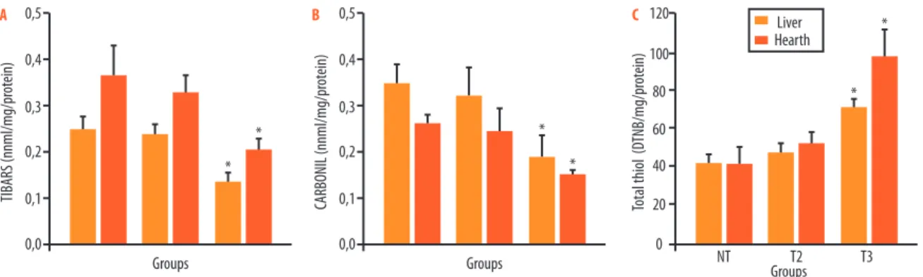
Figure 1A shows that the level of TBARS in the liver (0.13±0.02 nmol/mg/ protein) and in the heart (0.20±0.01 nmol/mg/protein) in the T3 group was lower than in the NT group (0.25±0.02 and 0.36±0.06 nmol/mg/protein). he results of protein carbonylation (Figure 1E) also resulted in a decrease in carbonyl content in the liver (0.19±0.049 nmol/mg/protein) and the heart (0.15±0.011 nmol/mg/protein) in the T3 group when compared with the NT group (0.35±0.041 and 0.26±0.017 nmol/mg/protein). However, training twice a week (T2) did not change TBARS levels or carbonyl contents in the liver (0.23±0.02 and 0.32±0.09) and the heart (0.32±0.03; 0.24±0.04 nmol/ mg/protein) when compared with the NT group. he results of total thiol (Figure 1C) showed a greater content in the liver (71.08±4.79 DTNB/mg/ protein) and the heart (97.7±14.2 DTNB/mg/protein) in the T3 group than in the NT group (41.7±4.07 and 41.6±8.3 DTNB/mg/protein). However, training twice a week did not afect this marker signiicantly (47.7±4.2 and 51.9±5.8 DTNB/mg/protein) when compared with the NT group.
he results of the analysis of antioxidant enzymes (Figure 2A) showed an increase of SOD in the liver (0.37±0.05U/mg/protein) and in the heart (0.23±0.02 U/mg/protein) in the T3 group when compared with the NT group (0.15±0.01 and 0.12±0.01 U/mg/protein). However, training twice a week (T2) did not afect this marker signiicantly (0.24±0.03 and 0.07±0.006 U/mg/protein). he results of the analysis of CAT (Figure 2B) showed an increase in the liver (0.17±0.03 U/mg/protein) and in the heart (0.45±0.04 U/ mg/protein) in the T3 group when compared with the NT group (0.07±0.02
Training frequency and oxidative stress Tromm et al.
and 0.02±0.001 U of CAT/mg/protein). Again, training twice a week (T2) did not signiicantly increase CAT activity in the liver (0.05±0.01) and in the heart (0.02±0.001 U/mg/protein) when compared with the NT group. he GPX activity (Figure 2C) in groups T2 and T3 in the liver (0.8±0.06 and 1.0±0.1 mM/mg/protein) and in the heart (0.7±0.1; 0.9±0.1 mM/mg/ protein) was not signiicantly diferent from that in the NT group (0.7±0.1 and 0.6±0.04 mM/mg/protein).
Figure 2. Superoxide dismutase (A), catalase (B) and glutathione peroxidase (C) in the liver and heart of mice 48 h after the last training session. Values were described as mean ± SEM and analyzed using ANOVA and the Tukey test. Superoxide dismutase and catalase were described in U/mg/protein, and glutathione peroxidase, in nM/min/mg/protein (p < 0.05 vs. NT).
DIsCUssION
Studies have shown that exhaustive physical exercise increases the produc-tion of ROS and, consequently, leads to OS in several organs and tissues6,7.
However, other studies found that moderate chronic exercise produces metabolic adaptations that may help to reduce OS in several organs, par-ticularly in special populations13,14.
In their analysis of physiological adaptations as a result of the practice of physical exercise in twice a week exercise sessions, Dalleck et al. 25 found
improvements in physiological training parameters (lactate and VO2max). However, the efects of biochemical responses on oxidative stress markers remain unclear. he results of this study showed that two training ses-sions per week is not a suicient frequency to promote improvements in OS parameters. he long interval (>72 h) between sessions may exceed the supercompensation phase and may inhibit the biochemical adaptive efect of training.
he association between OS and physical exercise is directly associ-ated with training intensity and duration2,26. herefore, training should
have a suicient intensity and duration to create an adaptive response of the organism in each session. Our results suggest that exercise sessions at least three times a week for eight weeks are necessary to reduce oxidative damage and to increase the activity of antioxidant enzymes in the liver and in the heart of animals.
ani-indings conirm the results of several studies that found a reduction in oxidative stress parameters ater similar training programs (three times a week)14,23.
he reduction of oxidative damage induced by physical training may be explained by at least three main mechanisms: irst, the increase of both the expression12 and the activity9 of antioxidant enzymes; second, the
re-duction of oxidant prore-duction13 and, also, the lower electron leakage from
mitochondria9; and third, the chronic exposure of tissue to ROS, induced
by training, which makes the organ more resistant to the efects that derive from the mechanisms of oxidative stress10.
ROS may afect amino acids in chain reactions by means of protein aggregates susceptible to proteolysis. During this process, some amino acids are converted into carbonyl derivates1. It is clearly established in the
literature that oxidized proteins are less degraded by proteasomes. hese intracellular proteases are responsible for 70% to 80% of degradation ater exposure to oxidants and play an essential role in the antioxidant system28. One of the possible mechanisms to reduce the levels of PC,
ac-cording to Radák et al.11,is that physical training increases proteasome
activity in adaptation to protein oxidation, which accelerates their repair (protein turnover).
Another important marker of protein oxidation is total thiol (TT) con-tent. Our study found an increase in TT only in the T3 group. he technique
used measured non-oxidized sulhydryls in amino acids20. he SH group
may be oxidized by free radicals, which compromises protein functioning. A possible explanation for these results is the increase of stress proteins (HSP) induced by exercise26. he function of these proteins is to control
cell homeostasis and to protect against excessive oxidation. However, HSP measurement was not carried out here, which is a limitation of our study.
he analysis of antioxidant enzymes showed that there was an increase in SOD and CAT only in the groups that received training three times a week. SOD converts superoxide radicals (O2•-) into hydrogen peroxide
(H2O2), which is, subsequently, catalyzed by CAT and converted into
water and molecular oxygen.Some studies have argued that physical
training has no efect on antioxidant enzymes in the liver and in the heart29. However, the results of our study, in agreement with other
ind-ings, showed that physical training increases SOD activity in the liver5,6
and in the heart15.
Ater physical training, SOD activity increases, probably in response to oxidative stress induced by exercise. his inding may be explained by the fact that regular physical training activated transcription factors, such as NF-kB, responsible for activating a variety of genes, and mito-chondrial SOD30.
Training frequency and oxidative stress Tromm et al.
trained rats8. Physical training activates transcription factors, such as
AMPK, which activate CATmRNA and stimulate its protein synthesis, which may increase its activity1,8. Moreover, the high activity of CAT
may be assigned to H2O2 formation by SOD. According to Halliwel and
Gutteridge1, the chemical interaction of H
2O2 in the active site of catalase
transfers a hydrogen atom from the irst to the second oxygen atom, which leads to heterolytic ission between atoms and forms water (non-deleterious molecule). his, in turn, explains the decrease of oxidative damage in tissues.
In contrast, the activity of glutathione peroxidase (GPX) was not sig-niicantly diferent between the experimental groups in this study. GPX
and CAT have similar functions in H2O2 decomposition. However, GPX
is more eicient when ROS concentrations are high, whereas CAT plays an important role when H2O2 is low3. Physical training three times a week
may promote an adaptive efect in the redox balance of antioxidants so that the low H2O2 concentrations are afected only by CAT, a hypothesis that may explain the results described above.
CONClUsION
he results of this study showed that treadmill training three times a week reduced oxidative damage and increased the eiciency of the enzyme an-tioxidant system in the liver and heart of mice.
REFERENCEs
1. Halliwell B, Gutteridge MC. Free radicals in biology and medicine. Oxford: Uni-versity Press; 2007
2. Finaud J, Lac G, Filaire E. Oxidative stress: relationship with exercise and training. Sports Med 2006;36:327-58.
3. Jenkins RR, Goldfarb A. Introduction: oxidant stress, aging, and exercise. Med Sci Sports Exerc 1993;25:210-2.
4. Aucello M, Dobrowolny G, Musarò A. Localized accumulation of oxidative stress causes muscle atrophy through activation of an autophagic pathway. Autophagy 2009;5:527-9.
5. Silva LA, Rosani MM, Souza PS, Severino JB, Fraga D, Streck EL, et al. Comparação do treinamento físico de quatro e oitro semanas sobre a atividade da cadeia transpor-tadora de elétrons e marcadores de estresse oxidativo em fígado de camundongos. Rev Bras Med Esporte 2010;16:126-9.
6. Navarro-Arevalo A, Sanchez-del-Pino MJ. Age and exercise-related changes in lipid peroxidation and superoxide dismutase activity in liver and soleus muscle tissues of rats. Mech Ageing Dev 1998;104:91-102.
7. Ogonovszky H, Sasvári M, Dosek A, Berkes I, Kaneko T, Tahara S, et al. he efects of moderate, stre nuous, and overtraining on oxidative stress markers and DNA repair in rat liver. Can J Appl Physiol 2005;30:186-95.
muscle. Eur J Appl Physiol 2009;105:861-7.
10. Pinho RA, Andrades ME, Oliveira MR, Pirola AC, Zago MS, Silveira PC, et al. Imbalance in SOD/CAT activities in rat skeletal muscles submitted to treadmill training exercise. Cell Biol Int 2006;30:848-53.
11. Radák Z, Tahara TK, Nakamoto H, Ohno H, Sasvári M, Nyakas C, et al. he efect of exercise training on oxidative damage of lipids, proteins, and DNA in rat skel-etal muscle: evidence for beneicial outcomes. Free Radic Biol Med 1999;27:69-74. 12. Frederico M, Luz G, Justo SL, Silva S, Medeiros C, Barbosa VA, et al. Exercise training provides cardioprotection via a reduction in reactive oxygen species in rats submitted to myocardial infarction induced by isoproterenol. Free Radic Res 2009;11:1-8.
13. Coelho BLP, Rocha LGC, Scarabelot KS, Schefer D, Rosani MM, Silveira PCL, et al. Physical exercise prevents the exacerbation of oxidative stress parameters in chronic kidney disease. J Ren Nutr 2010;47:16-19.
14. Nojima H, Watanabe H, Yamane K, Kitahara Y, Sekikawa K, Yamamoto H, et al. Efect of aerobic exercise training on oxidative stress in patients with type 2 diabetes mellitus. Metabolism 2008;57:170-6.
15. Hamilton KL, Powers SK, Sugiura T, Kim S, Lennon S, Tumer N, et al. Short-term exercise training can improve myocardial tolerance to I/R without elevation in heat shock proteins. Am J Physiol Heart Circ Physiol 2001;281:1346-52.
16. Olert E, Cross B, Mcwillians A. Guide to care and use of experimental animals. 2nd ed. Canadian Council on Animal, Ottawa; 1993.
17. Fernando P, Bonen A, Hofman-Goetz L. Predicting submaximal oxygen consump-tion during treadmill running in mice. Can J Physiol Pharmacol 1993;71:854-7. 18. Draper HH, Hadley M. Malondialdehyde determination as index of lipid
peroxida-tion. Meth Enzymol 1990;186:421-31.
19. Levine RL, Garland D, Oliver CN, Amici A, Climent I, Lenz AG, et al. Deter-mination of carbonyl content in oxidatively modiied proteins. Meth Enzymol 1990;186:464-78.
20. Aksenov MY, Markesberya WR. Changes in thiol content and expression of glu-tathione redox system genes in the hippocampus and cerebellum in Alzheimer’s disease. Neurosci Lett 2001;302:141-5.
21. Bannister JV, Calabrese L. Assay for SOD. Meth Biochem 1987;32:279-312. 22. Aebi H. Catalase in vitro. Meth Enzymol 1984;105:121-126.
23. Flohé l, Gunzler W. Assays of glutathione peroxidase. Meth Enzymol 1984;105:114-21. 24. Lowry OH, Rosebrough NJ, Farr AL, Randall RJ. Protein measurement with the
Folin phenol reagent. J Biol Chem 1951;193:265-7.
25. Dalleck L, Bushman TT, Crain RD, Gajda MM, Koger EM, Derksen LA. Dose-response relationship between interval training frequency and magnitude of improvement in lactate threshold. Int J Sports Med 2010;31:567-71.
26. Smolka, MB, Zoppi CC, Alves AA, Silveira LR, Marangoni S, Pereira-Da-Silva L, et al. HSP72 as a complementary protection against exercise induced oxidative stress in the soleus muscle of rats. Am J Physiology 2000;279:1539-45.
27. Karolkiewicz J, Michalak E, Pospieszna B, Deskur-Smielecka E, Nowak A, 28. Pilaczyńska-Szcześniak Ł. Response of oxidative stress markers and antioxidant
Training frequency and oxidative stress Tromm et al.
Endereço para correspondência
Camila Baumer Tromm Av. Universitária, 1105 – Bairro Universitário
88806-000 – Criciúma, SC. Brasil E-mail: [email protected] 29. Davies KJ, Shringarpure R. Preferential degradation of oxidized proteins by the
20S proteasome may be inhibited in aging and in inlammatory neuromuscular diseases. Neurology 2006;66:93-6.
30. Tiidus PM, Houston ME. Antioxidant and oxidative enzyme adaptations to vitamin E deprivation and training. Med Sci Sports Exerc 1994;26:354-9.