Research Article
Effect of Melatonin Intake on Oxidative Stress Biomarkers in
Male Reproductive Organs of Rats under Experimental Diabetes
Marina G. Gobbo,
1,2Carolina F. Pereira Costa,
1,2Danilo G. Humberto Silva,
1,3Eduardo A. de Almeida,
3and Rejane M. Góes
21Department of Biology, Institute of Biosciences, Humanities and Exact Sciences, Universidade Estadual Paulista (UNESP), Crist´ov˜ao Colombo 2265, 15054-000 S˜ao Jos´e do Rio Preto, SP, Brazil
2Department of Cell Biology, Institute of Biology, State University of Campinas (UNICAMP), Charles Darwin Street, Building N, 13083 863 Campinas, SP, Brazil
3Department of Chemistry and Environmental Sciences, Institute of Biosciences, Humanities and Exact Sciences, Universidade Estadual Paulista (UNESP), Cris´ov˜ao Colombo 2265, 15054-000 S˜ao Jos´e do Rio Preto, SP, Brazil
Correspondence should be addressed to Rejane M. G´oes; [email protected]
Received 9 December 2014; Revised 11 April 2015; Accepted 17 April 2015
Academic Editor: Silvana Hrelia
Copyright © 2015 Marina G. Gobbo et al. his is an open access article distributed under the Creative Commons Attribution License, which permits unrestricted use, distribution, and reproduction in any medium, provided the original work is properly cited.
his study investigated the antioxidant system response of male reproductive organs during early and late phases of diabetes and the inluence of melatonin treatment. Melatonin was administered to ive-week-old Wistar rats throughout the experiment, in
drinking water (10�g/kg b.w). Diabetes was induced at 13 weeks of age by streptozotocin (4.5 mg/100 g b.w., i.p.) and animals
were euthanized with 14 or 21 weeks old. Activities of catalase (CAT), glutathione-S-transferase (GST), glutathione peroxidase (GPx), and lipid peroxidation were evaluated in prostate, testis, and epididymis. he enzymes activities and lipid peroxidation were not afected in testis and epididymis ater one or eight weeks of diabetes. Prostate exhibited a 3-fold increase in GPx activity
at short-term diabetes and at long-term diabetes there were 2- and 3-fold increase in CAT and GST, respectively (� ≤ 0.01).
Melatonin treatment to healthy rats caused a 47% increase in epididymal GPx activity in 14-week-old rats. In prostate, melatonin administration normalized GST activity at both ages and mitigated GPx at short-term and CAT at long-term diabetes. he testis and epididymis were less afected by diabetes than prostate. Furthermore, melatonin normalized the enzymatic disorders in prostate demonstrating its efective antioxidant role, even at low dosages.
1. Introduction
Diabetes mellitus (DM) afects 8.3% of the world population and approximately 5.1 million people aged between 20 and 79 years died from diabetes in 2014 [1]. he high mortality and negative impact of diabetes on life quality are due to the progressive impairment of multiple organ systems, caused mainly by hyperglycemia and oxidative stress [2]. he oxida-tive status in diabetes is due to impairment of mitochondrial electron transfer, the activation of polyol pathways, the catal-ysis of cyclooxygenase intermediate products, and enhanced nonenzymatic glycation [3–5]. In turn, advanced glycation end-products (AGE) produced by nonenzymatic glycation lead to the generation of reactive oxygen species (ROS),
the activation of Bax, and expression of proapoptotic and proinlammatory genes, such as c-Jun N-terminal kinase [2,
6,7]. hus, as conirmed by cDNA microarray analysis, dia-betes can alter the expression of multiple genes, particularly those related to cell proliferation and diferentiation, oxida-tive stress biomarkers, DNA damage repair, and apoptosis [8]. he vast majority of patients with type 1 DM are diag-nosed before the age of 30 and a modest excess of cases occurs in males [9] where its negative inluence on reproductive function is relevant. Clinical evidence indicates that diabetes is associated with multiple impairment of male genital phys-iology, such as reduced androgen levels, erectile dysfunction, retrograde ejaculation, poor semen quality, and reduced fertility [10, 11]. Diabetes has also been associated with an
increased risk of numerous cancers, but the data concerning prostate cancer are inconsistent [12, 13]. Most evidence, including a meta-analysis [14] of 19 studies published between 1971 and 2005, has indicated an inverse correlation between diabetes and prostate cancer [15,16].
Most clinical data concerning the negative impact of diabetes on male reproductive physiology have been cor-roborated by experimental models of induced diabetes [17–
25]. he inluence of oxidative stress due to diabetes on the response of diferent genital organs has been previously inves-tigated in rats [26–32]. However, information concerning how these changes occur during disease progression is scarce. he neurohormone melatonin (N-acetyl-5-methoxytryp-tamine) is secreted rhythmically following a periodicity that is controlled by a circadian pacemaker located in the suprachiasmatic nucleus [33]. Melatonin (MLT) regulates several physiological functions, according to the light-dark daily cycle. It has been suggested that the rhythmicity of MLT action also controls the activity and gene expression of antioxidant enzymes [34]. MLT and its metabolites, N1-acetyl-N2-formyl-5-methoxykynuramine (AFMK), and N1-acetyl-5-methoxykynuramine (AMK) exhibit antioxidant activities that are related to the direct removal of hydroxyl radicals, nitric oxide, and peroxynitrite anions acting as free radical scavengers [35–38]. A few studies using cultured cells indicate that melatonin promoted the generation of ROS at pharmacological concentrations; however its prooxidant action in vivo remains to be elucidated [39]. Experimental evidence has shown that administration of MLT at doses of 5 mg to 150 mg/kg body weight ameliorates the oxidative sta-tus in the pancreas, liver, heart, kidneys, and testis [26,40,41]. Besides, the administration of low doses of MLT (25�g/mL) to rats fed with high fat-diet showed that this hormone was able to normalize the altered biochemical proinlammatory proile in these animals [42]. he consequences of MLT consumption at low doses during sexual maturation of the male genital organs and their oxidative status at adulthood are unknown. In addition, considering that MLT interferes with androgen production and afects androgen-dependent organs, which also occurs in diabetes, more information is necessary to better discriminate the putative and protective role of exogenous MLT in genital organs under diabetes and also to delineate the response of organs during disease progression. hus, this study comparatively examined the early and advanced responses of the antioxidant system in rat male genital organs subjected to experimental diabetes and the inluence of low MLT dose treatment prior to and concomitant with the disease in these systems.
2. Material and Methods
2.1. Experimental Design. Eighty male Wistar rats (Rattus norvegicus) were obtained from the breeding house of S˜ao Paulo State University (Botucatu, SP, Brazil). All experi-ments were performed in accordance with the Guide for the Care and Use of Laboratory Animals published by the US National Institutes of Health and acknowledged by the institutional ethical committee for animal experimentation
C1 M1 D1 MD1 C2 M2 D2 MD2
1st week 5th week 13th week 14th week 21st week
Untreated period Melatonin treatment
Experimental diabetes Short-term experiment
Long-term experiment
Diabetes/melatonin treatment
Figure 1: Experimental design of the study. Melatonin was ofered
in the drinking water (10�g/kg b.w), and diabetes was induced
by streptozotocin injection (4.5 mg/100 g b.w., i.p.). C1: one-week control; M1: week control treated with melatonin; D1: one-week diabetic; MD1: one-one-week diabetic treated with melatonin; C2: two-month control; M2: two-month control treated with melatonin; D2: two-month diabetic; MD2: two-month diabetic treated with
melatonin (� = 10animals/group). he euthanasia was performed
at 13 weeks of age for short-term experiment and at 21 weeks of age for long-term experiment.
(Protocol number 051/2011-CEEA). he animals were kept in polyethylene cages with wood shavings in a 12 : 12 light/dark cycle, at a temperature of about 22∘C and with free access to food (Presence, Invivo, Paulinia, SP, Brazil) and iltered water. Ater an adaptation period, the rats were weighed and randomly distributed into eight groups (Figure 1, � = 10 per group). he short-term experiment consisted of a control (C1), a control treated with MLT (M1), one-week-diabetic rats (D1), and one-week-one-week-diabetic rats treated with MLT (MD1). he long-term experiment consisted of a control (C2), a control treated with MLT (M2), two-month diabetic rats (D2), and two-month diabetic rats treated with MLT (MD2). he administration of MLT (Sigma Chemical Co., St Louis, MO, USA) followed the procedures established by Wolden-Hanson et al. [43]. his hormone was dissolved in ethanol and stored in aliquots at−70∘C. Rats in groups M1, M2, MD1, and MD2 were provided with MLT from ive to 14 weeks of age, via drinking water (10�g/kg body weight in ethanol 0.001%/day). he MLT intake per day in this investigation was based on mean daily water consumption of 80 mL/day/animal and a mean body weight of 350 g and was available to the animals in plastic bottles protected from light. hese conditions were standardized by the application of various consumption preference and aversion tests; therefore, this is an appropriate dosage for the induction of increased MLT levels during the night [43].
Mannheim, Germany). Only animals that showed blood glucose levels above 220 mg/dL were included in the diabetic groups. Because water consumption is higher for diabetic animals, the MLT dose was corrected for groups D1, D2, MD1, and MD2 following the diagnosis of diabetes. he C1, M1, D1, and MD1 groups were euthanized when with 14 weeks old and the C2, M2, D2, and MD2 groups were euthanized when 21 weeks old. he rats were euthanized using CO2inhalation and were subsequently decapitated for blood collection.
2.2. Activity of Antioxidant Enzymes. he antioxidant enzyme activity of all animals was assayed in the ventral prostate, testis, and epididymis and also in blood. Ater dissec-tion, these organs were weighed and homogenized in 1 : 4 volume of bufer with protease inhibitors (50 mM Tris-HCl, 1 mM EDTA, 1 mM DTT, 0.5 M sucrose, and 0.15 M KCl, 1 mM PMSF, pH 7.4) and centrifuged at 10.000 g for 20 min at 4∘C. he supernatant was then recentrifuged at 50.000 g for another 60 min at 2∘C and the supernatant fraction was removed and was used to measure the activity of catalase (CAT), glutathione-S-transferase (GST), and glutathione peroxidase (GPx).
he blood samples were collected in polyethylene tubes containing EDTA immediately ater decapitating the animals. For the determination of CAT activity, the blood samples were diluted 50 times in distilled water, whereas for the determination of GPx and GST activities, blood was diluted 20 times in a hemolyzing solution (7 mM 2-mercaptoetanol; 2 mM NADP; 0.27 M EDTA).
he CAT activity was quantiied at 240 nm for 1 min by the decomposition of 10 nm H2O2[44]. he total GST activity was determined by measuring the increase in absorbance at 340 nm for 1 min 40 s by an assay containing reduced glutathione (200 mM GSH) and 1-chloro-2,4-dinitrobenzene (200 mM CNDB) as substrates, according to Keen et al. [45]. he total GPx activity was evaluated by NADPH (0.2 M) oxidation, concomitant with GSSG reduction by excess glutathione reductase, causing a decrease in absorbance at 340 nm for 1 min, according to Sies et al. [46]. All tests were performed at room temperature. he total protein content (mg/mL) in the samples was determined using bovine serum albumin as a standard, by the modiied Lowry method [47]. he speciic molar extinction constant (�) was used to esti-mate the levels of enzyme activity in U/mg protein (�= 0.071 for CAT,�= 6.22 for GPx, and�= 9.6 for GST). he equation used for enzymatic activity was [Absorbance variation × 1000/� ×sample volume (�L)/total protein concentration of the sample (mg/mL)].
2.3. Determination of Lipid Peroxidation Levels. he levels of lipid peroxidation were evaluated in the same organs and in blood, by the quantiication of malondialdehyde (MDA) levels, an indicator of oxidation. For this, the presence of the colored derivative formed between MDA and 2-thiobarbituric acid (TBA) was detected via HPLC at 532 nm [48]. Quantiication of MDA in the tissues was performed with 100�L of homogenized tissue in bufer (1 : 4 v/v) and
100–200�L of plasma. hree hundred �L of 0.4% thiobar-bituric acid solution (diluted in 0.2 M HCl) was added to the tissue and plasma samples and they were incubated for 40 min at 90∘C in a dry block. he samples of TBA-MDA were extracted with 1 mL n-butanol and centrifuged at 890 g for 3 min at 3∘C. TBA-MDA (20�L) samples were directly injected in HPLC and monitored at 532 nm. he mobile phase consisted of 50 nM monobasic phosphate potassium solution, pH 7.0, 20% methanol, and a pumped isocratic low of 1.0 mL/min. he HPLC system (Shimadzu) consisted of two LC-10ADVP pumps, a SPD-M10ADVP UV-visible detector, and a SCL-10AVP controller and a LC-18 (150 × 4.6 mm, 5�m pore diameter) column was used. he MDA estimation was based on a standard calibration curve of tetramethoxypropane (TMP) previously prepared using the same procedure as that used for samples. he data were expressed as nmol/mg tissue and �mol/mL plasma. he equation to calculate the amount of MDA in samples was (peak area/slope of calibration curve)/C,C= [sample volume (�L)×injection volume (�L))/1000�L of n-butanol].
2.4. Statistical Analysis. Statistical analyses were performed among groups of the same experimental period (multiple comparison tests) and between both experimental periods for the same treatment (paired diference tests) using the Statistica 9.0 sotware (Statsot Inc., Tulsa, OK, USA). Data were tested for normality and homogeneity of variance assumptions according to the Shapiro-Wilk’s test and Levene’s test, respectively. Groups that met the assumptions (para-metric data) were compared by applying attest or one-way ANOVA followed by Tukey’s post hoc test. hose groups that did not meet the assumptions (nonparametric data) were compared using the Mann-Whitney or Kruskal-Wallis test followed by Dunn’s post hoc test. Data were expressed as mean ±standard deviation and� < 0.05 was considered statistically signiicant.
Correlation tests were conducted (Pearson’s test for para-metric data and Spearman’s test for nonparapara-metric data) between the levels of lipid peroxidation (MDA) and the activity of CAT, GST, and GPX, using Statistica 9.0 sotware (Statsot Inc.).
3. Results
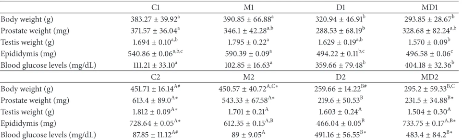
3.1. Biometric Parameters and Glycaemia. he body weights were, respectively,∼16 and∼42% lower (� ≤ 0.001) ater one week (D1) or two months of diabetes (D2), compared to the control groups (Table 1). he MLT treatment did not afect the body weight of normal rats or the body weight loss in diabetic groups (Table 1).
he ingestion of low doses of MLT also did not inluence the weight of the prostate of healthy rats (Table 1). Such treatment avoided the prostatic atrophy induced by short-term diabetes, but not by the long-short-term treatment (Table 1; � ≤ 0.05).
In both experiments, the testicular weight was not afected by diabetes independent of treatment with MLT
Table 1: he mean and standard deviation of body, prostate, testis, and epididymis wet weight and blood glucose levels of short- and long-term experimental groups.
C1 M1 D1 MD1
Body weight (g) 383.27±39.92a 390.85±66.88a 320.94±46.91b 293.85±28.67b
Prostate weight (mg) 371.57±36.04a 346.1±42.28a,b 288.53±68.19b 328.68±82.24a,b
Testis weight (g) 1.694±0.10a,b 1.795±0.22a 1.629±0.19a,b 1.570±0.09b
Epididymis (mg) 540.86±0.06a,b,c 590.39±0.09a 494.22±0.11b,c 496.58±0.06c
Blood glucose levels (mg/dL) 111.21±33.10a 102.85±16.63a 359.66±79.48b 404.18±32.36b
C2 M2 D2 MD2
Body weight (g) 451.71±16.14A# 450.57±40.72A,C∗ 259.66±14.22B# 295.2±59.33B,C
Prostate weight (mg) 613.4±89.0A∗ 543.33±67.58A∗ 219.6±50.53B 231.5±34.88B∗
Testis weight (g) 1.812±0.09A∗ 1.701±0.21A 1.603±0.24A 1.504±0.30A
Epididymis (mg) 728.64±0.05A∗ 612.35±0.15A,B 466.04±0.05B 733.75±0.17A,B∗
Blood glucose levels (mg/dL) 87.85±11.12A# 89±9.05A 491.16±56.55B∗ 483.4±84.2B∗
C1: one-week control; M1: one-week control treated with melatonin; D1: one-week diabetic; MD1: one-week diabetic treated with melatonin; C2: two-month control; M2: two-two-month control treated with melatonin; D2: two-two-month diabetic; MD2: two-two-month diabetic treated with melatonin (� = 10 animals/group).∗Diferent lowercase letters indicate statistical diferences among short-term experimental groups (parametric data: prostate and epididymis weight; nonparametric data: body and testis weight and blood glucose levels). Diferent uppercase letters indicate statistical diferences among long-term experimental groups (parametric data: prostate weight; nonparametric data: body, testis and epididymis weight, and blood glucose levels).∗Indicates a statistical diference between experimental periods (parametric data).#Indicates a statistical diference between experimental periods (nonparametric data).
compared to the diabetic groups (Table 1; � ≤ 0.004). Diabetes reduced the epididymal weight in both experiments
(Table 1; � ≤ 0.04). Despite the higher epididymal atrophy
conirmed in long-term diabetes, the MLT treatment pre-vented this atrophy (Table 1).
Animals showed blood glucose levels that were about three and six times higher ater short- or long-term diabetes, respectively, than that in the control groups (� ≤ 0.001), regardless of MLT treatment (Table 1). Administration of MLT did not afect the glucose level homeostasis of groups M1 and M2.
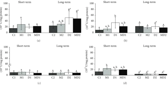
3.2. CAT Activity. he CAT activity in the blood (Figure 2(a)) increased by about 40% in 21-week-old rats in comparison to the 14-week-old rats but did not change among the groups in both experiments. In the ventral prostate, the CAT activity (Figure 2(b)) was unchanged in the groups of the irst experiment, but doubled ater two months of diabetes compared with the control group and this increase was prevented by MLT treatment. he levels of testicular CAT were higher in the groups of long-term experiment
(Figure 2(c),� ≤ 0.04). Melatonin had an inhibitory efect
on testis CAT activity in long-term diabetes (Figure 2(c)). Similar to the testis, the activity of CAT in the epididymis of 21-week-old rats was also higher in comparison to those of 14-week-old healthy rats (� ≤ 0.04;Figure 2(c)and� ≤ 0.01;
Figure 2(d), resp.).
3.3. GST Activity. GST was the only biomarker of oxidative stress that changed in the blood ater short-term diabetes
(Figure 3(a)). he GST activity increased during short-term
diabetes (� = 0.0042) and this rise was partially prevented in the MLT-treated group (Figure 3(a)); however, blood GST activity decreased (� = 0.0007) during long-term diabetes, regardless of the MLT treatment (Figure 3(a)). he blood
GST activity increased in 21-week-old groups in comparison to younger groups (� ≤ 0.001). Prostatic GST activity
(Figure 3(b)) also increased ater the onset of diabetes (� =
0.0186), and MLT administration prevented this increase only in the long-term experiment. he GST activity was not afected in testis and epididymis among the animals of short and long-term experimental groups (Figure 3(d)); however the activity of this enzyme was decreased in the epididymis of older healthy rats, regardless of MLT treatment (Figure 3(d)).
3.4. GPx Activity. he GPx activity in blood had an inverse behavior to GST (Figure 4(a)); that is, the activity was unchanged during short-term diabetes and increased twofold ater two months of diabetes compared to the control group, independent of MLT treatment. In the prostate gland, GPx activity increased∼70% one week ater the onset of diabetes and MLT normalized this value in the MD1 group; how-ever, it was unafected in groups of the longer experiment
(Figure 4(b)). he activity of this antioxidant enzyme in testis
was not altered neither in experimental diabetes nor mela-tonin treatment (Figure 4(c)). here was an increase in GPx activity in the epididymis of healthy rats ater MLT treatment in comparison to those in the control group (Figure 4(d); � = 0.007). Moreover, the epididymis GPx activity was very low in the two-month experiment (Figure 4(d)).
3.5. Lipid Peroxidation. In blood, the MDA levels were unchanged in the groups of the short-term experiment
(Figure 5(a)) and increased ater two months of untreated
diabetes (� = 0.038), regardless of MLT treatment. Diabetes did not afect lipid peroxidation in prostate (Figure 5(d)), either in the short-term or in long-term, but MLT treatment reduced by 50% the lipid peroxidation levels of long-term diabetic rats in comparison with untreated diabetic group
0 10 20 30 40
(U/m
g p
ro
tein)
Long-term Short-term
a a a
a
A
A∗ A∗
A∗
C1 M1 D1 MD1 C2 M2 D2 MD2
(a)
0 10 20 30 40
(U/m
g p
ro
tein)
Long-term Short-term
a a
a a
A A
B A, B∗
C1 M1 D1 MD1 C2 M2 D2 MD2
(b)
0 10 20 30 40
(U/m
g p
ro
tein)
Long-term Short-term
a a a a A∗
B# A, B∗
A, B∗
C1 M1 D1 MD1 C2 M2 D2 MD2
(c)
0 10 20 30 40
(U/m
g p
ro
tein)
Long-term Short-term
a a
a a
A A
A∗
A∗
C1 M1 D1 MD1 C2 M2 D2 MD2
(d)
Figure 2: Catalase activity (U/mg protein) in blood (a), prostate (b), testis (c), and epididymis (d) of short- and long-term experiments. C1: one-week control; M1: one-week control treated with melatonin; D1: one-week diabetic; MD1: one-week diabetic treated with melatonin; C2: two-month control; M2: two-month control treated with melatonin; D2: two-month diabetic; MD2: two-month diabetic treated with
melatonin (� = 10animals/group). Diferent lowercase letters indicate statistical diferences among short-term experimental groups
(parametric data: (a), (b), (c), and (d)). Diferent uppercase letters indicate statistical diferences among long-term experimental groups
(parametric data: (a) and (d); nonparametric data: (b) and (c)).∗Indicates a statistical diference between experimental periods (parametric
data). # Indicates a statistical diference between experimental periods (nonparametric data).
Short-term Long-term
a a
b
a, b B B
0 3 200 300 400
C1 M1 D1 MD1 C2 M2 D2 MD2
A# A∗
(10
2 U/m
g p
ro
tein)
(a)
Short-term Long-term
a a
a, b b
A A
B
0 100 200 300 400
C1 M1 D1 MD1 C2 M2 D2 MD2
A#
(10
2 U/m
g p
ro
tein)
(b)
Short-term Long-term
a a, b a, b b
A A A
A
0 100 200 300 400
C1 M1 D1 MD1 C2 M2 D2 MD2
(10
2U/m
g p
ro
tein)
(c)
0 100 200 300 400
C1 M1 D1 MD1 C2 M2 D2 MD2
Short-term Long-term
a
a a a A∗ A∗ A A
(10
2U/m
g p
ro
tein)
(d)
Figure 3: Glutathione-S-transferase activity (U/mg protein) in blood (a), prostate (b), testis (c), and epididymis (d) extracts. C1: one-week control; M1: one-week control treated with melatonin; D1: one-week diabetic; MD1: one-week diabetic treated with melatonin; C2: two-month control; M2: two-two-month control treated with melatonin; D2: two-two-month diabetic; MD2: two-two-month diabetic treated with melatonin (� = 10animals/group). Diferent lowercase letters indicate statistical diferences among short-term experimental groups (nonparametric data: (a), (b), (c), and (d)). Diferent uppercase letters indicate statistical diferences among long-term experimental groups (parametric data:
(c) and (d); nonparametric data: (a) and (b)).∗Indicates a statistical diference between experimental periods (parametric data). # Indicates
a a
a a A A, B
B#
B#
Long-term Short-term
0 20 40 60 80 100
C1 M1 D1 MD1 C2 M2 D2 MD2
(10
2U/m
g p
ro
tein)
(a)
a a, b b
a, b A
A A A
#
0 20 40 60 80 100
C1 M1 D1 MD1 C2 M2 D2 MD2
Long-term Short-term
(10
2U/m
g p
ro
tein)
(b)
a a a a A A A A
0 10 40 60 80 100
C1 M1 D1 MD1 C2 M2 D2 MD2
Long-term Short-term
(10
2 U/m
g p
ro
tein)
(c)
0
20 a
b
a, b a, b 40
60 80 100
C1 M1 D1 MD1 C2 M2 D2 MD2
A# A# A# A#
Long-term Short-term
(10
2 U/m
g p
ro
tein)
(d)
Figure 4: Glutathione peroxidase activity (U/mg protein) in blood (a), ventral prostate (b), testis (c), and epididymis (d) of rats. C1: one-week control; M1: one-one-week control treated with melatonin; D1: one-one-week diabetic; MD1: one-one-week diabetic treated with melatonin; C2: two-month control; M2: two-two-month control treated with melatonin; D2: two-two-month diabetic; MD2: two-two-month diabetic treated with melatonin (� = 10animals/group). Diferent lowercase letters indicate statistical diferences among short-term experimental groups (parametric data: (c); nonparametric data: (a), (b), and (d)). Diferent uppercase letters indicate statistical diferences among long-term experimental groups (parametric data: (b), (c), and (d); nonparametric data: (a)). # Indicates a statistical diference between experimental periods (nonparametric data).
in all groups of the long-term experiment compared to the short-term groups (� ≤ 0.04). he testicular levels of MDA were lower (� ≤ 0.01) in the groups of the long-term experiment (Figure 5(c)) whereas in the epididymis, the peroxidation levels were high in these groups (Figure 5(d); � ≤ 0.04).
3.6. Correlation Tests. here was an inverse correlation between lipid peroxidation and GST activity in both short-(� = −0.477; � < 0.05) and long-term experiments (� = −0.669; � < 0.05), in blood (Figures 3(a)and 5(a)). he increase in GPx activity was directly proportional to the increase in plasma levels of MDA ater two months of diabetes (� < 0.05and� = 0.74; Figures4(a)and5(a)).
In the prostate, the rise in GST activity ater one week of diabetes correlated inversely with the decrease in prostatic MDA levels (� = −0.604;� = 0.022, Figures3(b)and5(b)).
4. Discussion
In the present study, we delineate the comparative oxidative status in three important reproductive organs in terms of acute and chronic response to streptozotocin-induced dia-betes, based on the activity of the major antioxidant enzymes and quantiication of lipid peroxidation. In addition, we analyzed the efects of preadministration and prolonged use of low MLT doses on the antioxidant system of these organs and its inluence on alterations caused by experimentally
induced diabetes. However it is worthwhile to mention that the provision of melatonin in the drinking water, although less stressful and adequate for long-term experiments, was a limitation for this study since there is no way to know the exact amount of MLT that was ingested by each animal as well as the changes in the daily rhythm of this hormone throughout the experiment. Rasmussen et al. using a similar experimental protocol [49] veriied that rats drank more than 90% of their total daily water during the dark period resulting in higher melatonin levels during the night (150.5 ± 19.2pg/mL against24.1±8.8pg/mL of control) but not in day-time (14.1 ± 2.6pg/mL against11.5 ± 0.00pg/mL of control). As we used the same model of melatonin administration as Rasmussen et al. [49], we assume that the animals exhibited a comparable pattern of melatonin consumption and probably exhibited elevation in the levels of this hormone in dark period.
a a a
a B B∗
∗
A# A, B#
0 1 10 15 20
C1 M1 D1 MD1 C2 M2 D2 MD2
(𝜇
mo
l/mL p
lasm)
Long-term Short-term
(a)
a a a
a B
∗
B∗
A∗
A, B∗ 0
2 10 15 20
C1 M1 D1 MD1 C2 M2 D2 MD2
(nmo
l/m
g tis
su
e)
Long-term Short-term
(b)
a
a a a
B#
A#
0 5 10 15 20
C1 M1 D1 MD1 C2 M2 D2 MD2
(nmo
l/m
g tissue)
A, B# A, B#
Long-term Short-term
(c)
0 5 10 15 20
C1 M1 D1 MD1 C2 M2 D2 MD2
(nmo
l/m
g t
issue)
a a
a a
A
A∗
A∗
A#
Long-term Short-term
(d)
Figure 5: Lipid peroxidation quantiied by MDA levels in plasm ((a),�mol/mL plasm) and in extracts (U/mg protein) of prostate (b),
testis (c), and epididymis (d). C1: one-week control; M1: one-week control treated with melatonin; D1: one-week diabetic; MD1: one-week diabetic treated with melatonin; C2: month control; M2: month control treated with melatonin; D2: month diabetic; MD2:
two-month diabetic treated with melatonin (� = 10animals/group). Diferent lowercase letters indicate statistical diferences among short-term
experimental groups (parametric data: (b), (c), and (d); nonparametric data: (a)). Diferent uppercase letters indicate statistical diferences
among long-term experimental groups (parametric data: (d); nonparametric data: (a), (b), and (c)).∗Indicates a statistical diference between
experimental periods (parametric data). # Indicates a statistical diference between experimental periods (nonparametric data).
increase in CAT activity was accompanied by a decrease in GST and GPx activity. Taken together these indings might explain the higher susceptibility to oxidative stress of the prostate during aging, in comparison to testis and epididymis as indicated by the increase in MDA levels. hese data also reinforce the organ speciicity of antioxidant defense and the existence of compensatory mechanisms during aging.
he experimental protocol used here for the induction of type I diabetes is widely accepted, and most diabetic rats exhibited glucose levels above 360 mg/dL. As expected, a drastic reduction in body weight was observed, mainly in animals with chronic untreated diabetes. he body weight or blood glucose levels of normal rats were not afected by MLT treatment. Even in short-term diabetes, MLT ingestion did not normalize the glycemia or ameliorate the severe weight loss, as previously observed with doses above 10 mg/kg b.w. [28,50,51]. Some studies with experimental diabetes, includ-ing those with high doses of MLT, showed a normalization of blood glucose levels [40,52]; however, this efect of MLT has been clearly reported for obese rodents in which there is an improvement in insulin sensitivity [43,53]. Furthermore, MLT increases the metabolic activity of brown adipose tissue [54] which might favor weight loss in diabetic animals.
Streptozotocin-induced diabetes caused atrophy in the prostate and epididymis. he atrophy of these androgen-dependent organs was expected, since diabetes leads to androgen withdrawal [55, 56] and has been reported in previous studies using similar protocols for diabetes induc-tion [28, 32, 57]. Some reports indicate a decrease in
the relative testicular weight within three weeks of experi-mentally induced diabetes, but thereater, testicular atrophy is no longer conspicuous [24,57] and a similar variation was observed here, although this was not statistically signiicant. Treatment with MLT mitigated prostate atrophy induced by diabetes in the short-term, whereas an opposite efect was observed for the epididymis, where atrophy ater short-term diabetes was not prevented, but maintenance of wet weight for long-term diabetic rats was observed. Results of our laboratory demonstrated that maintenance of epididymal weight by MLT ater long-term diabetes was due to higher sperm counts in this organ (unpublished data).
the prostate antioxidant system is vulnerable to androgen regulation, as shown by in vitro studies [60], and also to the hyperglycemic status [32].
Our indings emphasize that GST is an important com-ponent in the defense against oxidative damage in the prostate. hese indings agree with previous data that report a pivotal role of GST isoforms in the healthy prostate and in disease progression [61,62]. he GSTP1 gene, which encodes the pi-class glutathione S-transferase, is a defense against oxidative damage to the genome and is expressed in high levels by epithelial cells in proliferative inlammatory Atrophy (PIA) [61–63], which are considered to be precursors of premalign and malign prostate lesions. he expression of GSTP1 is impaired in prostatic epithelial neoplasia (PIN) and neoplastic lesions, due to somatic “CpG island” DNA methylation changes [60,63–65]. Such cells become vulner-able to oxidants and electrophiles, which result in genome damage. Furthermore, studies with transplants of tumor-cell lines demonstrated that the use of Gst-pi-siRNA suppressed the cell proliferation rate and high levels of intracellular ROS occurred in the Gst-pi knockout [63]. Experimental data from our laboratory have shown that the progression of aloxan-induced diabetes can lead to prostatic atrophy and neoplastic lesions in rats [18]. Previous studies regarding medium-term experimental diabetes showed that GST levels increased in the diabetic group and were reduced by vitamin C supplementation, which also restored rates of apoptosis in the prostate [32]. In this context, MLT treatment during diabetes prevented the increase in prostate GST, which might indicate a protective action of this neurohormone in the gland, even at low doses.
he assessment of blood-stress biomarkers was per-formed, to infer the systemic oxidative status. As expected, the lipid peroxidation rate indirectly demonstrated a rise in reactive oxygen species in chronic diabetes. Lipid peroxi-dation culminates in reduced membrane luidity, increased nonspeciic permeability, and the activation of membrane enzymes [66]. he results here and previous data [32] indicate that blood CAT activities are not altered by diabetes. In addition, they demonstrate the involvement of GST in the short-term systemic response and the suppression of its action at later stages of disease, whereas GPx exhibited the opposite behavior, with a more important role at later stages. Both GST and GPx appeared to be efective in avoiding the increase in oxidative stress due to high glucose levels, and, therefore, the levels of plasma lipid peroxidation correlated with the activity of these biomarkers although this inverse correlation was weak for MDA and GST levels.
As previously mentioned, unlike in the prostate, the response of the epididymis and testes to diabetes did not involve an increase in antioxidant enzyme activity. hese results difered from data of Shrilatha and Muralidhara [28], who observed signiicant changes in testicular antioxidant enzymes in diabetic rats on the ith day of exposure to the disease. hese discrepancies are presumably due to small diferences in streptozotocin doses, the age of the animals used, and the duration of the experiment, since this study consisted of eight weeks of experimental diabetes, compared with six weeks in that of Shrilatha and Muralidhara [28].
Surprisingly, lipid peroxidation was not afected in the testis ater two months of diabetes. An increase in MDA levels was reported by Shrilatha and Muralidhara [28] in testicular mitochondria during the progression of diabetes, but no such increase for the testicular microsomal fraction was observed. his diference can be explained by the protective efect of the hematotesticular barrier and also by the existence of other oxidative stress protection mechanisms. Furthermore, our method of MDA extraction was performed using total testis homogenates, not in mitochondria and microsoma as in Shrilatha and Muralidhara [28]. Spermatozoa are very vulnerable to oxidative stress, as its polyunsaturated fatty acids in the cell membrane and nuclear and mitochondrial DNA are susceptible to oxidization [67, 68]. Furthermore, spermatozoa are very poor in free radicals scavengers [31]. he process of steroidogenesis produces ROS largely from the mitochondrial respiration chain and the catalytic reactions of the steroidogenic cytochrome P450 enzymes [69, 70]. Otherwise, within in the testis, sperm is reasonably protected from oxidative stress by the microenvironment generated by the Sertoli cells [71]. For these reasons, it is reasonable to assume that the testis provides an environment that is relatively well protected against oxidative stress, as observed here.
Sperm maturation in the epididymis necessitates a certain level of oxidation, because ROS appear to be key modulators of the early signal transduction mechanisms that lead to capacitation [72]. hus, a ine equilibrium between benei-cial oxidation and detrimental oxidative damage has to be maintained in the epididymal environment [73]. Previous reports indicated that CAT does not appear to be a major participant in the control of oxidative stress in this organ [74], whereas GPx have been implicated in this process [75] and its expression is regulated by androgens [76]. Except for the increase of GPx activity in epididymis of healthy rats treated with MLT for 9 weeks, our data indicated no marked variations in activity of antioxidant enzymes and lipid peroxidation levels under diabetes and reinforce the importance of ine adjustment of epididymal antioxidant system [72,73] and its resilience to experimental diabetes.
Several studies have demonstrated the protective action of MLT against ROS during aging in various organs [77–
79]; however, there is no information concerning organs of the male genital system. hese biochemical assays indicated that treatment with low MLT doses did not afect changes in the antioxidant system during aging, except for a discrete reduction in CAT activity in the epididymis.
defense in the ventral prostate, corroborating our previous data on medium-term diabetes. Melatonin normalized the activities of antioxidant enzymes in the prostate, even at low doses, which demonstrates its efective antioxidant role in this organ.
Conflict of Interests
he authors declare that there is no conlict of interests regarding the publication of this paper.
Acknowledgments
he authors are grateful for the technical assistance of Mr. Luiz Roberto Faleiros Jr., Mr. Guilherme Henrique Tamarindo, and Ms. Viviane Sanches Masiteli. he work was supported by National Research Council (CNPq), Fellowship for R. M. G´oes (no. 306258/2011-0), and S˜ao Paulo State Research Foundation (FAPESP), Fellowships no. 2011/19467-0 for Marina G. Gobbo and no. 22011/19467-012011/19467-0/22011/19467-0756-4 for Carolina F. Pereira Costa.
References
[1] International Diabetes Federation (IDF),Diabetes Atlas, 2014,
http://www.idf.org/diabetesatlas/6e/Update2014.
[2] M.-P. Wautier, O. Chappey, S. Corda, D. M. Stern, A. M. Schmidt, and J.-L. Wautier, “Activation of NADPH oxidase by AGE links oxidant stress to altered gene expression via
RAGE,” he American Journal of Physiology—Endocrinology
and Metabolism, vol. 280, no. 5, pp. E685–E694, 2001.
[3] P. L. Montilla, J. F. Vargas, I. F. T´unez, M. C. Mu˜noz, M. E. D. Valdelvira, and E. S. Cabrera, “Oxidative stress in diabetic rats induced by streptozotocin: protective efects of melatonin,”
Journal of Pineal Research, vol. 25, no. 2, pp. 94–100, 1998. [4] J. S. Jang, J. S. Lee, J. H. Lee et al., “Hispidin produced from
Phellinus linteusprotects pancreatic �-cells from damage by
hydrogen peroxide,”Archives of Pharmacal Research, vol. 33, no.
6, pp. 853–861, 2010.
[5] W. I. Sivitz and M. A. Yorek, “Mitochondrial dysfunction in diabetes: from molecular mechanisms to functional
signii-cance and therapeutic opportunities,”Antioxidants and Redox
Signaling, vol. 12, no. 4, pp. 537–577, 2010.
[6] X. Du, K. Stockklauser-F¨arber, and P. R¨osen, “Generation of
reactive oxygen intermediates, activation of NF-�B, and
induc-tion of apoptosis in human endothelial cells by glucose: role of
nitric oxide synthase?”Free Radical Biology and Medicine, vol.
27, no. 7-8, pp. 752–763, 1998.
[7] L. J. Buccellato, M. Tso, O. I. Akinci, N. S. Chandel, and G. R. S. Budinger, “Reactive oxygen species are required for hyperoxia-induced Bax activation and cell death in alveolar epithelial cells,”
he Journal of Biological Chemistry, vol. 279, no. 8, pp. 6753– 6760, 2004.
[8] C. Ye, X. Li, Y. Wang et al., “Diabetes causes multiple genetic alterations and downregulates expression of DNA repair genes
in the prostate,”Laboratory Investigation, vol. 91, no. 9, pp. 1363–
1374, 2011.
[9] I. Weets, J. van Autreve, B. J. van der Auwera et al., “Male-to-female excess in diabetes diagnosed in early adulthood is
not speciic for the immune-mediated form nor is it HLA-DQ restricted: possible relation to increased body mass index,”
Diabetologia, vol. 44, no. 1, pp. 40–47, 2001.
[10] R. C. Kolodny, C. B. Kahn, H. H. Goldstein, and D. M. Barnett,
“Sexual dysfunction in diabetic men,”Diabetes, vol. 23, no. 4,
pp. 306–309, 1974.
[11] N. Pitteloud, M. Hardin, A. A. Dwyer et al., “Increasing insulin resistance is associated with a decrease in Leydig cell
testosterone secretion in men,”Journal of Clinical Endocrinology
and Metabolism, vol. 90, no. 5, pp. 2636–2641, 2005.
[12] U. Smith and E. A. M. Gale, “Cancer and diabetes: are we ready
for prime time?”Diabetologia, vol. 53, no. 8, pp. 1541–1544, 2010.
[13] C. Garc´ıa-Jim´ezes, J. M. Garc´ıa-Martinez, A. Chocarro-Calvo, and A. de la Vieja, “A new link between diabetes and cancer:
enhanced WNT/�-catenin signaling by high glucose,”Journal
of Molecular Endocrinology, vol. 52, no. 1, pp. R51–R66, 2013. [14] J. S. Kasper, Y. Liu, and E. Giovannucci, “Diabetes mellitus and
risk of prostate cancer in the health professionals follow-up
study,”International Journal of Cancer, vol. 124, no. 6, pp. 1398–
1403, 2009.
[15] E. L. Turner, J. A. Lane, J. L. Donovan et al., “Association of dia-betes mellitus with prostate cancer: nested case—control study
(Prostate testing for cancer and Treatment study),”International
Journal of Cancer, vol. 128, no. 2, pp. 440–446, 2011.
[16] O. H. Y. Yu, W. D. Foulkes, Z. Dastani, R. M. Martin, R. Eeles, and J. B. Richards, “An assessment of the shared allelic architecture between type II diabetes and prostate cancer,”
Cancer Epidemiology Biomarkers and Prevention, vol. 22, no. 8, pp. 1473–1475, 2013.
[17] D. L. Ribeiro, E. J. Caldeira, E. M. Cˆandido, A. J. Manzato, S. R. Taboga, and V. H. Cagnon, “Prostatic stromal
microen-vironment and experimental diabetes,” European Journal of
Histochemistry, vol. 50, no. 1, pp. 51–60, 2006.
[18] D. L. Ribeiro, S. F. G. Marques, S. Alberti et al., “Malignant lesions in the ventral prostate of alloxan-induced diabetic rats,”
International Journal of Experimental Pathology, vol. 89, no. 4, pp. 276–283, 2008.
[19] D. L. Ribeiro, S. R. Taboga, and R. M. G´oes, “Diabetes induces stromal remodelling and increase in chondroitin sulphate
proteoglycans of the rat ventral prostate,”International Journal
of Experimental Pathology, vol. 90, no. 4, pp. 400–411, 2009. [20] W. R. Scarano, A. G. Messias, S. U. Oliva, G. R. Klinefelter, and
W. G. Kempinas, “Sexual behaviour, sperm quantity and qual-ity ater short-term streptozotocin-induced hyperglycaemia in
rats,”International Journal of Andrology, vol. 29, no. 4, pp. 482–
488, 2006.
[21] E. Guneli, K. Tugyan, H. Ozturk, M. Gumustekin, S. Cilaker, and N. Uysal, “Efect of melatonin on testicular damage
in streptozotocin-induced diabetes rats,” European Surgical
Research, vol. 40, no. 4, pp. 354–360, 2008.
[22] F. O. Arcolino, D. L. Ribeiro, M. G. Gobbo, S. R. Taboga, and R. M. Goes, “Proliferation and apoptotic rates and increased frequency of p63-positive cells in the prostate acinar epithelium
of alloxan-induced diabetic rats,”International Journal of
Exper-imental Pathology, vol. 91, no. 2, pp. 144–154, 2010.
[23] M. G. Gobbo, D. L. Ribeiro, S. R. Taboga et al., “Short-term stromal alterarions in the rat ventral prostate following
alloxan-induced diabetes and inluence of insulin replacement,”Micron,
vol. 43, no. 2, pp. 326–333, 2011.
Miralles-Garc´ıa, “Efect of experimental diabetes and STZ on
male fertility capacity. Study in rats,”Journal of Andrology, vol.
31, no. 6, pp. 584–592, 2010.
[25] M. Yono, S. M. Mane, A. Lin, R. M. Weiss, and J. Latifpour, “Diferential efects of diabetes induced by streptozotocin and that develops spontaneously on prostate growth in Bio Breeding
(BB) rats,”Life Sciences, vol. 83, no. 5-6, pp. 192–197, 2008.
[26] A. Armagan, E. Uz, H. R. Yilmaz, S. Soyupek, T. Oksay, and N. Ozcelik, “Efects of melatonin on lipid peroxidation and antioxidant enzymes in streptozotocin-induced diabetic rat
testis,”Asian Journal of Andrology, vol. 8, no. 5, pp. 595–600,
2006.
[27] M. S¨onmez, A. Y¨uce, and G. T¨urk, “he protective efects of melatonin and Vitamin E on antioxidant enzyme activities and epididymal sperm characteristics of homocysteine treated male
rats,”Reproductive Toxicology, vol. 23, no. 2, pp. 226–231, 2007.
[28] B. Shrilatha and Muralidhara, “Occurrence of oxidative impair-ments, response of antioxidant defences and associated bio-chemical perturbations in male reproductive milieu in the
Streptozotocin-diabetic rat,”International Journal of Andrology,
vol. 30, no. 6, pp. 508–518, 2007.
[29] B. Shrilatha, “Early oxidative stress in testis and epididymal sperm in streptozotocin-induced diabetic mice: its progression
and genotoxic consequences,”Reproductive Toxicology, vol. 23,
no. 4, pp. 578–587, 2007.
[30] I. Amiri, J. Karimi, H. Piri et al., “Association between nitric oxide and 8-hydroxydeoxyguanosine levels in semen of diabetic
men,”Systems Biology in Reproductive Medicine, vol. 57, no. 6,
pp. 292–295, 2011.
[31] R. Bal, G. T¨urk, M. Tuzcu et al., “Protective efects of
nanos-tructures of hydrated C60fullerene on reproductive function in
streptozotocin-diabetic male rats,”Toxicology, vol. 282, no. 3, pp.
69–81, 2011.
[32] M. G. Gobbo, D. L. Ribeiro, S. R. Taboga, E. A. de Almeida, and R. M. G´oes, “Oxidative stress markers and apoptosis in the prostate of diabetic rats and the inluence of vitamin C
treatment,”Journal of Cellular Biochemistry, vol. 113, no. 7, pp.
2223–2233, 2012.
[33] R. J. Reiter, D. X. Tan, and L. Fuentes-Broto, “Melatonin: a
multitasking molecule,”Progress in Brain Research, vol. 181, pp.
125–151, 2010.
[34] C. Rodriguez, J. C. Mayo, R. M. Sainz et al., “Regulation of
antioxidant enzymes: a signiicant role for melatonin,”Journal
of Pineal Research, vol. 36, no. 1, pp. 1–9, 2004.
[35] Z. Matuszak, K. J. Reszka, and C. F. Chignell, “Reaction of melatonin and related indoles with hydroxyl radicals: EPR and
spin trapping investigations,”Free Radical Biology and Medicine,
vol. 23, no. 3, pp. 367–372, 1997.
[36] M.-D. Huang, X. Sun, X. Cao, Q.-Y. Hu, M.-H. Zhao, and Y.-Q. Yu, “he protective efect of melatonin on auditory cortex
toxicity induced by cis-platinum,”Zhongguo Ying Yong Sheng Li
Xue Za Zhi, vol. 25, no. 4, pp. 539–542, 2009.
[37] R. J. Reiter, L. C. Manchester, and D.-X. Tan, “Neurotoxins:
free radical mechanisms and melatonin protection,” Current
Neuropharmacology, vol. 8, no. 3, pp. 194–210, 2010.
[38] A. Galano, D. X. Tan, and R. J. Reiter, “On the free radical scavenging activities of melatonin’s metabolites, AFMK and
AMK,”Journal of Pineal Research, vol. 54, no. 3, pp. 245–257,
2013.
[39] H. M. Zhang and Y. Zhang, “Melatonin: a well-documented
antioxidant with conditional pro-oxidant actions,”Journal of
Pineal Research, vol. 57, no. 2, pp. 131–146, 2014.
[40] V. S. N. Rao, F. A. Santos, R. M. Silva, and M. G. Teixiera, “Efects of nitric oxide synthase inhibitors and melatonin on
the hyperglycemic response to streptozotocin in rats,”Vascular
Pharmacology, vol. 38, no. 3, pp. 127–130, 2002.
[41] N. Aksoy, H. Vural, T. Sabuncu, and S. Aksoy, “Efects of melatonin on oxidative-antioxidative status of tissues in
streptozotocin-induced diabetic rats,” Cell Biochemistry and
Function, vol. 21, no. 2, pp. 121–125, 2003.
[42] P. Cano Barquilla, E. S. Pagano, V. Jim´enez-Ortega, P. Fern´andez-Mateos, A. I. Esquiino, and D. P. Cardinali, “Mela-tonin normalizes clinical and biochemical parameters of mild inlammation in diet-induced metabolic syndrome in rats,”
Journal of Pineal Research, vol. 57, no. 3, pp. 280–290, 2014. [43] T. Wolden-Hanson, D. R. Mitton, R. L. McCants et al., “Daily
melatonin administration to middle-aged male rats suppresses body weight, intraabdominal adiposity, and plasma leptin and insulin independent of food intake and total body fat,”
Endocrinology, vol. 141, no. 2, pp. 487–497, 2000.
[44] E. Beutler, “Catalase,” inRed Cell Metabolism. A Mannual of
Biochemical Methods, E. Beutler, Ed., pp. 105–106, Grune and Stratton, New York, NY, USA, 1982.
[45] J. H. Keen, W. H. Habig, and W. B. Jakoby, “Mechanism for the
several activities of the glutathione S-transferases,”he Journal
of Biological Chemistry, vol. 251, no. 20, pp. 6186–6188, 1976. [46] H. Sies, O. R. Koch, E. Martino, and A. Boveris, “Increased
biliary glutathione disulide release in chronically
ethanol-treated rats,”FEBS Letters, vol. 103, no. 2, pp. 287–290, 1979.
[47] G. L. Peterson, “A simpliication of the protein assay method
of Lowryet al.which is more generally applicable,”Analytical
Biochemistry, vol. 83, no. 2, pp. 346–356, 1977.
[48] E. A. Almeida, A. C. D. Bainy, A. P. M. Loureiro, M. H. G. Medeiros, and P. Di Mascio, “DNA and lipid damage in the
brown musselPerna pernafrom a contaminated site,”Bulletin
of Environmental Contamination and Toxicology, vol. 71, no. 2, pp. 270–275, 2003.
[49] D. D. Rasmussen, B. M. Boldt, C. W. Wilkinson, S. M. Yellon, and A. M. Matsumoto, “Daily melatonin administration at middle age suppresses male rat visceral fat, plasma leptin, and
plasma insulin to youthful levels,”Endocrinology, vol. 140, no. 2,
pp. 1009–1012, 1999.
[50] N. Klepac, Z. Rudeˇs, and R. Klepac, “Efects of melatonin on plasma oxidative stress in rats with streptozotocin induced
diabetes,”Biomedicine & Pharmacotherapy, vol. 60, no. 1, pp. 32–
35, 2006.
[51] M. Akmali, I. R. Ahmad, and M. Vassal, “Pre- and post-treatment of Streptozotocin administered rats with melatonin: efects on some hepatic enzymes of carbohydrate metabolism,”
Archives of Iranian Medicine, vol. 13, no. 2, pp. 105–110, 2010. [52] E. J. Sudnikovich, Y. Z. Maksimchik, S. V. Zabrodskaya
et al., “Melatonin attenuates metabolic disorders due to
streptozotocin-induced diabetes in rats,”European Journal of
Pharmacology, vol. 569, no. 3, pp. 180–187, 2007.
[53] R. Zanuto, M. A. Siqueira-Filho, L. C. Caperuto et al., “Mela-tonin improves insulin sensitivity independently of weight loss
in old obese rats,”Journal of Pineal Research, vol. 55, no. 2, pp.
156–165, 2013.
[54] D.-X. Tan, L. C. Manchester, L. Fuentes-Broto, S. D. Paredes, and R. J. Reiter, “Signiicance and application of melatonin in the regulation of brown adipose tissue metabolism: relation to
human obesity,”Obesity Reviews, vol. 12, no. 3, pp. 167–188, 2011.
reproductive tract axis of the adult rat,”Journal of Urology, vol. 138, no. 1, pp. 190–194, 1987.
[56] R. W. Steger, A. Amador, E. Lam, J. Rathert, J. Weis, and M. S. Smith, “Streptozotocin-induced deicits in sex behavior and
neuroendocrine function in male rats,”Endocrinology, vol. 124,
no. 4, pp. 1737–1743, 1989.
[57] G. S. A. Fernandes, C. D. B. Fernandez, K. E. Campos, D. C. Damasceno, J. A. Anselmo-Franci, and W. D. G. Kempinas, “Vitamin C partially attenuates male reproductive deicits in
hyperglycemic rats,”Reproductive Biology and Endocrinology,
vol. 9, article 100, 2011.
[58] M. Murata, A. Takahashi, I. Saito, and S. Kawanishi, “Site-speciic DNA methylation and apoptosis: induction by
diabeto-genic streptozotocin,”Biochemical Pharmacology, vol. 57, no. 8,
pp. 881–887, 1999.
[59] B. R. Fulmer and T. T. Turner, “A blood-prostate barrier restricts cell and molecular movement across the rat ventral prostate
epithelium,”Journal of Urology, vol. 163, no. 5, pp. 1591–1594,
2000.
[60] J. H. Pinthus, I. Bryskin, J. Trachtenberg et al., “Androgen induces adaptation to oxidative stress in prostate cancer:
impli-cations for treatment with radiation therapy,”Neoplasia, vol. 9,
no. 1, pp. 68–80, 2007.
[61] J. K. Parsons, C. P. Nelson, W. R. Gage, W. G. Nelson, T. W. Kensler, and A. M. de Marzo, “GSTA1 expression in normal,
preneoplastic, and neoplastic Human prostate Tissue,”Prostate,
vol. 49, no. 1, pp. 30–37, 2001.
[62] A. M. De Marzo, Y. Nakai, and W. G. Nelson, “Inlammation,
atrophy, and prostate carcinogenesis,”Urologic Oncology:
Semi-nars and Original Investigations, vol. 25, no. 5, pp. 398–400, 2007. [63] T. Naiki, M. Asamoto, N. Toyoda-Hokaiwado et al., “Organ spe-ciic Gst-pi expression of the metastatic androgen independent
prostate cancer cells in nude mice,”Prostate, vol. 72, no. 5, pp.
533–541, 2012.
[64] J. D. Hayes and D. J. Pulford, “he glutathione S-transferase supergene family: regulation of GST and the contribution of the isoenzymes to cancer chemoprotection and drug resistance,”
Critical Reviews in Biochemistry and Molecular Biology, vol. 30, no. 6, pp. 445–600, 1995.
[65] Z. Arsova-Sarainovska, A. Eken, N. Matevska et al., “Increased oxidative/nitrosative stress and decreased antioxidant enzyme
activities in prostate cancer,”Clinical Biochemistry, vol. 42, no.
12, pp. 1228–1235, 2009.
[66] H.-W. Gil, M.-H. Oh, K.-M. Woo, E.-Y. Lee, and S.-Y. Hong, “Relationship between pulmonary surfactant protein and lipid peroxidation in lung injury due to Paraquat intoxication in rats,”
Korean Journal of Internal Medicine, vol. 22, no. 2, pp. 67–72, 2007.
[67] E. Chabory, C. Damon, A. Lenoir et al., “Epididymis seleno-independent glutathione peroxidase 5 maintains sperm DNA
integrity in mice,”he Journal of Clinical Investigation, vol. 119,
no. 7, pp. 2074–2085, 2009.
[68] R. J. Aitken and A. J. Koppers, “Apoptosis and DNA damage in
human spermatozoa,”Asian Journal of Andrology, vol. 13, no. 1,
pp. 36–42, 2011.
[69] V. Peltola, I. Huhtaniemi, T. Metsa-Ketela, and M. Ahotupa, “Induction of lipid peroxidation during steroidogenesis in the
rat testis,”Endocrinology, vol. 137, no. 1, pp. 105–112, 1996.
[70] D. B. Hales, “Another piece in the maddening puzzle of
declin-ing steroidogenesis in agdeclin-ing Leydig cells,”Journal of Andrology,
vol. 23, no. 3, pp. 327–328, 2002.
[71] F. Bauch´e, M. H. Fouchard, and B. J´egou, “Antioxidant system
in rat testicular cells,”FEBS Letters, vol. 349, no. 3, pp. 392–396,
1994.
[72] R. J. Aitken, M. Paterson, H. Fisher, D. W. Buckingham, and M. Van Duin, “Redox regulation of tyrosine phosphorylation in human spermatozoa and its role in the control of human sperm
function,”Journal of Cell Science, vol. 108, no. 5, pp. 2017–2025,
1995.
[73] A. Noblanc, M. Peltier, C. Damon-Soubeyrand et al., “Epi-didymis response partly compensates for spermatozoa
oxida-tive defects in snGPx4 and GPx5 double mutant mice,”PLoS
ONE, vol. 7, no. 6, Article ID e38565, 15 pages, 2012.
[74] P. Vernet, N. Fulton, C. Wallace, and R. J. Aitken, “Analysis of reactive oxygen species generating systems in rat epididymal
spermatozoa,”Biology of Reproduction, vol. 65, no. 4, pp. 1102–
1113, 2001.
[75] V. Schwaab, J. J. Lareyre, P. Vernet et al., “Characterization, reg-ulation of the expression and putative roles of two glutathione
peroxidase proteins found in the mouse epididymis,”Journal of
reproduction and fertility. Supplement, vol. 53, pp. 157–162, 1998. [76] N. Ezer and B. Robaire, “Gene expression is diferentially
regulated in the epididymis ater orchidectomy,”Endocrinology,
vol. 144, no. 3, pp. 975–988, 2003.
[77] K. Kleszczynski and T. W. Fischer, “Melatonin and human skin
aging,”Dermato-Endocrinology, vol. 4, no. 3, pp. 245–252, 2012.
[78] T. Lord, B. Nixon, K. T. Jones, and R. J. Aitken, “Melatonin prevents postovulatory oocyte aging in the mouse and extends
the window for optimal fertilization in vitro,”Biology of
Repro-duction, vol. 88, no. 3, article 67, 2013.
[79] P. K. Manikonda and A. Jagota, “Melatonin administration diferentially afects age-induced alterations in daily rhythms of lipid peroxidation and antioxidant enzymes in male rat liver,”
Submit your manuscripts at
http://www.hindawi.com
Stem Cells
International
Hindawi Publishing Corporation
http://www.hindawi.com Volume 2014
Hindawi Publishing Corporation
http://www.hindawi.com Volume 2014
INFLAMMATION
Hindawi Publishing Corporation
http://www.hindawi.com Volume 2014
Behavioural
Neurology
Endocrinology
International Journal ofHindawi Publishing Corporation
http://www.hindawi.com Volume 2014
Hindawi Publishing Corporation
http://www.hindawi.com Volume 2014
Disease Markers
Hindawi Publishing Corporation
http://www.hindawi.com Volume 2014
BioMed
Research International
Oncology
Journal ofHindawi Publishing Corporation
http://www.hindawi.com Volume 2014
Hindawi Publishing Corporation
http://www.hindawi.com Volume 2014
Oxidative Medicine and Cellular Longevity
Hindawi Publishing Corporation
http://www.hindawi.com Volume 2014
PPAR Research
The Scientiic
World Journal
Hindawi Publishing Corporationhttp://www.hindawi.com Volume 2014
Immunology Research
Hindawi Publishing Corporation
http://www.hindawi.com Volume 2014 Journal of
Obesity
Journal ofHindawi Publishing Corporation
http://www.hindawi.com Volume 2014
Hindawi Publishing Corporation
http://www.hindawi.com Volume 2014 Computational and Mathematical Methods in Medicine
Ophthalmology
Journal ofHindawi Publishing Corporation
http://www.hindawi.com Volume 2014
Diabetes Research
Journal ofHindawi Publishing Corporation
http://www.hindawi.com Volume 2014
Hindawi Publishing Corporation
http://www.hindawi.com Volume 2014
Research and Treatment
AIDS
Hindawi Publishing Corporation
http://www.hindawi.com Volume 2014
Gastroenterology Research and Practice
Hindawi Publishing Corporation
http://www.hindawi.com Volume 2014
Parkinson’s
Disease
Evidence-Based Complementary and Alternative Medicine
Volume 2014 Hindawi Publishing Corporation