Biopolymers Regulate Silver Nanoparticle
under Microwave Irradiation for Effective
Antibacterial and Antibiofilm Activities
Palaniyandi Velusamy1*, Chia-Hung Su2, Govindarajan Venkat Kumar1,
Shritama Adhikary1, Kannaiyan Pandian3, Subash C. B. Gopinath4,5, Yeng Chen6, Periasamy Anbu7
1Department of Biotechnology, School of Bioengineering, SRM University, Kancheepuram, Tamil Nadu, India,2Department of Chemical Engineering, Ming Chi University of Technology, Taishan, Taipei, Taiwan,
3Department of Inorganic Chemistry, University of Madras, Guindy Campus, Chennai, Tamil Nadu, India,
4Institute of Nano Electronic Engineering, Universiti Malaysia Perlis, Kangar, Perlis, Malaysia,5School of Bioprocess Engineering, Universiti Malaysia Perlis, Arau, Perlis, Malaysia,6Department of Oral Biology and Biomedical Sciences, Faculty of Dentistry, University of Malaya, Kuala Lumpur, Malaysia,7Department of Biological Engineering, College of Engineering, Inha University, Incheon, Republic of Korea
Abstract
In the current study, facile synthesis of carboxymethyl cellulose (CMC) and sodium alginate capped silver nanoparticles (AgNPs) was examined using microwave radiation and aniline as a reducing agent. The biopolymer matrix embedded nanoparticles were synthesized under various experimental conditions using different concentrations of biopolymer (0.5, 1, 1.5, 2%), volumes of reducing agent (50, 100, 150μL), and duration of heat treatment (30 s
to 240 s). The synthesized nanoparticles were analyzed by scanning electron microscopy, UV-Vis spectroscopy, X-ray diffraction, and Fourier transform infrared spectroscopy for identification of AgNPs synthesis, crystal nature, shape, size, and type of capping action. In addition, the significant antibacterial efficacy and antibiofilm activity of biopolymer capped AgNPs were demonstrated against different bacterial strains,Staphylococcus aureus
MTCC 740 andEscherichia coliMTCC 9492. These results confirmed the potential for pro-duction of biopolymer capped AgNPs grown under microwave irradiation, which can be used for industrial and biomedical applications.
Introduction
Nanotechnology is currently a rapidly expanding area of science because of the wide potential applications of nanoparticles in various disciplines [1–6]. Among different soluble and insolu-ble nanoparticles, metal nanoparticles including silver (Ag), gold (Au), and copper (Cu) play important role in nanotechnological studies and exhibit a wide range of interesting optical and electronic properties in the biological sciences because of their brilliant colors and inherent antibacterial activity [7–9]. In addition, silver nanoparticles (AgNPs) have become very impor-tant owing to their unique and remarkable electronic, optical, catalytic, mechanical, heat
a11111
OPEN ACCESS
Citation:Velusamy P, Su C-H, Venkat Kumar G, Adhikary S, Pandian K, Gopinath SCB, et al. (2016) Biopolymers Regulate Silver Nanoparticle under Microwave Irradiation for Effective Antibacterial and Antibiofilm Activities. PLoS ONE 11(6): e0157612. doi:10.1371/journal.pone.0157612
Editor:Yogendra Kumar Mishra, Institute for Materials Science, GERMANY
Received:March 26, 2016
Accepted:June 1, 2016
Published:June 15, 2016
Copyright:© 2016 Velusamy et al. This is an open access article distributed under the terms of the
Creative Commons Attribution License, which permits unrestricted use, distribution, and reproduction in any medium, provided the original author and source are credited.
Data Availability Statement:All relevant data are within the paper.
transfer, and conductivity, and their potential for use in biomedical applications [10–14]. Solu-ble silver has been reported to have antimicrobial activity and used in treatment of nicotine addiction, mental illness, gastroenteritis, epilepsy, and diseases including gonorrhea and syphi-lis. The antimicrobial activity of AgNPs makes them useful in cosmetics, textiles, household appliances, electronics, food products, diagnosis, and drug-delivery. Because of the dissolved oxygen AgNPs in aqueous systems are more toxic to the cells than bulk silver [15]. Due to sta-bility and the solusta-bility nature of silver, it has also been used as a replacer in agrochemicals [16].
Several methods have been demonstrated for preparation of silver nanoparticles via chemi-cal reduction, photochemichemi-cal reduction, reverse micelles processes, electrochemichemi-cal, reflux sonochemical methods, UV photolysis, microwave dielectric heating reduction, ultrasonic irra-diation radiolysis, solvothermolysis, and green synthesis of metal salts [17–19]. Silver nanopar-ticles prepared using such routes must have good dispersibility and thermal stability, which are important factors for use in industrial applications. Utilization by reducing and protecting molecules or organic capping of molecules during synthesis of nanoparticles is a common approach to synthesis of stable nanoparticles without aggregation problems [20–22]. Currently, green synthesis of silver nanoparticles has opened up a new route for production of different sized and shaped nanoparticles without use of harmful chemical reducing agents. In this regard, plant extracts, natural antioxidants, green solvents, amino acids, and glucose and its derivatives are commonly used as reducing agents [23–25]. The silver nanoparticles produced by use of this technique have been utilized to study antimicrobial activity [26], dye decolora-tion, and electrochemical degradation of chloro-organic compounds [27]. Microwave synthesis of silver nanoparticles is another green method for their production [28].
Three important factors that must be considered during green synthesis of AgNPs are (i) use of green solvents, (ii) use of an ecological, benign reducing agent, and (iii) use of a safe material as a stabilizer. One green method for preparation of AgNPs is the polysaccharide method, which employs H2O as a green solvent and polysaccharides as capping agents.
Raveendran et al. [7] reported on the first absolutely green synthesis of AgNPs with H2O,
starch and D-glucose as the solvent, reducing and capping agent, respectively. Based on modifi-cation of this method, production of AgNPs has been demonstrated using different sugars and biopolymers (starch, gelatin) as reducing agents [29,30].
Sodium alginate (SA), which is derived from brown marine algae, is a naturally occurring poly-anionic polysaccharide consisting of 1, 4-linkedβ-D-mannuronic andα-L-guluronic
resi-dues in different proportions. Carboxymethyl cellulose (CMC) macromolecules are composed of chemically altered chains of cellulose comprising carboxyl and reducing groups. The non-reducing and anionic features support the use of CMC and SA as non-reducing and stabilizing agents for synthesis of AgNPs. Solubilized CMC and SA negative charges assist in the attraction of positively charged cations of silver to chains of polymer followed by reduction with available reducing groups. Both are economical, biocompatible, and environmentally non-threatening biopolymers with various applications in the biotechnology industry as non-toxic food preser-vatives, condensing agents, crystallizing agents, and colloidal stabilizers [31,32]. Alginate has wide applications in wound curative materials, bioactive agent delivery, and cell transplanta-tion due to its structural similarities to extracellular conditransplanta-tions of living tissues [9].
Microwave synthesis is a rapid and time saving green synthesis method for preparation of nanoparticles. In the current study, we used the microwave method to synthesize AgNPs because of the different advantages of this approach and its capacity for rapid synthesis of nanoparticles. The microwave synthesis method also ensures uniform heating of the reaction mixture, and herein it was employed in the aqueous synthesis of AgNPs using various biopoly-mers as stabilizing agents in the presence of trace amounts of aniline as reducing agents. In
addition, the synergistic effects of biopolymer capped AgNPs in the presence of trace amounts of polyaniline as the oxidation product were tested for antibacterial behavior against Gram-negative (Escherichia coli) and Gram-positive (Staphylococcus aureus).E.coliandS.aureusare more common nosocomial pathogens and show resistance to a majority of commercially avail-able antibiotics [33]. The emerging nanomaterial based approach to control of biofilm forma-tion has shown promise as they are less resistant against metal nanoparticles than commercial antibiotics.
Materials and Methods
Materials
Silver nitrate (AgNO399.0%), carboxymethyl cellulose (CMC) sodium salt (Mw ~90,000),
sodium alginate (SA), and aniline were obtained from Sigma-Aldrich (USA). Clinical patho-gens includingEscherichia coliMTCC 9492 andStaphylococcus aureusMTCC 740 were obtained from Microbial Type Culture Collection (MTCC), Chandigarh, India. Muller-Hinton broth and agar were purchased from Himedia, India. Deionized Milli-Q H2O was used during
the course of the experiments.
Microwave synthesis of biopolymer capped AgNPs
The experiment was performed under different experimental conditions, with varying concen-trations of biopolymer (0.5, 1, 1.5 and 2%), volumes of reducing agent (aniline) (50, 100, 150μL) and times of heat treatment (30 s to 240 s). In a typical experiment, 32 mL of water, 1 mL of 0.04 M AgNO3and 1.6 mL of CMC/SA at different concentrations and different
vol-umes of aniline were mixed at room temperature and heated in a microwave at 80% power for different time intervals. The samples were then cooled to room temperature. Following optimi-zation, the samples were characterized.
Characterization
Samples were analyzed by scanning electron microscopy (SEM) (Quanta FEI 200, USA) to exam-ine the morphological and structural features of the capped AgNPs at 20.00 kV. UV-visible spec-tra of coated nano-silver hydrogel were measured in the range of 200 to 1000 nm using an UV-visible spectrophotometry (Shimadzu, Japan). X-ray diffraction analysis was performed using an X’PertPro A Analytical X-ray diffractometer with Cu Kαradiation (k = 1.54056 Å) in the range
of 30 to 80 (2θvalues) at 40 keV and compared with the JCPDS. Fourier transform infrared spectroscopy (FTIR) from Perkin-Elmer (USA) was performed for analysis of the functional groups on the coated AgNPs in the range of 400 to 4000 cm-1at a resolution of 4 cm-1.
Antibacterial assay
Antibacterial activity of the capped AgNPs was tested against Gram-positive bacteria (S. aureus) and Gram-negative bacteria (E.coli) using the disc agar diffusion method [34]. The bacterial suspensions (106CFU/ml) were swabbed on Mueller-Hinton agar media plates using
sterile cotton buds, followed by placement of 6 mm sterile discs on the plates. Biopolymer capped AgNPs suspensions were then loaded into the discs at different volumes (6, 9, 12, 15μL) with incubation of the plates at 37°C for 24 h. Following incubation, the zone of inhibi-tion was measured using the HI-antibiotic zone scaleTM.
concentration of 106CFU/mL, and 2 mL bacterial suspensions were pipetted into polystyrene
24 well plates in the presence of different concentrations (8, 16, 32, 64, and 128μg/mL) of CMC@AgNPs and (SA@AgNPs. Chloramphenicol (20μg/mL) was used as a known antibiotic and double sterilized millipore water used as a control. Bacteria were allowed to grow aerobi-cally for 48 h at 37°C, followed by removal of the planktonic culture and the wells were washed thoroughly with 1X PBS (phosphate-buffer saline) to discard non-adherent bacterial cells; 1 mL of crystal violet dye (0.1%) was added and mixed in each well with incubation at room tem-perature for 15–20 min for staining of the adhered biofilm. The dye was then discarded and the wells were washed thoroughly two times with millipore water for removal of excess unbound dye, followed by addition of 1 mL of 33% acetic acid to solubilize the dye bound to the biofilm. The intensity of the stained suspension was measured at 575 nm absorbance using 33% glacial acetic acid as a control.
Results and Discussion
Biopolymers have many applications including catalysis [11], antibacterial studies, and sensors [14,35], and they are non-toxic, inexpensive, and biocompatible [36,37]. This enhanced biocompatibility of synthesized enables their use in various biomedical applications [3]. Com-pared to other chemical and physical methods for synthesis of AgNPs, microwave assisted syn-thesis of biopolymer capped AgNPs is an easy and effective method for increasing their stability and for manipulation of particle size and growth rate [38,39]. The advantage of micro-wave-mediated synthesis over other physical treatment is rapid initial heating which improves the kinetics of the reaction by one or two orders of magnitude, homogenous heating, and higher yields. In addition, the microwave synthesis approach is simple, fast, and environment-friendly. Hence, the method described herein is considered a green chemistry approach for synthesis of AgNPs for large scale production. A summary of the comparison of microwave assisted methods with other physical methods and their morphological size is shown in
Table 1. The major objective of this work was to synthesize stable AgNPs and determine their antibacterial behavior and synergistic properties. Two different biopolymer capped AgNPs were synthesized under different experimental conditions, and we examined the inherent anti-bacterial activity of biopolymer capped AgNPs. The synergistic effects observed in this study could lead to eradication of some antibiotic resistant bacteria.
Microwave synthesis of biopolymer capped AgNPs
A stable dispersion of biopolymer capped AgNPs in aqueous medium was observed using the microwave irradiation method. A sudden color change was observed within a few minutes of heating, indicating the formation of biopolymer capped AgNPs. A yellow colored solution was obtained when SA was used as a stabilizing agent, which became a dark brownish orange upon cooling. For CMC@AgNPs, the solution turned yellow after standing at room temperature. Synthesis was repeated under various experimental conditions to optimize the experimental
Table 1. Comparison of the microwave assisted methods with other physical methods and summary of their morphological size.
Method of synthesis AgNPs Size of the AgNPs Morphology of the AgNPs References
Microwave annealing <50 nm Spherical shape [40]
Thermal annealing 5–45 nm; 7–20 nm Spherical shape [41,42]
Ion irradiation 5–25 nm;<10 nm Almost spherical shape [43,44]
UV irradiation <50 nm Spherical or ellipsoidal or triangular [45]
γ-irradiation <20nm;<30nm Mostly spherical<10nm [46,47]
procedures and observe the formation of characteristic AgNPs. This biopolymer capped AgNPs were isolated by concentration using lyophilization. Intrinsic properties of AgNPs gen-erated with polymer are influenced by the size, shape, and structure as reported by Bonnemann and Richards [40]. The interplay mechanism is mainly due to the morphologies forming cluster nucleation, its growth and assembly during synthesis [41]. Theoretically the repulsion energy among nanoparticles decreases with the concomitant increment in ionic strength. Surface and bulk quantity and surface atoms are the predominant considerations for the size influences on the thermodynamic properties of silver or other nanoparticles. The dried, powdered nanoparti-cles were studied by FTIR, XRD, and SEM analysis.
Characterization
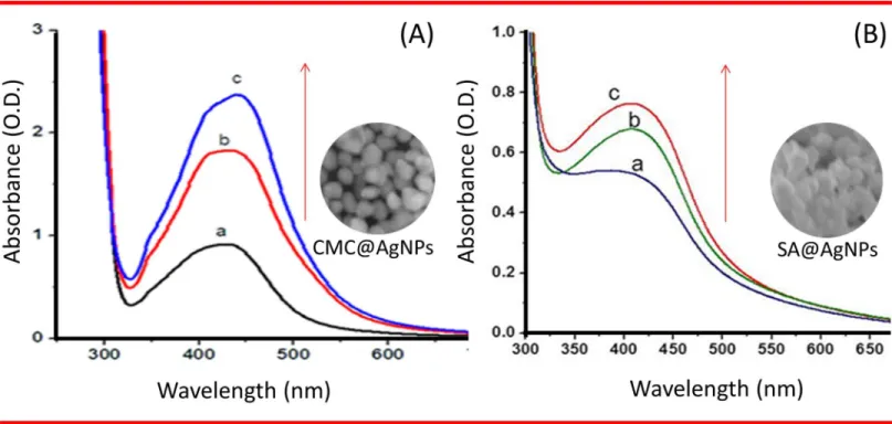
In CMC@AgNPs, characteristic peaks were observed at 440, 430, and 425 nm for different vol-umes of aniline (150, 100, and 50μL, respectively) as shown inFig 1A. The absorption bands may be represented by the surface plasmon resonance (SPR) of AgNPs. The broad nature of the peaks indicated the formation of different shaped AgNPs, which was further confirmed by the SEM images. Based on the results, 100μL of aniline was considered the optimal volume. Evaluation of SA@AgNPs showed absorbance peaks at 430, 425, and 415 nm for 150, 100, and 50μL of aniline, respectively (Fig 1B). The characteristic bands confirm the synthesis of silver crystals. A gradual increase in the peaks was observed, inferring an increase in the size of the crystals. Based on these findings, 150μL of aniline was considered the optimal volume for SA@AgNPs.
Characteristic peaks at 415 nm indicating surface plasma resonance of AgNPs were obtained for all concentrations of CMC. Peaks were observed at 415, 435, 440, and 445 nm for 0.5, 1, 1.5, and 2% CMC, respectively (Fig 2A). These peaks signified the SPR of the AgNPs.
Fig 1. Ultraviolet–Visible spectroscopy.(A) CMC@AgNPs at (a) 50μl, (b) 100μl, (c) 150μl and (B) SA@AgNPs at 50μl (a), 100μl (b) and 150μl (c) of
aniline. Arrow indicates direction of spectral changes.
There was a gradual decrease in absorbance, which implied an increase in size and decrease in the rate of synthesis of the biopolymer capped AgNPs. Hence, 1% (w/v) was the minimum per-centage of CMC required to coat AgNPs. As shown inFig 2B, the maximum absorbance was obtained for both 0.5% and 1% SA. Based on these findings, 1% (w/v) SA was considered the optimum percentage for coating AgNPs. The broad nature of the peaks suggested different shaped nanoparticles, which was validated by the SEM images. The UV-Vis spectral data of CMC@AgNPs and SA@AgNPs (Fig 3A & 3B) for different heating times (30 s to 240 s) showed that the absorbance also increased with increasing time of heat treatment. These findings implied that the quantity of nanoparticles synthesized increased with the increase in heating time. Based on these results, heating for 240 s was considered optimal for synthesis of biopoly-mer capped AgNPs.
Following optimization, the biopolymer capped AgNPs were synthesized and characterized by SEM for morphological analysis. The SEM images indicated that the polygon shaped CMC@AgNPs had a size of 50–100 nm. For SA@AgNPs, nanoparticles with encapsulation of SA were observed (Fig 4A & 4B) in the range of 100–150 nm. No aggregation of AgNPs was observed in both cases. The distribution patterns were analyzed by scanning using ImageJ soft-ware, which showed a spacious distribution for CMC@AgNPs, whereas close distribution was observed for SA@AgNPs (Lower panels inFig 4). Synthesis of AgNPs usually involves a two-stage process, formation of an atom and atom polymerization. Initially, biopolymers reduce the percentage of metal ions in the solution and catalyze with the reduction of remaining metal ions, where the atoms formed act as a nucleation core. Consequently, the atoms amalgamate, leading to metal cluster formation, with formation of larger particles as the surface ions that are continuously reduced until high standards of nucleation are attained.
Fig 2. Ultraviolet–Visible spectroscopy.(A) CMC@AgNPs at (a) 0.5%, (b) 1%, (c) 1.5%, and (d) 2% (B) CMC. (II) SA@AgNPs at (a) 0.5%, (b) 1%, (c)
1.5%, and (d) 2% SA. Arrow indicates direction of spectral changes.
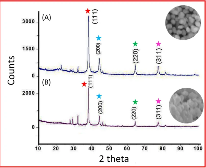
In addition, the polymer interacts with the metal particles, which stabilizes and prevents additional coalescence [42–50]. The results of XRD analysis data for CMC@AgNPs and SA@AgNPs are shown inFig 5. Distinct characteristic peaks at 38.1°, 44.3°, 64.5°, and 77.4° were obtained representing the crystallographic planes (111), (200), (220), and (311), respectively, of the silver nanocrystals [43,46]. The lattice constant calculated from the diffracted spectrum was a =4.0857 Å and the resultant data matched that in JCPDS file no. 01-087-0717. The mean crys-tallite diameter (D) of the biopolymer capped AgNPs formed in the reduction process is deter-mined using Scherrer’s equation Dp= Kλ/β1/2cosθand is estimated as 87.82 nm and 117.09 nm
for CMC@AgNPs and SA@AgNPs, respectively, in which K is the shape dependent Scherrer’s constant (0.94),λis X-ray wavelength (1.5406 Å),β1/2is X-ray line full width at half maximum,
andθis the Bragg angle. It remains the same with altering experimental conditions. As previously reported by Campi et al. [41] spatial correlations between primary particles and the dynamic fractal geometry with time evolution influenced the polymer-silver matrix and determined the mechanism of aggregation and the morphological features of the nanostructures formed. This might be the reason for the intense diffraction line of Ag with polymer aggregation.
The peak at 1432 cm-1was absent from CMC@AgNPs, indicating involvement of symmet-ric COO−in formation of the coating. The peak shift to 1598 cm-1from 1630 cm-1was due to
involvement of C = C in CMC hydrogel formation on the surface of the AgNPs (Fig 6A). In SA@AgNPs, the FTIR spectra (Fig 6B) showed peaks at 3377 cm-1and 3407 cm-1
correspond-ing to OH groups, as well as a peak at 1614 cm-1corresponding to COO−groups. A blue shift
was observed from 1401 cm-1to 1386 cm-1, which confirmed participation of the CO group in
creation of the coating on the surface of the AgNPs. A peak shift from 1113 cm-1to 1097 cm-1 confirmed involvement of the CO group in formation of the biopolymer network. These find-ings are in agreement with those of previous studies [8,37].
Fig 3. Ultraviolet–Visible spectroscopy.(A) CMC@AgNPs at (a) 30s, (b) 60s, (c) 90s, (d) 120s, (e) 150s, (f) 180s, (g) 210s and (h) 240s. (B) SA@AgNPs
at (a) 30s, (b) 60s, (c) 90s, (d) 120s, (e) 150s, (f) 180s, (g) 210s and (h) 240s. Arrow indicates direction of spectral changes.
Antibacterial assay of biopolymer capped AgNPs
The bacterial cell membrane consists of sulfur-containing proteins and phosphorus containing nuclear elements, which are the target sites for AgNPs as Ag has a higher tendency to bind with these elements [51,52]. AgNPs form a region of low molecular weight in the core of the bacte-rial cell and attack the respiratory chain process, resulting in cell death. AgNPs further release silver ions from bacterial cells, increasing bactericidal activity [53–56].
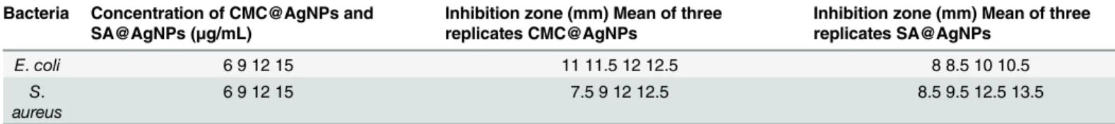
Biopolymer capped AgNPs exerted effective antibacterial activity against both Gram-posi-tive bacteria (S.aureus) and Gram-negative bacteria (E.coli). The inhibition zones (mm) around each well for different concentrations of AgNPs are shown inTable 2. Comparison of the maximum inhibitory zones for both Gram-negative and Gram-positive bacteria for differ-ent biopolymer capped AgNPs against gdiffer-entamicin (15μg/mL) found that CMC@AgNPs and SA@AgNPs were effective in inhibiting growth of Gram-negative and Gram-positive bacteria. CMC@AgNPs showed more activity toward Gram-negative than Gram-positive bacteria.
Fig 4. Scanning electron microscopic images.(A) CMC@AgNPs and (B) SA@AgNPs.
Several mechanisms of action by silver ions were thought to include structural changes in the cell wall of bacteria and interactions with thiol groups in proteins and enzymes and interrup-tion of DNA replicainterrup-tion due to damage of the DNA [22].
Similarly, in the biofilm assay CMC@AgNPs was more active againstE.coliand SA@AgNPs were more active againstS.aureus(Fig 7A & 7B). Importantly, the increase in the concentra-tion of CMC@AgNPs and SA@AgNPs caused a significant (P<0.05) increase in biofilm inhi-bition relative to that grown in the negative control. The higher resistance observed in Gram-negative bacteria compared with Gram-positive bacteria was due to differences in cell mem-brane polarity. Gram-positive bacteria have a thicker peptidoglycan layer consisting of chains of short peptide cross linked linear polysaccharide, which restricts the entry of CMC@AgNPs into cells, preventing cell damage. However, for Gram-negative bacteria, CMC@AgNPs attach easily to the thinner peptidoglycan layer causing rupture of the cell wall, resulting in cell death.
Fig 5. X-ray diffraction analysis.(A) CMC@AgNPs and (B) SA@AgNPs. Peak comparison is indicated by respective colored stars.
SA@AgNPs showed more activity toward Gram-positive than Gram-negative bacteria, which might have been due to the cell membrane polarity response to SA@AgNPs. Shilpa Sharma et al. [57] reported that Gram-negative bacteria have a higher negative surface charge com-pared with positive bacteria, in turn the alginate AgNPs will interact less with Gram-negative bacteria compared with Gram-positive bacteria. CMC and SA reportedly have no antibacterial inhibition activity [58,59], however they direct AgNPs to attack bacteria. Several studies elucidating the antibacterial effect have been proposed. Different antibacterial mecha-nisms of action include interference with cell wall synthesis, inhibition of protein synthesis, interference with nucleic acid synthesis, and inhibition of a metabolic pathway [60–63]. Fig 6. Fourier transform infrared spectra.(A) CMC—(a) Pure CMC and (b) CMC@AgNPs. (B) SA—(a) Pure SA and (b) SA@AgNPs.
doi:10.1371/journal.pone.0157612.g006
Table 2. Antibacterial activities of different concentrations of CMC@AgNPs and SA@AgNPs by disc agar diffusion method.
Bacteria Concentration of CMC@AgNPs and SA@AgNPs (μg/mL)
Inhibition zone (mm) Mean of three replicates CMC@AgNPs
Inhibition zone (mm) Mean of three replicates SA@AgNPs
E.coli 6 9 12 15 11 11.5 12 12.5 8 8.5 10 10.5
S. aureus
6 9 12 15 7.5 9 12 12.5 8.5 9.5 12.5 13.5
Conclusion
The microwave method is a green approach to synthesis of different biopolymer capped AgNPs in a specific minute, instead of hours or even days. The microwave method is currently considering a rapid increase in acceptance as a technique for enhancing facile synthesis of AgNPs using CMC and SA. Formation of polymer capped AgNPs under different experimental conditions was observed by UV-Vis spectroscopy and optimized. XRD analysis confirmed the purity of the samples. Successful capping of the AgNPs with the biopolymers was confirmed by comparing the FTIR spectra of pure biopolymers to those of biopolymer capped AgNPs. The size of the synthesized CMC@AgNPs and SA@AgNPs was approximately 100–150 nm. As shown in our results, polymer capping on the silver showed transition changes due to alter-ations in absorbance, and, upon changing the conditions, clear transition changes were observed between alginate and cellulose capping on AgNPs. Different biopolymer capped AgNPs showed potent antibacterial activity against both Gram-negative and Gram-positive bacteria. However, CMC@AgNPs were more potent at inhibiting the growth of Gram-negative bacteria than positive bacteria, and SA@AgNPs showed more inhibition toward Gram-positive than Gram-negative bacteria. These biopolymers would protect humans from the toxicity of AgNPs if used for biomedical applications. In the case of industrial applications, bio-polymer capped AgNPs can be used as surface coatings to prevent bacterial adhesion, especially in food packing applications. In addition, the heating methods can address the problems of heating in homogeneity in conventional thermal techniques, as it provides increased reaction kinetics, rapid initial heating, clean reaction products with rapid consumption of starting mate-rials, and higher yields. Hence, biopolymer capped AgNPs can be used for industrial food and biomedical applications.
Fig 7. (A) Antibiofilm efficacy ofCMC@AgNPsand (B)SA@AgNPsagainst bothE.coliandS.aureus.Error bars represent the standard deviation.* Significantly different (P<0.05) from the positive control.
Acknowledgments
We sincerely thank the Nanotechnology Research Centre, Interdisciplinary School of Indian System of Medicine and Department of Physics and Nanotechnology of SRM University for performing SEM, FTIR, and XRD analysis, respectively. We also thank the School of Bioengi-neering, SRM University for providing the facilities for conduct of these studies. The author would like to thank Inha University Research grant for proof reading support.
Author Contributions
Conceived and designed the experiments: PV KP CHS. Performed the experiments: PV KP CHS SA GVK. Analyzed the data: PV KP SCBG YC PA GVK. Contributed reagents/materials/ analysis tools: PV KP. Wrote the paper: PV SCBG PA.
References
1. Petros RA, DeSimone JM (2010) Strategies in the design of nanoparticles for therapeutic applications. Nat Rev Drug Discov 9: 615–27. doi:10.1038/nrd2591PMID:20616808
2. Lunhong A, Jing J (2013) Catalytic reduction of 4-nitrophenol by AgNPs stabilized on environmentally benign macroscopic biopolymer hydrogel. Bioresour Technol 132: 374–377. doi:10.1016/j.biortech. 2012.10.161PMID:23206807
3. Li N, Huang J, Dufresne A (2012) Preparation, properties and applications of polysaccharide nanocrys-tals in advanced functional nanomaterials: a review. Nanoscale 4: 3274–3294. doi:10.1039/
c2nr30260hPMID:22565323
4. Gopinath SCB, Awazu K, Fons P, Tominaga J, Kumar PKR (2009) A sensitive multilayered structure suitable for biosensing on the BioDVD platform. Anal Chem 81: 4963–4970. doi:10.1021/ac802757z PMID:19453160
5. Parlak O, Seshadri P, Lundstrom I, Turner APF, Tiwari A (2014) Two-dimensional gold-tungsten disul-phide biointerface for high-throughput electrocatalytic nano-bioreactors. Adv Mater Interfaces 1: 1400136.
6. Wahab R, Dwivedi S, Khan F, Mishra YK, Hwang IH, Shin HS, et al. (2014). Statistical analysis of gold nanoparticle-induced oxidative stress and apoptosis in myoblast (C2C12) cells. Colloids Surf B 123: 664–672.
7. Raveendran P, Fu J, Wallen SL (2003) Completely Green synthesis and stabilization of metal nanopar-ticles. J Am Chem Soc 125: 13940–13941. PMID:14611213
8. Xihui Z, Yanzhi X, Qun L, Xiaomei M, Fengyu Q, Cunzhen G, et al (2014) Microwave-assisted synthesis of AgNPs using sodium alginate and their antibacterial activity. Colloids Surf A 444: 180–188.
9. Lee KY, Mooney DJ (2012) Alginate: properties and biomedical applications. Progr Polym Sci 37: 106–
126.
10. Avasthi DK, Mishra YK, Kabiraj D, Lalla NP, Pivin JC (2007) Synthesis of metal–polymer nanocompo-site for optical applications. Nanotechnology 18: 125604.
11. Yang J, Pan J (2012) Hydrothermal synthesis of AgNPs by sodium alginate and their applications in surface-enhanced Raman scattering and catalysis. Acta Mater 60: 4753–4758.
12. Mohapatra S, Mishra YK, Ghatak J, Kabiraj D, Avasthi DK (2008) Surface plasmon resonance of Ag nanoparticles embedded in partially oxidized amorphous Si matrix. J Nanosci Nanotechnol 8: 4285–
4289. PMID:19049219
13. Dwivedi S, Saquib Q, Al-Khedhairy AA, Ahmad J, Siddiqui MA, Musarrat J (2015) Rhamnolipids functio-nalized AgNPs-induced oxidative stress and modulation of toxicity pathway genes in cultured MCF-7 cells. Colloids Surf B 132: 290–8.
14. Kalipada B, Dipanwita Maity MD, Masud RM (2014) Antibacterial activity of Ag–Au alloy NPs and chem-ical sensor property of Au NPs synthesized by dextran, Carbohydr Polym 10: 151–157.
15. Fard JK, Jafari S, Eghbal MA (2015) A review of molecular mechanisms involved in toxicity of nanopar-ticles. Adv Pharm Bull 5: 447–454. doi:10.15171/apb.2015.061PMID:26819915
16. Sarmast MK, Salehi H. Silver nanoparticles: An influential element in plant nanobiotechnology. Mol Bio-technol. 2016 (In Press)
18. Bhui DK, Pyne S, Sarkar P, Bar H, Sahoo GP, Misra A (2011) Temperature controlled synthesis of sil-ver nanostructures of variable morphologies in aqueous methyl cellulose matrix. J Mol Liq 158: 170–
174.
19. Darroudi M, Ahmad MB, Abdullah AH, Ibrahim NA (2011) Green synthesis and characterization of gela-tin-based and sugar-reduced AgNPs. Int J Nanomed 6: 569–574.
20. Mohan YM, Raju KM, Sambasivudu K, Singh S, Sreedhar B (2007) Preparation of acacia-stabilized AgNPs: a green approach. J Appl Polym Sci 3375–3381.
21. Li S, Zhang YY, Xu XJ, Zhang LN (2011) Triple helical polysaccharide induced good dispersion of AgNPs in water. Biomacromolecules 12: 2864–2871. doi:10.1021/bm2001439PMID:21542604
22. Velusamy P, Das J, Pachaiappan R, Vaseeharan B, Pandian K (2015) Greener approach for synthesis of antibacterial silver nanoparticles using aqueous solution of neem gum (Azadirachta indicaL.). Ind Crop Prod 66: 103–109.
23. Moulton MC, Braydich-Stolle LK, Nadagouda MN, Kunzelman S, Hussain SM, Varma RS (2010) Syn-thesis, characterization and biocompatibility of‘‘green”synthesized AgNPs using tea polyphenols, Nanoscale 2010; 2: 763–770. doi:10.1039/c0nr00046aPMID:20648322
24. Kou J, Varma RS (2013) Speedy fabrication of diameter-controlled Ag nanowire using glycerol under microwave irradiation conditions, Chem Commun 49: 692–694.
25. Li SM, Jia N, Zhu N, Ma JF, Xu F, Wang B, et al. (2011) Rapid microwave assisted preparation and characterization of cellulose-silver nanocomposites. Carbohydr Polym 83: 422–429.
26. Shankar S, Rhim JW (2015) Amino acid mediated synthesis of silver nanoparticles and preparation of antimicrobial agar/silver nanoparticles composite films. Carbohydr Polym 130: 353–363. doi:10.1016/ j.carbpol.2015.05.018PMID:26076636
27. Rastogi PK, Ganesan V, Krishnamoorthi S (2012) Microwave assisted biopolymer stabilized synthesis of AgNPs and its application in the degradation of environmental pollutants. Mater Sci Eng B 177: 456–
461.
28. Ali K, Ahmed B, Dwivedi S, Saquib Q, Al-Khedhairy AA, Musarrat J (2015) Microwave accelerated green synthesis of stable silver nanoparticles withEucalyptus globulusleaf extract and their antibacte-rial and antibiofilm activity on clinical isolates. PLoS One 10: e0131178. doi:10.1371/journal.pone. 0131178PMID:26132199
29. Wei DW, Sun WY, Qian WP, Ye YZ, Ma XY (2009) The synthesis of chitosan-based AgNPs and their antibacterial activity. Carbohydr Res 344: 2375–2382. doi:10.1016/j.carres.2009.09.001PMID: 19800053
30. Dipak KB, Ajay M (2012) Synthesis of worm like AgNPs in methyl cellulose biopolymeric matrix and its catalytic activity. Carbohydr Polym 89: 830–835. doi:10.1016/j.carbpol.2012.04.017PMID:24750868
31. Carneiro-da-Cunha MG, Cerqueira MA, Souza BWS, Carvalho S (2010) Physical and thermal proper-ties of a chitosan/alginate nanolayered PET film. Carbohydr Polym 82: 153–159.
32. Hebeish AA, El-Rafie MH, Abdel-Mohdy FA, Abdel-Halim ES, Emam HE (2010) Carboxymethyl cellu-lose for green synthesis and stabilization of silver nanoparticles. Carbohydr Polym 82: 933–941.
33. Dwivedi S, Wahab R, Khan F, Mishra YK, Musarrat J, Al-Khedhairy AA (2014) Reactive Oxygen Spe-cies Mediated Bacterial Biofilm Inhibition via Zinc Oxide Nanoparticles and Their Statistical Determina-tion. PLoS One 9: e111289. doi:10.1371/journal.pone.0111289PMID:25402188
34. Bauer AW, Kirby WMM, Sherris JC, Turck M (1996) Antibiotic susceptibility testing by a standardized single disk method. Am J Clin Pathol 36: 493–496.
35. Deepika H, Jacob L, Mallikarjuna NN, Rajender SV (2013) Greener techniques for the synthesis of AgNPs using plant extracts, enzymes, bacteria, biodegradable biopolymers, and microwaves. ACS Sustainable Chem Eng 1: 703–712.
36. Abdel-Halim ES, Al-Deyab SS (2011) Utilization of hydroxypropyl cellulose for green and efficient syn-thesis of AgNPs, Carbohydr Polym 86: 1615–1622.
37. Hebeish A, Hashem M, El-Hady M, Sharaf S (2013) Development of CMC hydrogels loaded with AgNPs for medical applications. Carbohydr Polym 92: 407–413. doi:10.1016/j.carbpol.2012.08.094 PMID:23218313
38. Kumar M, Reddy GB (2016) Stability-Inspired Entrapment of Ag Nanoparticles in ZrO2 Thin films. Plas-monics 11: 261–267.
39. Kumar M, Reddy GB (2010) Effect of atmospheric exposure on the growth of citrate-capped silver nanoparticles. Physica E 42: 1940–1943.
41. Campi G, Mari A, Pifferi A, Amenitsch H, Fratini M, Suber L (2011) Control of silver-polymer aggregation mechanism by primary particle spatial correlations in dynamic fractal-like geometry. Nanoscale 3: 3774–3779. doi:10.1039/c1nr10474hPMID:21826362
42. Zhao X, Xia Y, Li Q, Ma X, Quan F, Geng C, et al. (2014) Microwave-assisted synthesis of silver nano-particles using sodium alginate and their antibacterial activity, Colloids Surf A 444: 180–188.
43. Kumar M, Reddy GB (2009) Ag:ZrO2nanocomposite thin films derived using a novel sol–gel technique.
Phys Status Solidi B 246:2232–2237.
44. Kumar M, Suchand Sandeep CS, Kumar G, Mishra YK, Philip R, Reddy GB (2014) Plasmonic and Non-linear Optical Absorption Properties of Ag:ZrO2Nanocomposite Thin Films. Plasmonics 9: 129–136. 45. Kumar M, Kulriya PK, Pivin JC, Avasthi DK (2011) Evolution and tailoring of plasmonic properties in Ag:
ZrO2nanocomposite films by swift heavy ion irradiation. J Appl Phys 109: 044311.
46. Mishra YK, Mohapatra S, Kabiraj D, Mohanta B, Lalla NP, Pivin JC, et al. (2007) Synthesis and charac-terization of Ag nanoparticles in silica matrix by atom beam sputtering. Scripta Mater 56: 629–632.
47. Liangbao Y, Yuhua S, Anjian X, Baocheng Z (2007) Facile Size-Controlled Synthesis of Silver Nano-particles in UV-Irradiated Tungstosilicate Acid Solution. J Phys Chem C 111: 5300–5308.
48. Rao YN, Banerjee D, Datta A, Das SK, Guin R, Saha A (2010) Gamma irradiation route to synthesis of highly re-dispersible natural polymer capped silver nanoparticles. Radiat Phys Chem 79: 1240–1246.
49. Sadanand P, Gopal KG, Karuna KN (2012) Green synthesis of biopolymer–silver nanoparticle nano-composite: An optical sensor for ammonia detection. Int J Biol Macromol 51: 583–589. doi:10.1016/j. ijbiomac.2012.06.033PMID:22750580
50. Seo SY, Lee GH, Lee SG, Jung SY, Lim JO, Choi JH (2012) Alginate-based composite sponge contain-ing AgNPs synthesized in situ. Carbohydr Polym 90: 109–115. doi:10.1016/j.carbpol.2012.05.002 PMID:24751017
51. Won KS, Ji HY, Won HP (2006) Antimicrobial cellulose acetate nanofibers containing AgNPs. Carbo-hydr Polym 65: 430–434.
52. Shu-Ming L, Ning J, Ming-Guo M, Zhe Z, Qing-Hong L, Run-Cang S (2011) Cellulose–silver nanocom-posites: Microwave-assisted synthesis, characterization, their thermal stability, and antimicrobial prop-erty, Carbohydr Polym 86: 441–447.
53. Feng QL, Wu J, Chen GQ, Cui FZ, Kim TN, Kim JO (2000) A mechanistic study of the antibacterial effect of silver ions onEscherichia coliandStaphylococcus aureus. J Biomed Mater 52: 662–668.
54. Morones JR, Elechiguerra JL, Camacho A, Ramirez JT (2005) The bactericidal effect of silver nanopar-ticles. Nanotechnology 16: 2346–2353. doi:10.1088/0957-4484/16/10/059PMID:20818017
55. Sondi I, Salopek-Sondi B (2007) Silver nanoparticles as antimicrobial agent: a case study onE.colias a model for Gram-negative bacteria. J Colloid Interface 275: 177–182.
56. Hsueh Y-H, Lin K-S, Ke W-J, Hsieh C-T, Chiang C-L, Tzou D-Y, et al. (2015) The Antimicrobial proper-ties of silver nanoparticles inBacillus subtilisare mediated by released Ag+ ions. PLoS One 10: e0144306. doi:10.1371/journal.pone.0144306PMID:26669836
57. Shilpa S, Pallab S, Arun C, Siddhartha SG (2012) Fabrication of antibacterial silver nanoparticle—
sodium alginate–chitosan composite films. RSC Adv 2: 5837–5843.
58. Kai L, Xinxing L, Lihui C, Liulian H, Shilin C, Huangwei W (2013) Preparation of microfibrillated cellu-lose/chitosan−benzalkonium chloride biocomposite for enhancing antibacterium and strength of sodium alginate films. J Agric Food Chem 61: 6562–6567. doi:10.1021/jf4010065PMID:23750871
59. Guzman M, Dille J, Godet S (2012) Synthesis and antibacterial activity of silver nanoparticles against Gram-positive and gram-negative bacteria. Nanomedicine 8: 37–45. doi:10.1016/j.nano.2011.05.007 PMID:21703988
60. Feng QL, Wu J, Chen GQ, Cui FZ, Kim TN, Kim JO (2000) A mechanistic study of the antibacterial effect of silver ions onE.coliandS.aureus. J Biomed Mater Res 52: 662–668. PMID:11033548
61. Benjamin LO, Francesco S (2015) Antibacterial activity of silver nanoparticles: A surface science insight. Nano Today 10: 339–354.
62. Vishvanath T, Manoj KK, Monalisa T, Swati B, Manish K (2014) Anti-bacterial activity of polyvinyl pyrro-lidone capped silver nanoparticles on the carbapenem resistant strain ofAcinetobacter baumannii. J Nanomed Nanotechnol 5: 6.