Article
ISSN 0102-695X
doi: 10.1590/S0102-695X2011005000016 Received 24 Feb 2010
Accepted 1 Sep 2010 Available online 4 Feb 2011
Betania Barros Cota,
*,1Susana Johann,
1Djalma M. Oliveira,
2Ezequias P. Siqueira,
1Elaine M. Souza-Fagundes,
3Patrícia S.
Cisalpino,
4Tânia M. A. Alves,
1Carlos Leomar Zani
11Laboratório de Química de Produtos Naturais, Centro de Pesquisas René
Rachou-Fiocruz, Brazil,
2Universidade Estadual do Sudoeste de Bahia, Campus Jequié, Brazil,
3Departamento de Fisiologia e Biofísica, Instituto de Ciências Biológicas,
Universidade Federal de Minas Gerais, Brazil,
4Departamento de Microbiologia, Instituto de Ciências Biológicas,
Universidade Federal de Minas Gerais, MG, Brazil.
Abstract: Organic extracts from leaves and stems of Stillingia oppositifolia
Baill. ex Müll. Arg., Euphorbiaceae, were screened for antifungal and cytotoxic properties. The extracts presented Minimum Inhibitory Concentration values
around 250 μg.mL-1 against Candida krusei and Candida tropicalis, and around
63 μg.mL-1 for Paracoccidioides brasiliensis. They were tested on three
human cell lines (UACC-62, MCF-7, and TK-10), disclosing GI50 values, (concentration able to inhibit 50% of the cell growth) ranging from 50 to 100
μg.mL-1. Organic extract from stems furnished hexanic, dichloromethanic and
aqueous phases after partition. Chromatographic fractionation of the hexanic
soluble phase of the stems yielded aleuritolic acid 3-acetate, β-sitosterol, 3-epi-β-amyrin, β-amyrone and palmitic acid. These compounds showed antifungal
and cytotoxic activities in the same range as the organic crude extract and low toxic effect against mononuclear cells obtained from human peripheral blood. This is the first report on chemical and biological potential of S. oppositifolia. Keywords:
antifungal cytotoxic Euphorbiaceae
Stillingia oppositifolia
terpenoids
Introduction
Stillingia oppositifolia Baill. ex Müll. Arg., popularly known as “leiterinho” in Brazil, grows at altitudes ranging from 600 to 1000 m in Araucaria forests of Minas Gerais and Rio Grande do Sul states
in Brazil (Rogers, 1951). The genus Stillingia belongs
to the Euphorbiaceae family, one of the largest families of flowering plants, with approximately 300 genera and 8000 species (Webster, 1994; Radcliffe-Smith, 2001).
Species from this family are known for producing a large amount of diterpenes and triterpens, some of them with important pharmacological activities such as antitumor and anti-inflammatory properties (Aylward et al., 2001; Aylward & Parsons,
2002). Only few works on the chemistry of Stillingia
species have been reported in the literature. Toxic and irritant daphnane and tigliane diterpene esters were isolated from the roots of S. sylvatica (Adolf & Hecker, 1980), while pimarane, kaurane, atisane and tonantzitlolone diterpenes were isolated from the roots of S. sanguinolenta, a plant used to prepare a poultice applied after childbirth (Draeger et al., 2007).
In an effort to expand the spectrum of active agents from natural resources, we screened extracts
from stems and leaves of Stillingia oppositifolia for
antifungal and cytotoxic properties. We report herein the first biological investigation on extracts and the
isolation of five compounds from S. oppositifolia.
Material and Methods
General experimental procedures
Nuclear Magnetic Resonance experiments (1H
and 13C, DEPT, HMQC and HMBC) were recorded on a
Brucker DRX 400 spectrometer using standard Bruker pulse sequences and conditions. Electron impact (70 eV) low-resolution mass spectra (EI-MS) were obtained on a Shimadzu QP5050A equipped with a direct insertion
probe. Centrifugal Thin-Layer Chromatography (CTLC)
on 1 mm thick silica gel 60 PF254 layer was performed
on a 7924T model Chromatotron device (Harrison Research). Medium pressure liquid chromatography
(MPLC) was performed on a 250 x 20 mm Büchi glass column with the mobile phases pumped at 5 mL.min-1
flow rate. Column chromatography (CC) was carried
out on silica gel (Merck). Thin-Layer Chromatography (TLC) was run in precoated commercial plates on
pre-saturated chamber using solvents mixtures in different proportions of: a) chloroform:methanol:water
(CHCl3:MeOH:H2O), b) hexane:dichloromethane
(Hex:DCM) or c) hexane:ethyl acetate (Hex:EtOAc). The spots were visualized under visible, UV light at 254 nm and 360 nm, and after spraying the plate with
an ethanol solution of vanilin-H2SO4 and heating with a
heated plate.
Plant collection
Leaves and stems from Stillingia oppositifolia Baill. ex Müll. Arg., Euphorbiaceae, were collected in São Gonçalo do Rio Preto, Minas Gerais, Brazil, on October 2003. A voucher specimen (BHCB 87513) was deposited in the BHCB Herbarium at the Instituto de Ciências Biológicas, Universidade Federal de Minas Gerais, Belo Horizonte, Brazil.
Extract preparation and isolation of compounds
Leaves and stems of S. oppositifolia were macerated in DCM:MeOH (1:1) at room temperature and the solutions were concentrated under vacuum in a rotary evaporator at temperatures below 45 °C. The residual solvent was removed in a vacuum centrifuge at 40 °C. An aliquot of organic crude extract from stems
(23 g) was suspended in MeOH:H2O (1:1) and extracted
successively with Hex and DCM. This procedure afforded the phases denominated Hex (3.8 g), DCM (4.2 g) and Aqueous (11.2 g). Hex phase (3.5 g) was subjected to CC (70-230 mesh) using Hex, Hex:DCM, DCM:MeOH mixtures as eluent to yield 27 subfractions.
Subfraction 13 (120 mg) was subjected to MPLC on
silica gel (25-40 mesh) using a gradient system from Hex to Hex:EtOAc (80:20) and furnished compounds
1 (100 mg) and 2 (4 mg). Subfraction 17 (140 mg)
was fractionated by successive CC using mixtures of
Hex:DCM and CHCl3:EtOAc, to afford compound 3
(15 mg). Subfractions 19-20 were grouped (150 mg)
and subjected to centrifugal TLC on a Chromatotron
system using Hex, DCM and MeOH mixtures and gave a material (25 mg) that after crystallization on
hexane furnished compound 4 (3 mg). Subfraction
21 (840 mg) was fractionated on a Sephadex LH-20
column using ethanol as eluent to afford 10 groups of fractions. Groups 6-8 weighting 313 mg was further chromatographed on silica gel CC (70-230 mesh), eluted in Hex, Hex:DCM, and Hex:EtOAc at different proportions. This procedure resulted in the isolation of
5 mg of compound 5.
Analysis of volatiles by Gas Chromatography-Mass Spectrometry (GC-MS)
The volatiles present in the hexanic soluble
phase of S. oppositifolia were adsorbed on a Solid
Phase Micro-Extraction (SPME) sampling device and analyzed by GC-MS. Briefly, 1 mg of the Hex phase
was transferred to a 2 mL glass vial, closed with a cap
sealed with a teflon coated septum (Supelco, USA) and placed in a heat block adjusted to 90 °C. A SPME fiber (PDMS/DVB TM 65 µm, Supelco, USA) was inserted through the septum and left in the headspace during 5 min. Before use, the fiber was preconditioned at 230 °C during 30 min in the GC injector port. GC-MS analysis were performed on a Shimadzu QP-5050A (SHIMADZU, JP) instrument, equipped with a PTE-5™ column (30 m, 0.25 mm, 0.25 µm, Supelco, USA). The following conditions were employed for analysis: helium at 22.3
mL.min-1, as carrier gas; injector temperature, 230 °C;
column temperature, 3 min at 80 °C, 80-300 °C at 7 °C.min-1, 5 min at 300 °C. The split valve was closed
during the first minute of injection and then opened, with a 1:10 ratio. The mass detector was set to scan from 50 to 500 atomic mass unit, at a rate of 2 scans.sec-1. Data acquisition and handling was done via CLASS
5000 Shimadzu software. Raw data files were analyzed by Automated Mass Deconvolution and Identification System software (AMDIS), version 2.1, supplied by National Institute of Standards and Technology (NIST, USA). The compounds identification were performed by comparison of the experimental spectra with those stored in the NIST/EPA/NIH library version 2.0 using the NIST Mass Spectral Search Program.
Cytotoxicity assays with human cancer cell lines
The organic crude extracts, phases and compounds assays were performed using the tumor cell lines UACC-62 (human melanoma cancer), MCF-7 (human breast cancer) and TK-10 (human renal cancer). These cell lines were purchased from the National Cancer Institute, Maryland, USA. The cell toxicity assays were run according to the protocols established at NCI using the sulphorhodamine colorimetric assay (Monks et al., 1991). The samples were dissolved in DMSO aqueous
0.1% at concentrations of 200 μg.mL-1.
All assays were run in triplicate wells and
repeated at least once. Etoposide at 1.6 μg.mL-1, culture
medium without samples and culture medium with DMSO 0.1% were controls. Results were expressed in terms of the growth inhibition percentage (%), where the sample tested was considered cytostatic from 0-99% and cytocidal from 100-200%.
Fungal strains
(ATCC, Rochville, MD, USA) were used: Candida
albicans ATCC 18804, C. krusei ATCC 20298, C.
tropicalis ATCC 750, C. parapsilosis ATCC 22019, C.
glabrata ATCC 2001, Cryptococcus neoformans ATCC
32608 and Paracoccidioides brasiliensis (Pb18), (from
the fungi collection of the Faculty of Medicine of the Universidade de São Paulo, São Paulo, SP, Brazil). The fungi strains were maintained on Sabouraud Dextrose Agar (SDA, Oxoid, Basingstoke, UK) and YPD (Yeast, Peptone and Dextrose).
Antifungal assays
The minimal inhibitory concentration (MIC) was determined on synthetic RPMI medium (Sigma, St
Louis, MO, USA) containing L-glutamine and buffered to pH 7.0 with 0.165 mol.L-1) morpholine propanesulfonic
acid (MOPS; Sigma). Final inocula of 1.5 x 10³ CFU.
mL-1 was prepared using the spectrophotometric dilution
method. Broth microdilution testing was performed in
accordance with the guidelines in the CLSI M27-A2 document (NCCLS, 2002), with modifications proposed
by Johann et al. (2007). Amphotericin B (Sigma, St
Louis, USA) was included as positive antifungal control
being the stock solutions prepared in DMSO. RPMI medium was used without compounds or solvents as a control for growth and sterility. Solvent DMSO at the same volumes used in the assay was used as control for toxicity. After inoculation of fungal strains, the
plates were incubated at 35 °C for 48 h for the Candida
species, at 35 °C for 72 h for C. neoformans and 37 °C
for 72 h for P. brasiliensis. The tests were performed in triplicate. The endpoints were determined visually by comparing the growth in the test wells with that the growth in the drug-free control wells. MIC values
were expressed in μg.mL-1. Extracts and phases were
considered active when they exhibited MIC values less
than or equal to 500 μg.mL-1, while compounds were
considered active when they exhibited MIC values less
than or equal to 100 μg.mL-1.
Cytotoxicity assays with human peripheral blood mononuclear cells
Peripheral blood mononuclear cells (PBMC) were obtained using the protocol described by Gazzinnelli et al. (1983). Briefly, peripheral venous blood from healthy adult volunteers was collected in heparinized vials and the cells separated by centrifugation on Ficoll-Hypaque gradient. Mononuclear cells were collected and washed three times in RPMI-1640 before further processing. The cell suspensions were adjusted to 2x10⁶ cells.mL-1 (2.0x10⁵ cells per well).
All cultures were carried out in RPMI-1640 medium (GIBCO, Grand Island, NY), supplemented with 5%
(v/v) heat-inactivated, pooled human sera type AB
(Flow Laboratories, Royaune, UN) and L-glutamine (2 mM, GIBCO, Grand Island, NY). An antibiotic/
antimicotic solution containing 100 U.mL-1 penicillin, 100 μg.mL-1 streptomycin and 25 μg.mL-1 fungisone (SIGMA, St. Louis, MO) was added. Cells were
incubated with the isolated compounds for 48 h at 37
°C in a humidified atmosphere containing 5% CO2. The
cell viability was determined using the MTT (methyl thiazolyl tetrazolium) based colorimetric assay (Jiang & Xu, 2003). The results were expressed as percent inhibition of the cell viability in relation to the control without test compounds. The results were expressed in terms of percentage of cells prolipheration, where the sample tested was considered inactive from 100-70%; moderate active from 69-40% and active from 39-0%.
Results and Discussion
Antifungal and cytotoxic activities of crude extracts
Few species of Stillingia genus were available
for biological activity. Latex obtained from stems of
S. patagonica, Euphorbiaceae, presented proteolytic
activity showing its potential source for digestive enzymes and anti inflammatory agents (Sequeiros et al., 2003). In the present study we investigated antifungal and cytotoxic activities of the crude extracts
from stems and leaves of S. oppositifolia. The crude
extract of leaves showed, in general, similar activity of that from stems. Organic extracts from stems and leaves inhibited the growth of C. krusei, C. tropicalis
(MIC value of 250 μg.mL-1) and P. brasiliensis (MIC value of 63 μg.mL-1) (Table 1). The crude extracts
presented very low cytostatic activity (around 30%)
at 20 μg.mL-1 against UACC-62 and MCF-7 cell lines.
Nevertheless, the extracts showed cytotoxic activity in a concentration-dependent manner doses (GI50
value of 50-100 μg.mL-1, Table 2) on three cancer
cell lines recommended by NCI. To access if these extracts are toxic to normal human cells they were
tested on leukocytes in the ex-vivo assays. The results
demonstrated that they are not toxic at 20 μg.mL-1
(Table 1).
Table 2. Extracts and compounds concentration (μg.mL-1)
required to inhibit cell growth by 50% (GI50), (means of three determinations).
Extracts/Compounds UACC-62 MCF-7 TK-10
1 > 200 128±29 >200
3 > 200 139±33 >200
4 100±17 88±21 100±13
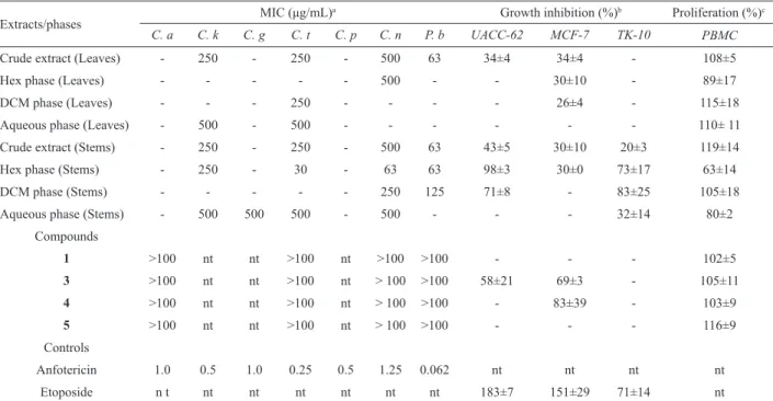
Table 1. Antifungal and cytotoxic activities of extracts, phases and compounds of Stillingia oppositifolia Baill. ex Müll. Arg., Euphorbiaceae.
Extracts/phases MIC (μg/mL)
a Growth inhibition (%)b Proliferation (%)c
C. a C. k C. g C. t C. p C. n P. b UACC-62 MCF-7 TK-10 PBMC
Crude extract (Leaves) - 250 - 250 - 500 63 34±4 34±4 - 108±5
Hex phase (Leaves) - - - 500 - - 30±10 - 89±17
DCM phase (Leaves) - - - 250 - - - - 26±4 - 115±18
Aqueous phase (Leaves) - 500 - 500 - - - 110± 11
Crude extract (Stems) - 250 - 250 - 500 63 43±5 30±10 20±3 119±14
Hex phase (Stems) - 250 - 30 - 63 63 98±3 30±0 73±17 63±14
DCM phase (Stems) - - - 250 125 71±8 - 83±25 105±18
Aqueous phase (Stems) - 500 500 500 - 500 - - - 32±14 80±2
Compounds
1 >100 nt nt >100 nt >100 >100 - - - 102±5
3 >100 nt nt >100 nt > 100 >100 58±21 69±3 - 105±11
4 >100 nt nt >100 nt > 100 >100 - 83±39 - 103±9
5 >100 nt nt >100 nt > 100 >100 - - - 116±9
Controls
Anfotericin 1.0 0.5 1.0 0.25 0.5 1.25 0.062 nt nt nt nt
Etoposide n t nt nt nt nt nt nt 183±7 151±29 71±14 nt
The minus symbol (-) means inactive, nt: not tested, ± variation coeficient. (a): C.a: Candida albicans; C. k: Candida krusei; C. g: Candida glabrata; C. p: Candida parapsilosis; C. t: Candida tropicalis; C. n: Cryptococcus neoformans; P. b: Paracoccidioides brasiliensis. Anfotericin was tested in the range from 30 µg.mL-1 to 0.031 µg.mL-1; (b): UACC-62: human melanoma cancer; MCF-7: human breast cancer; TK-10: human renal cancer. All
samples tested at 20 µg.mL-1. Etoposide was tested at 1.6 µg.mL-1. (c): PBMC: peripheral blood mononuclear cells. All samples tested at 20 µg.mL-1.
Antifungal and cytotoxic activities of phases
The crude extracts of S. oppositifolia were
concentrated and subjected to solvent partitioning to furnish Hex, DCM and aqueous soluble phases.
The hexanic soluble phase (Hex) from stems of S.
oppositifolia exhibited outstanding antifungal activity
against C. neoformans and P. brasiliensis among all
phases (Table 1), indicating that this phase has a good potential as antifungal. Our results showed that Hex phase of stems exhibited the strongest activity against C. tropicalis (MIC value of 30 µg.mL-1), C. neoformans (MIC value of 63 µg.mL-1), P. brasiliensis (MIC 63 µg.mL-1). Dichloromethanic phase (DCM) of stems
presented moderated activity against P. brasiliensis
(MIC value of 125 µg.mL-1). Hex and DCM phases were
cytostatic against melanoma (98 and 71%, respectively) and renal (73 and 83%, respectively) cell lines. Hex phase
presented moderated in vitro cytotoxity effect (around 37%
of inhibition) on freshly isolated PBMC at 20 µg.mL-1.
Aqueous phase presented better activity against C. krusei,
C. glabrata (MIC value of 500 µg.mL-1), than DCM
subfraction, that did not show activity against species of Candida tested and minor activity against C. neoformans
(MIC value of 500 µg.mL-1).
Compounds isolation
Due to interesting biological activity displayed by the hexanic soluble phase from stems, it was chromatographed on a silica gel open column to afford 27 groups. All groups were screened on cytotoxic assays and exhibited the best cytostatic effect against
melanoma cell line (UACC-62) at 20 µg.mL-1 (data not
shown). The groups were chosen to be purified based on the results of cytotoxic assays and their fingerprint
on TLC. To our knowledge, there is not any report
about phytochemical of S. oppositifolia.
Subfractions of Hex phase from stems were
purified by Si gel, Sephadex LH-20, CC or MPLC and
by crystallization to afford three triterpenes: 3-epi -β-amyrin (1), β-amyrone (2), aleuritolic acid 3-acetate (3), one steroid: β-sitosterol (4), and a fatty acid: palmitic acid (5). The spectral properties, including 1H NMR and
13C NMR data, were identical to those previously reported
in the literature (Martin et al., 1984; Mahato & Kundu,
1994; De-Eknamkul & Potduang, 2003; Lima et al., 2004).
Compounds 1 and 2 are oleanane triterpenes and compound
3 is friedooleanane triterpene.
All isolated compounds were inactive against C.
albicans, C. tropicalis and P. brasiliensis at concentration
of 100 µg.mL-1 and showed cytotoxic activity in the same
range than crude extracts against tumoral lines (GI50 values
≥88 μg.mL-1, Table 2). The compounds demonstrated no
toxicity against human leukocytes after 48 h of incubation
Epi-β-amyrin (1) was previously isolated from
leaves of Sebastiania adenophora, Euphorbiaceae
(Macias-Rubalcava et al., 2007) and from bark of
Gelonium multilorum (Row & Rao, 1969). This compound
was able to inhibit Mycobacterium tuberculosis growth
(MIC value of 12.2 μg.mL-1) and showed cytotoxicity against Vero cells (IC50 127.2 μg.ml-1) (Woldemichael et
al., 2004). Compound 2 (β-amyrenone) was isolated from
light petrol extracts of the stems of Macaranga tanarius,
Euphorbiaceae (Hui et al., 1975) and epicuticular wax of Euphorbia cyparissias L., Euphorbiaceae (Hemmers & Gülz, 1989). Crude extract of stems of S. oppositifolia
showed be a good source of epi-β-amyrin while
β-amyrenone, a related isomeric compound possessing
the cetone group at C-3, was isolated as minor compound. Epi-β-amyrin could be a chemical marker of extracts of S. oppositifolia since it is not is frequently isolated on Euphorbiaceae family.
Acetyl aleuritolic acid was obtained from several
Euphorbiaceae species as Jatropha macrorhiza (Torrance
et al., 1977), Croton tonkinensis (Pham & Pham, 2002),
C. cajucara (Maciel et al., 2006), Discoglypremna
caloneura (Nyasse et al., 2006). This triterpene showed tumor-inhibitory properties toward the P-388 lymphocytic leukemia (Torrance et al., 1977). It was active against
against Staphylococcus aureus, Salmonella typhy, Vibrio
cholera, Escherichia coli and Shigella dysentery in
microdilution method (MIC value of 50 μg.mL-1) and
was not cytotoxic to Vero cell lines in vitro (IC50 of 400
μg.mL-1, Mathabe et al., 2008).
Many Euphorbiaceae species belonging to the
genus Acalypha (Wang et al., 2008; Tauiq-Yap et al.,
2000), Bridelia (Yadav & Nigam, 1975), Croton (Palmeira et al., 2006; Santos et al., 2008), Euphorbia (Kong & Min, 1996; Ekpo & Pretorius, 2007; El-Fiky et al., 2008) and Glochidium (Hui & Fung, 1969; Hui & Li, 1976) have
furnished β-sitosterol as a chemical constituent. This
compound showed to induce the macrophage tumoricidal activity, stimulate the lymphocyte blastogenesis (Park et al., 2003) and showed therapeutic angiogenic effects on damaged blood vessels (Choi et al., 2002). This compound showed hypocholesterolemic activity (Day,
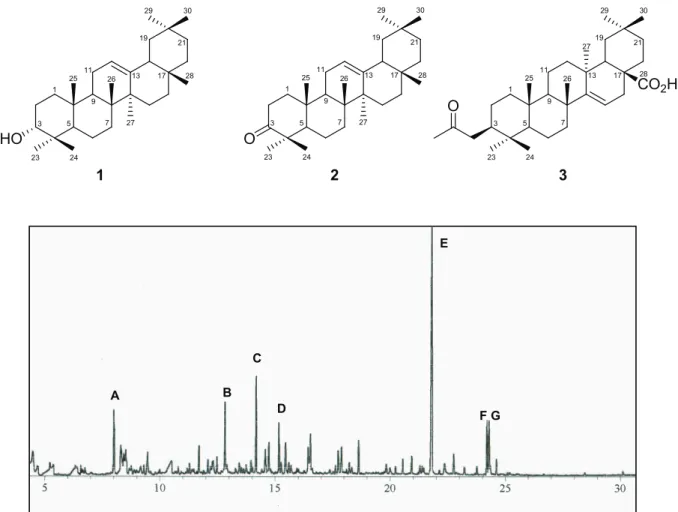
Figure 1. Total ion chromatogram of the hexanic phase from Stillingia oppositifolia, Euphorbiaceae. A: decahydro 2,6-dimethyl naphthalene (6.78%), B: tetradecane (5.25%), C: decahydro1,1,7, trimethyl,4 methylene-1H, cycloprop[e]azulene (8.74%), D: dodecanoic acid methyl ester (4.20%), E: hexadecanoic acid methyl ester (5, 31.93%), F: 9-dodecenoic acid methyl ester (4.53%) and G: octadecanoic acid methyl ester (4.99%).
A
C
B
D
E
F G A
C
B
D
E
F G
HO O
CO2H O
3 1
5
23 24
29 30
19 21
28 17 13
27 26 11
9 25
7 3
1
5
23 24
29 30
19 21
28 17 13
27 26 11
9 25
7 3
1
5
23 24
29 30
19 21
28 17 13 27
26 11
9 25
7
1991), inhibitory activity on human platelet-type
12(S)-lipoxygenase [12(S)-LOX] (Schneider et al., 2004)
and was active against the mutagenicity of N
-methyl-N-nitrosourea and 2-aminoanthracene (Lawson et al., 1989).
Palmitic acid has been detected by GC/MS from oil seeds from various Euphorbiaceae species (Agarwal et al., 1995; Augustus et al., 2002; Mohan, 2009). In according with our observations, palmitic acid was not cytotoxic to three cell lines and human lymphocytes at
20 μg.mL1. Harada et al. (2002) demonstrated that at concentrations ranging from 12.5 to 50 μg.mL-1, palmitic
acid shows selective cytotoxicity to human leukemic cells
(Molt-4, HL-60, K-562), but no cytotoxicity to normal dermal ibroblasts cells. Carballeira (2008) suggests that
the cytotoxicity of palmitic acid to cancer cell lines could be linked to the inhibition of topoisomerase I, since it inhibited this enzyme only in high concentrations.
Gas chromatography/mass spectrometry analysis
The isolated compounds from Hex phase of S.
oppositifolia showed cytotoxic activities similar the crude
extract and did not present antifungal activity at 100 g.mL-1.
Hex phase was analyzed by gas chromatography coupled to mass spectrometry detection (GC-MS) to access the chemical composition of volatiles. The GC-MS analysis of the Hex phase revealed the presence of decahydro 2,6-dimethyl naphthalene (A, 6.78%), tetradecane (B, 5.25%), decahydro1,1,7, trimethyl,4 methylene-1H, cycloprop[e] azulene (C, 8.74%), dodecanoic acid methyl ester (D, 4.20%), hexadecanoic acid methyl ester (E, 31.93%), 9-dodecenoic acid methyl ester (F, 4.53%) and octadecanoic acid methyl ester (G, 4.99%) as main compounds (Figure 1). Hexadecanoic acid methyl ester (E) (palmitic methyl ester), was detected in the hexanic soluble phase (Figure 1) and crude extract one (data not shown) as the most abundant component.
Most GC-MS analyses are performed to reveal the composition of essential oils obtained from hydro-distillation, a conventional extraction procedure (Bakkali et al., 2008). In this work, we explore the analysis of the
volatile from hexanic phase of S. oppositifolia by
CG-MS-SPME technique. This methodology can be successfully applied to polar and non-polar compounds in gas, liquid and solid samples and avoid that some analytes be masked by the solvent, since it is a solvent-free technique (Cuevas-Glory et al., 2007). The conditions of sample preparation (Hex phase) and the methodology used to analyze the volatiles compounds are factors that explain the absence of usual volatiles compounds that are present in essential oils, however our results has shown that several substances are closely related in this genus. Analyses of the fatty acids of
the total lipids of stems of Stillingia texuna by gas-liquid
chromatography-mass spectrometry revealed the presence
of 2,4-decadienoic acid (Heimermann & Holman, 1972)
and from roots of Stillingia sylvatica were isolated
diterpene esters carrying saturated, polyunsaturated or hydroxilated fatty acids (Adolf & Hecker, 1980).
It is worthwhile to mention that, in this study, the
lowest polar phase, i.e. hexane, was the most active, and
this activity could be related to volatiles compounds. The Hex phase presented a large amount of saturated fatty acid methyl esters (45.65%), according to GC-MS analysis. Fatty acid methyl esters can disturb the lipid environment
and induce an elevation in membrane luidity (Avis &
Bélanger, 2001). This fact could explain the in vitro
activity presented by Hex phase. Regarding the biological assays results, it can be suggested that a synergic effect of constituents from the extract could be responsible for the inhibitory activity observed against fungi and cell lines.
Previous reports have shown that S. oppositifolia
is the most important in natural regeneration in a Mixed Ombrophila Forest at São Francisco de Paula National
Forest, Brazil (Narvaes et al., 2005). This is the irst report
about biological potential and isolation of compounds from this species, which contributes with their phytochemical knowledge.
Conclusion
At our knowledge this is the irst report concerning
the chemical and biological potential S. oppositifolia
extracts. Our results demonstrated that S. oppositifolia
extracts have antifungal activity and cytotoxic effects on breast, renal and melanoma cell lines, recommended by NCI. The Hex phase presented activity against microorganisms tested, and this activity can be associated with synergic effect between constituents of the extract.
This phase presented to be a source of triterpene 3-epi
-β-amyrin.
Acknowledgements
We are grateful to the Fundação Oswaldo Cruz
and FAPEMIG for inancial support. We are also grateful
to Daniela Nabak Bueno Maia and Patrícia Monteiro de Freitas Teixeira Fernandes for technical assistance.
References
Adolf W, Hecker E 1980. New irritant diterpene-esters from roots of Stillingia sylvatica L. (Euphorbiaceae). Tetrahedron Lett 21: 2887-2890.
Agarwal R, Mustafa J, Gupta A, Osman SM 1995. Oil rich Euphorbiaceae seeds with high contents of linoleic acid.
Fett Wiss Technol 97: 526-527.
Augustus GDPS, Jayabalan M, Seiler GJ 2002. Evaluation and bioinduction of energy components of Jatropha curcas. Biomass Bioenergy 23: 161-164.
antifungal fatty acid cis-9-heptadecenoic acid produced by Pseudozyma locculosa. Appl Environ Microbiol 67: 956-960.
Aylward JH, Parsons PG 2002. Diterpenes obtained from Euphorbiaceae for the treatment of prostate cancer. PCT InternationalApplications, 120 p.
Aylward JH, Parsons PG, Suhrbier A, Turner KA 2001. Euphorbiaceae macrocyclic diterpenes for the treatment
of inlammation (Peplin Research Pty. Ltd., Australia).
PCT International Applications, 172 p.
Bakkali F, Averbeck S, Averbeck D, Idaomar M 2008. Biological effects of essential oils - A review. Food Chem Toxicol 46: 446-475.
Carballeira NM 2008. New advances in fatty acids as antimalarial, antimycobacterial and antifungal agents. Prog Lipid Res 47: 50-61.
Choi S, Kim KW, Choi JS, Han ST, Park YI, Lee SK, Kim JS, Chung MH 2002. Angiogenic activity of β-sitosterol in
the ischaemia/reperfusion-damaged brain of mongolian gerbil. Planta Med 68: 330-335.
Cuevas-Glory LF, Pino JA, Santiago LS, Sauri-Duch E 2007. A
review of volatile analytical methods for determining the botanical origin of honey. Food Chem 103: 1032-1043.
Day CE 1991. Hypocholesterolemic activity of β-sitosterol in
cholesterol fed sea quail. Artery 18: 125-132.
De-Eknamkul W, Potduang B 2003. Biosynthesis of β-sitosterol
and stigmasterol in Croton sublyratus proceeds via a mixed origin of isoprene units. Phytochemistry 62: 389-398.
Draeger G, Jeske F, Kunst E, Lopez EG, Sanchez HV, Tsichritzis
F, Kirschning A, Jakupovic J 2007. Tonantzitlolone and other diterpenes from Stillingia sanguinolenta. European J Org Chem 30: 5020-5026.
Ekpo OE, Pretorius E 2007. Asthma, Euphorbia hirta and its
anti-inlammatory properties. S Afr J Sci 103: 201-203. El-Fiky F, Asres K, Gibbons S, Hammoda H, Badr J, Umer S
2008. Phytochemical and antimicrobial investigation of latex from Euphorbia abyssinica J.F. Gmel. Nat Prod Commun 3: 1505-1508.
Gazzinelli G, Katz N, Rocha RS, Colley DG 1983. Immune response during human schistosomiasis mansoni X. Production and standartization of an antigen-induced mitogenic activity by peripheral blood mononuclear cells from treated but not active cases of schistosomiasis. J Immunol 130: 2891-2895.
Harada H, Yamashita U, Kurihara H, Fukushi E, Kawabata J, Kamei Y 2002. Antitumor activity of palmitic acid found as a selective cytotoxic substance in a marine red alga.
Anticancer Res 22: 2587-2590.
Heimermann WH, Holman RT 1972. Highly optically active triglycerides of Sebastiana ligustrina and related species.
Phytochemistry 11: 799-802.
Hemmers H, Gülz PG 1989. Tetra- and pentacyclic triterpenoids from epicuticular wax of Euphorbia cyparissias L.,
Euphorbiaceae. Z Naturforsch, C, J Biosci 44: 563-567.
Hui WH, Li MM, Ng KK 1975. Euphorbiaceae of Hong Kong.
X. Terpenoids and steroids from Macaranga tanarius. Phytochemistry 14: 816-17.
Hui WH, Li MM 1976. An examination of the Euphorbiaceae of Hong Kong. Part 11. Lupene triterpenoids from
Glochidion eriocarpum. Phytochemistry 15: 561-562.
Hui WH, Fung ML 1969. Examination of the Euphorbiaceae of
Hong Kong. VI. Isolation and structure of glochidonol, a new triterpene ketol from Glochidion wrightii. J Chem Soc C 13: 1710-1712.
Jiang J, Xu Q 2003. Immunomodulatory activity of the aqueous extract from rhizome of Smilax glabra in the later phase of adjuvant-induced arthritis in rats. J Ethnopharmacol 85: 53-59.
Johann S, Pizzolatti MG, Donnici CL, Resende MA 2007.
Antifungal properties of plants used in Brazilian traditional medicine against clinically relevant fungal pathogens. Braz J Microbiol 38: 632-637.
Kong LY, Min ZD 1996. Studies on chemical constituents of roots
of Euphorbia pekinensis. Yao Xue Xue Bao 31: 524-529.
Lawson T, Nunnally J, Walker B, Bresnick E, Wheeler D, Wheeler
M 1989. Isolation of compounds with antimutagenic activity from savoy chieftain cabbage. J Agric Food Chem 37: 1363-1367.
Lima MP, Braga PAC, Macedo ML, Silva MFGF, Ferreira
AG, Fernandes JB, Vieira PC 2004. Phytochemistry of
Trattinnickia burserifoli, T. rhoifolia, and Dacryodes hopkinsii: Chemosystematic implications. J Braz Chem Soc 15: 385-394.
Macias-Rubalcava ML, Hernandez-Bautista BE, Jimenez-Estrada M, Cruz-Ortega R, Anaya AL 2007. Pentacyclic
triterpenes with selective bioactivity from Sebastiania adenophora leaves, Euphorbiaceae. J Chem Ecol 33: 147-156.
Maciel MAM, Castro-Dantas TN, Câmara JKP, Pinto AC, Veiga Jr VF, Kaiser CR, Pereira NA, Carneiro CMTS, Vanderlinde
FA, Lapa AJ, Agner AR, Cóllus IMS, Echevarria-Lima J,
Grynberg NF, Esteves-Souza A, Pissinate K, Echevarria
A 2006. Pharmacological and biochemical proiling
of lead compounds from traditional remedies: the case of Croton cajucara. In Khan MTH & Ather A (org.)
Advances in Phytomedicine (vol. 2) (Lead molecules
from natural products, Discovery and New Trends), 1. ed. Amsterdam: Elsevier, p. 229-257.
Mahato SB, Kundu AP 1994. 13C NMR spectra of pentacyclic
triterpenoids-A compilation and some salient features.
Phytochemistry 37: 1517-1575.
Martin F, Canet D, Marchal JP 1984. In vivo natural abundance
13C NMR studies of the carbohydrate storage in
ectomycorrhizal fungi. Physiol Veg 22: 733-743. Mathabe MC, Hussein AA, Nikolova RV, Basson AE, Meyer
JJM, Lall N 2008. Antibacterial activities and cytotoxicity
of terpenoids isolated from Spirostachys africana. J Ethnopharmacol 116: 194-197.
Mohan S 2009. Fatty acid composition of Baccaurea courtallensis
Muell. Arg seed oil. An endemic species of western Ghats, India. J Am Oil Chem Soc 86: 1017-1019. Monks A, Scudiero D, Skehan P, Shoemaker R, Paull K, Vistica
D, Hose C, Langley J, Cronise P, Vaigro-Wolff A,
Gray-Goodrich M, Campbell H, Mayo J, Boyd M 1991.
Feasibility of a high-lux anticancer drug screen using a
diverse panel of cultured human tumor cell lines. J Natl Cancer Inst 83: 757-766.
Narvaes IS, Brena DA, Longhi SJ 2005. Estrutura da regeneração natural em loresta ombróila mista na loresta nacional
de São Francisco de Paula, RS. Ci Fl 15: 331-342.
Standards. Reference Method for Broth Dilution Antifungal Susceptibility Testing of Yeasts; Approved Standard, M27-A2.
Nyasse B, Ngantchou I, Nono JJ, Schneider B 2006. Antiilarial
activity in vitro of polycarpol and 3-O-acetyl aleuritolic acid from cameroonian medicinal plants against
Onchocerca gutturosa. Nat Prod Res 20: 391-397.
Palmeira Junior SF, Alves VL, Moura FS, Vieira LFA, Conserva LM, Lemos RPL 2006. Chemical constituents from the
leaves and stems of Croton sellowii (Euphorbiaceae).
Rev Bras Farmacogn 16: 397-401.
Park DS, Choi SZ, Ki, KR, Lee SM, Lee KR, Pyo S 2003.
Immunomodulatory activity of triterpenes and phenolic compounds from Viscum album L. J Appl Pharmacol 11: 1-4.
Pham THM, Pham HN 2002. Isolation and identiication of some
triterpenoid compounds in Croton tonkinensis Gagnep Euphorbiaceae. Tap Chi Duoc Hoc 12: 8-9.
Radcliffe-Smith A 2001. Genera Euphorbiacearum. England: Royal Botanic Gardens, 455 p.
Rogers DJ 1951. A revision of Stillingia in the New World. Ann Mo Bot Gard 38: 207-259.
Row LR, Rao CS 1969. Crystalline constituents of Euphobiaceae:
Part X- The triterpenes of Gelonium multilorum Bark.
Indian J Chem 7: 207-209.
Santos HS, Mesquita FMR, Lemos TLG, Monta FJQ, Braz-Filho
R 2008. Casbane diterpenes and acetophenones of Croton nepetaefolius (Euphorbiaceae). Quim Nova 31: 601-604. Schneider I, Gibbons S, Bucar F 2004. Inhibitory activity of
Juniperus communis on 12(S)-HETE production in human platelets. Planta Med 70: 471-474.
Sequeiros C, López LMI, Cafinia NO, Nataluccia CL 2003.
Proteolytic activity in some Patagonian plants from Argentina. Fitoterapia 74: 570-577.
Tauiq-Yap YH, Peh TH, Ee GCL, Rahmani M 2000. Chemical
investigation of Acalypha indica (Euphorbiaceae). Orient J Chem 16: 249-251.
Torrance SJ, Wiedhopf RM, Cole JR 1977. Antitumor agents from Jatropha macrorhiza (Euphorbiaceae). III: Acetylaleuritolic acid. J Pharm Sci 66: 1348-1349. Wang X, Yu K, Peng S 2008. Chemical constituents of aerial part
of Acalypha australis. Zhongguo Zhong Yao Za Zhi 33: 1415-1417.
Webster GL 1994. Classiication of the Euphorbiaceae. Ann Mo Bot Gard 81: 3-32.
Woldemichael GM, Gutierrez-Lugo MT, Franzblau SG, Wang
Y, Suarez E, Timmermann BN 2004. Mycobacterium tuberculosis growth inhibition by constituents of Sapium haematospermum. J Nat Prod 67: 598-603.
Yadav N, Nigam SK 1975. Chemical examination of Bridelia montana (Euphorbiaceae) leaves. Q J Crude Drug Res 13: 127-128.
*Correspondence
Betania Barros Cota
Laboratório de Química de Produtos Naturais, Centro de Pesquisas René Rachou-Fiocruz, Avenida Augusto de Lima,
1715, 30190-002 Belo Horizonte-MG, Brazil
betania@cpqrr.i ocruz.br