J of Evolution of Med and Dent Sci/ eISSN- 2278-4802, pISSN- 2278-4748/ Vol. 4/ Issue 38/ May 11, 2015 Page 6682
LHERMITTE-DUCLOS DISEASE-INTRAOPERATIVE CYTOLOGICAL FINDINGS
OF A RARE ENTITY
Monika Bansal1, Rupinderjeet Kaur2, Harsh Malik3
HOW TO CITE THIS ARTICLE:
Monika Bansal, Rupinderjeet Kaur, Harsh Malik. Lhermitte-Duclos Disease-Intraoperative Cytological Findings of a Rare Entity . Journal of Evolution of Medical and Dental Sciences 2015: Vol. 4, Issue 38, May 11:
Page: 6682-6686, DOI: 10.14260/jemds/2015/968
ABSTRACT: OBJECTIVE: A rare case of LDD confirmed by biopsy is being reported. Review of the published cases shows that the disease usually manifests by the signs of increased intracranial pressure with inconstant cerebellar symptoms. MRI is the imaging modality of choice by now. As such, clinical problems after gross total or complete removal have not been reported so far, but herein we stress the importance of intraopeartive diagnosis of lesions that have no distinct border between the lesion and normal tissue. METHODS: After subtotal resection of lesion, small representative tissue was sent to pathology department intraoperatively in saline for squash smear cytology and postoperatively, whole tissue was sent in formalin for histopathology. The squash smears were stained with MGG, H&E, and PAP stain and report was rendered within 30 minutes. The morphological details as observed in the smears were correlated with Histopathology findings.
RESULTS: Squash smears showed a fibrillary background in most of the smears with scattered ganglion cells having vesicular nuclei and prominent nucleoli. Some of the smears showed small dark granular cells representing internal granular cell layer. The corresponding histopathological findings showed widening of the molecular layer with abnormal myelination that is occupied by abnormal ganglion cells, absence of the Purkinje cell layer and hypertrophy of the granular cell layer.
CONCLUSION: Squash smear preparation is a fairly accurate, simple and reliable tool for rapid intraoperative diagnosis of CNS lesions. It is helpful in defining the limits of resection in cases like LDD where margins are not well defined.
KEYWORDS: Squash, cytology, LDD, cerebellum, CNS.
INTRODUCTION: Lhermitte-Duclos disease, also known as dysplastic cerebellar gangliocytoma, is an uncommon tumor of uncertain origin, commonly seen in adults. It is classified as World Health Organization (WHO) grade I tumor.[1] After Lhermitte and duclos, Ambler et al in1969 and Ferrer et al in 1979 published their study on this rare entity followed by few other authors.[2,3] The lesion usually manifest as a slowly growing mass formed by abnormal neurons that appear more malformative than tumoral. The abnormal neurons are usually hypertrophied with heavily myelinated axons. The transition from normal to abnormal cortex is gradual and accompanied by disappearance of purkinje cells and core of the central white matter in the axis of cerebellar folia.[4]
J of Evolution of Med and Dent Sci/ eISSN- 2278-4802, pISSN- 2278-4748/ Vol. 4/ Issue 38/ May 11, 2015 Page 6683
CASE REPORT: A 25 yrs old female presented with headache for 3 months and loss of appetite for 15 days. The review of history was unremarkable. General examination and neurological examination was within normal limits. Magnetic resonance imaging (MRI) was taken which revealed a T2 hyperintensity in the left cerebellar hemisphere with preserved cerebellar architecture and alternating hypo and hyperintense stripes (tiger-striped). Contrast study was done which showed no significant enhancement. The lesion was producing mild brainstem compression. Subtotal resection was done due to poor demarcation of lesion from surrounding tissue and the tissue was sent to pathology department intraoperatively in saline for squash smear cytology and postoperatively in formalin for histopathology.
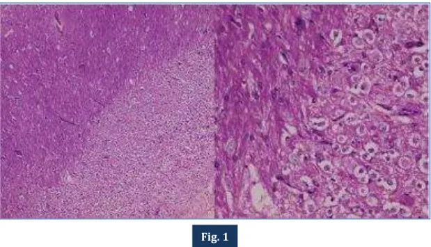
The squash smears stained with MGG, H&E and PAP stain, showed a fibrillary background in most of the smears with scattered ganglion cells having vesicular nuclei and prominent nucleoli (Fig. 2). Some of the smears showed small dark granular cells representing internal granular cell layer (Fig. 3). A provisional diagnosis of Lhermitte duclos disease was given keeping in view the increased number of large neurons and the MRI findings. The histopathology of the excised tissue was confirmatory showing two layers in the enlarged folia. The internal granular cell neuron layer was enlarged, separated by increased neuropil with scanty Purkinje cells. The cerebellar molecular layer was thickened with an abundance of radial processes. There were no mitoses or multinucleated cells. Foci of calcification was also seen (Fig. 1). Postoperatively the patient developed few cerebellar symptoms with only partial recovery after 4 years. MRI revealed no recurrent disease.
DISCUSSION: Lhermitte-Duclos disease was first described in 1920 in the literature under the names of Purkinjeoma, granular cell hypertrophy of the cerebellum, hamartoma of the cerebellum, dysplastic gangliocytoma, ganglioneuroma, and gangliomatosis of the cerebellum. It is a rare disorder of uncertain pathogenesis characterized by distortion of the normal cerebellar laminar cytoarchitecture.[6] Gross autopsy specimen show expanded cerebellar folia. Microscopic findings include thickening and hypermyelination of the molecular (outermost) layer and large pleomorphic cells that replace the Purkinje (middle) and granular (innermost) layers.[6,7] The association of Lhermitte-Duclos disease and Cowden syndrome, an autosomal dominant phakomatosis, has been described, suggesting that Lhermitte-Duclos disease fits the concept of Cowden syndrome as a hamartoma-neoplasia syndrome. Most cases of Lhermitte-Duclos disease occur in adults, usually in the third and fourth decades. There is no definite sex preference.[8] In our case, the age of the patient is 25 yrs.
Characteristic MR imaging findings are a non-enhancing mass with an alternating striated pattern. It is usually located in the cerebellar hemisphere with special preference for the left side and occasionally extends to the vermis. Hydrocephalus may be present due to mass effect along with compression of the fourth ventricle. CT demonstrates a non-calcified low-attenuation cerebellar mass and occasionally a mixed-attenuation cerebellar mass with occasionally scattered calcification. However, CT findings lack specific features to suggest the diagnosis. Correlation of the MR imaging findings with histologic and gross pathologic features of Lhermitte-Duclos disease shows that the characteristic striations demonstrated on MR images represent the abnormally thickened cerebellar folia.[6,9]
J of Evolution of Med and Dent Sci/ eISSN- 2278-4802, pISSN- 2278-4748/ Vol. 4/ Issue 38/ May 11, 2015 Page 6684
absence of tumor limits in the depth of the cerebellar hemisphere constitutes the major technical problem.[10] Intraoperative cytological opinion from the margin of lesion is helpful in such cases as the surgeon can know as to how far he should proceed. The best cytological details were seen with MGG stain and the best nuclear details including nuclear membrane outline and chromatin pattern was seen with PAP stain. Similar findings have been reported by Mouriquand et al and Kontozoglou et al who found a combination of MGG and PAP stains to reveal complementary information and better images as compared to H & E stain.[11,12]
The lesion, despite some similarities (clusters of voluminous neurons with thick bundles of nerve cell processes) is completely different from a tumoral ganglioneuroma or a cerebellar ganglioglioma in which variable architecture discloses proliferation of both abnormal neurons and glial tumor cells.[4]
Thus, the disease should be suspected preoperatively, when MRI shows a large, poorly limited, and partially calcified lesion of a cerebellar hemisphere without contrast enhancement in a young adult complaining of slowly progressive instability while walking and headaches. Intraoperative squash smear cytology showing increased number of large neurons in a fibrillary background of glial cells may be helpful in rendering a provisional diagnosis, but in any case, confirmation is done by histopathology only.
REFERENCES:
1. Louis DN, Ohgaki H, Wiestler OD, Cavenee WK, Burger PC, Jouvet A: The 2007 WHO classification of tumours of the central nervous system. Acta Neuropathol 114:97-109, 2007. 2. Ambler M, Pogacar S, Sidman R: Lhermitte-Duclos disease (granule cell hypertrophy of the
cerebellum). Pathological analysis of the first familial case. J Neuropathol Exp Neurol 28: 622-647, 1969.
3. Ferrer l, Isamat F, AcebesJ: A golgi and electron microscopic study of a dysplastic
gangliocytoma of the cerebellum. Acta Neuropathol (Berl) 47: 163-165, 1979.
4. Reznik M, Schoenen J: Lhermitte-Duclos Disease. Acta Neuropathol (Berl) 59: 88-94, 1983. 5. Williams RS: Phenotypic variability of the Purkinje cell: a golgi study of human cerebellar
malformations. J Neuropathol Exp Neurol 40: 307, 1981.
6. Meltzer CC, Smirniotopoulos JG, Jones RV: The striated cerebellum: an MR imaging sign in
Lhermitte-Duclos disease (dysplastic gangliocytoma). Radiology 194:699-703, 1995.
7. Lantos PL, Vandenberg SR, Kleihues P: Tumours of the nervous system. In: Graham DI, Lantos
PL, eds. Greenfield′s neuropathology. th ed. London, England: Arnold 83-879, 1997.
8. Albrecht S, Haber RM, Goodman JC, Duvic M: Cowden syndrome and Lhermitte-Duclos disease.
Cancer 70:869-876, 1992.
9. Grand S, Pasquier B, Le Bas JF, Chirossel JP: Case report: magnetic resonance imaging in
Lhermitte-Duclos disease. Br J Radiol 67:902-905, 1994.
10.Prestor B: Dysplastic gangliocytoma of the cerebellum (Lhermitte-Duclos disease). J Clin Neurosci 13:877-81, 2006.
11.Mouriquand C, Benabid AL, Breyton M: Stereotaxic cytology of brain tumors: Review of an eight-year experience. Acta Cytologica: 31: 756-64, 1987.
J of Evolution of Med and Dent Sci/ eISSN- 2278-4802, pISSN- 2278-4748/ Vol. 4/ Issue 38/ May 11, 2015 Page 6685
Fig. 1: Histopathology section showing widening of the molecular layer, absence of the Purkinje cell layer and hypertrophy of the granular cell layer containing dysplastic ganglion cells. Foci of calcification also seen. (H&E, 10x & 40x).
Fig. 2: Squash smears showing scattered large neurons with vesicular nuclei and prominent nucleoli alongwith small dark granular cells in a fibrillary background. (PAP, 40x & H&E, 40x).
Fig. 1
J of Evolution of Med and Dent Sci/ eISSN- 2278-4802, pISSN- 2278-4748/ Vol. 4/ Issue 38/ May 11, 2015 Page 6686
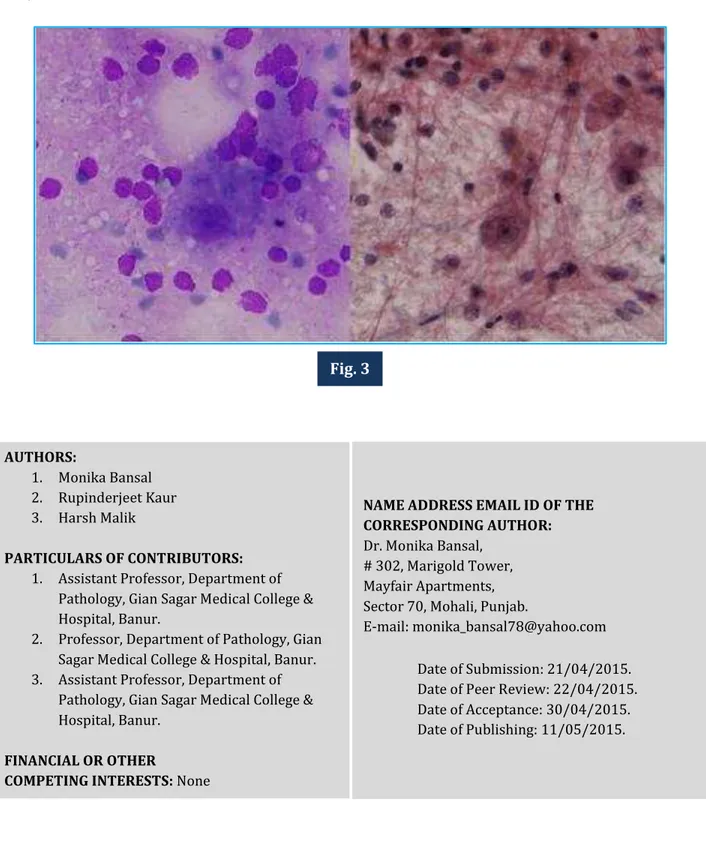
Fig. 3: Squash smears showing large ganglion cells having vesicular nuclei, prominent nucleoli and thick cytoplasm along with small dark granular cells in a fibrillary background. (MGG, 100x & PAP, 100x).
AUTHORS:
1. Monika Bansal 2. Rupinderjeet Kaur 3. Harsh Malik
PARTICULARS OF CONTRIBUTORS:
1. Assistant Professor, Department of Pathology, Gian Sagar Medical College & Hospital, Banur.
2. Professor, Department of Pathology, Gian Sagar Medical College & Hospital, Banur. 3. Assistant Professor, Department of
Pathology, Gian Sagar Medical College & Hospital, Banur.
FINANCIAL OR OTHER
COMPETING INTERESTS: None
NAME ADDRESS EMAIL ID OF THE CORRESPONDING AUTHOR:
Dr. Monika Bansal, # 302, Marigold Tower, Mayfair Apartments, Sector 70, Mohali, Punjab.
E-mail: monika_bansal78@yahoo.com
Date of Submission: 21/04/2015. Date of Peer Review: 22/04/2015. Date of Acceptance: 30/04/2015. Date of Publishing: 11/05/2015.