einstein. 2013;11(1):132
LEARNING BY IMAGES
Plasma cell leukemia
Leucemia de células plasmocitárias
Clarissa Lima e Moura de Souza1, Guilherme Fleury Perini1, Nelson Hamerschlak1,
Paulo Augusto Achucarro Silveira1
1 Hospital Israelita Albert Einstein, São Paulo, SP, Brazil.
Corresponding author: Clarissa Lima e Moura de Souza – Avenida Albert Einstein, 627/701 – Morumbi – Zip code: 05652-900 – São Paulo, SP, Brazil – Phone: (55 11) 2151-5555 – E-mail: [email protected]
Received on: Dec 6, 2011 – Accepted on: Dec 5, 2012
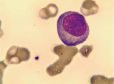
Figure 1. Plasma cell in peripheral blood Figure 2. Plasma cell
A 74-year-old woman with renal failure and lytic lesions in the spine was admitted in our hospital. She was diagnosed with IgG kappa multiple myeloma. In addition, monosomy of chromosome 13 and IgH infusion by FISH were seen.
The patient underwent five cycles of VMP (bortezomib, melphalan and prednisone) that achieved complete response, followed by maintenance with bortezomib. However, 18 months later, the patient was readmitted with asthenia and leucocytosis. Atypical cells with plasmacytoid characteristics were detected in peripheral blood smears, consistent with plasma cell leukemia (PCL) (Figures 1 and 2).
PCL is a rare lymphoproliferative disorder featured by the presence of more than 20% of plasma cells in peripheral blood(1). This disease can be primary, but in
40% of cases there is the diagnosis of multiple myeloma. Morphologically, plasma cells are oval-shaped with large basophilic cytoplasm, round eccentric nucleus
and spoke wheel appearance of chromatin without nucleolus. More immature forms can present dispersed chromatin, prominent nucleoli and high nucleo-cytoplasmic relationship.
Prognosis of PCL patients is poor, and the mean survival is 18-20 months mainly in secondary disease cases in which cytogenetic changes of poor prognosis with del (17p) and p53 loss are often found(2).
The patient underwent rescue chemotherapy, without response. After that, her family opted for palliative treatment.
REFERENCES
1. McKenna RW, Kyle RA, Kuehl WM,Gtogan TM, Harris NL, Coupland RW. Plasma cell neoplasm. In: Swerdlow SH, Campo E, Harris NL, Jaffe ES, Pileri SA, Stein H, et al. editors. World Health Organization classification of tumors of haematopoietic and lymphoid tissues. 4th ed. Lyon: IARC Press; 2008. p.200-13. 2. García-Sanz R, Orfão A, González M, Tabernero MD, Bladé J, Moro MJ, et al.