A Novel Large-Scale Deletion of The Mitochondrial
DNA of Spermatozoa of Men in North Iran
Maryam Gholinezhad Chari, M.Sc.1, 2*, Abasalt Hosseinzadeh Colagar, Ph.D.3, Ali Bidmeshkipour, Ph.D.2
1. Fatemehzahra Infertility and Reproductive Health Research Center, Babol University of Medical Sciences, Babol, Iran
2. Department of Biology, Faculty of Basic Sciences, Razi University, Kermanshah, Iran 3. Department of Biology, Faculty of Basic Sciences, University of Mazandaran, Babolsar, Iran
Abstract
Background:To investigate the level of correlation between large-scale deletions of the mitochondrial DNA (mtDNA) with defective sperm function.
Materials and Methods: In this analytic study, a total of 25 semen samples of the nor-mozoospermic infertile men from North of Iran were collected from the IVF center in an infertility clinic. The swim-up procedure was performed for the separation of sperma-tozoa into two groups; (normal motility group and abnormal motility group) by 2.0 ml of Ham’s F-10 medium and 1.0 ml of semen. After total DNA extraction, a long-range polymerase chain reaction (PCR) technique was used to determine the mtDNA deletions in human spermatozoa.
Results:The products of PCR analysis showed a common 4977 bp deletion and a novel
4866 bp deletion (lanked by a seven-nucleotide direct repeat of 5΄-ACCCCCT-3΄ within the deleted area) from the mtDNA of spermatozoa in both groups. However, the frequency of
mtDNA deletions in abnormal motility group was signiicantly higher than the normal motil -ity group (56, and 24% for 4866 bp-deleted mtDNA and, 52, and 28% for 4977 bp-deleted mtDNA, respectively).
Conclusion:It is suggested that large-scale deletions of the mtDNA is associated with poor sperm motility and may be a causative factor in the decline of fertility in men.
Keywords: Mitochondrial DNA, Large Deletions, Sperm Motility
Citation: Gholinezhad Chari M, Hosseinzadeh Colagar A, Bidmeshkipour A. A novel large-scale deletion of the mitochondrial DNA of spermatozoa of men in north Iran. Int J Fertil Steril. 2015; 8(4): 453-462.
Received: 24 Feb 2013, Accepted: 18 Nov 2013
* Corresponding Address: P.O. Box: 47135-547, Fatemehzahra Infertility and Reproductive Health Research Center, Babol Univer-sity of Medical Sciences, Babol, Iran
Email: [email protected] Royan Institute
International Journal of Fertility and Sterility
Introduction
Sperm motility is one of the key indicators of
fertility in men. Spermatozoa require enormous amount of energy for their survival and fast speed
of lagella during fertilization (1, 2). There are
~22-80 mitochondria in the midpiece of a single mature mammalian spermatozoon (2-4). Mito-chondria facilitate sperm’s rigorous demand for energy through oxidative phosphorylation (OX-PHOS) via the electron transport chain (ETC) in
eukaryotic cells. This process is accomplished by
the respiratory chain and ATP synthesis, which comprise a series of protein complexes that are
en-coded by both nuclear and mitochondrial genomes (nDNA and mtDNA respectively) (2, 4). Mito-chondria possess their own unique genome, which is compartmentalized away from the nDNA. Hu-man mtDNA is a 16569 base pair double-stranded circular DNA molecule that codes 13 polypeptide subunits of respiratory chain complexes, along with the 22 tRNAs and 2 rRNAs (12S and 16S) (5). Mutation rates of mtDNA are generally 10-100 times higher than those of nDNA (6) because
proofread-ing and it also lacks the protection of histones or
DNA-binding proteins (7). Furthermore, mtDNA is attached, at least transiently to the mitochondrial inner membrane where ROS (reactive oxygen spe-cies) are generated as byproducts of OXPHOS in the ETC (8, 9). Several types of mtDNA point
mu-tations and deletions have been identiied in the af -fected tissues of patients with overt mitochondrial diseases (10-15). Large-scale deletions of mtDNA
were irst observed in the skeletal muscle of pa -tients with mitochondrial myopathy (16). This type of DNA rearrangement has later been shown to occur frequently in the muscle of patients with chronic progressive external ophthalmoplegia (CPEO), Kearns-Sayre syndrome (KSS) and Pear-son’s marrow-pancreas syndrome and other multi-systemic disorders and male infertility (17, 18).
The accumulation of mtDNA with the common 4977 bp and 7436 bp large-scale deletions are well recognized to be associated with aging in various human tissues (19, 20). The 4977 bp deletion has been established to be the most common mtDNA mutation in affected tissues of about 40% of pa-tients with mitochondrial myopathy (17, 18). Kao
et al. irst demonstrated the association of the 4977
bp deletion of mtDNA with low motility of the hu-man spermatozoa. Several studies have also dem-onstrated that multiple mtDNA deletions are asso-ciated with defective sperm function and diminish fertility in men (14, 21-25). It has been suggested that these mutations cause infertility by affecting sperm motility. However, low levels of mtDNA
deletions have been identiied in human spermato -zoa and studies have not found a clear relationship between large-scale mtDNA deletions and male
infertility. Therefore, the identiication of mtDNA
mutations in the pathophysiology of human sper-matozoa dysfunction is considered to be important better understanding the etiology of idiopathic
in-fertility in men.
Materials and Methods
Study subjects and semen analysis
In this analytic study, a total of 25 semen samples were provided from the normozoospermic infertile patients ages 24-38 years attending the Infertil-ity Clinic of the Fatemehzahra Hospital in Babol, Iran, in 2010. This study was conducted with the approval of the Medical Research Ethics Commit-tee of the Faculty of Medical Sciences of Babol University. An informed written consent was ob-tained from all the subjects participating in the
study. Individuals with a signiicant medical his -tory, signs of defective androgenisation, testicular trauma, chromosomal disorders, cryptorchidism,
vasectomy, endocrine disorders, leukocytosper
-mic and, cigarette smoking and alcohol consump -tion were excluded from this study. The samples were collected into sterile containers after 3 days
of abstinence and were allowed to liquefy at 37˚C
for 30 minutes. Routine semen analysis was per-formed within 1 hour according to World Health Organization guidelines (26).
In order to remove much of the debris and
contami-nating leukocytes from the ejaculate and purify the
spermatozoa according to motility, each sample un-derwent separation into two sections using the swim-up method. Then, the 50 samples that were obtained
from the swim-up method were classiied into two
groups, the normal motility group (including motile spermatozoa) and abnormal motility group (includ-ing immobile spermatozoa). After, the motility and morphology of the spermatozoa were assessed us-ing microscopic examination. The morphology of the spermatozoa was reported according to Kruger,s criteria in which morphology <14% was considered abnormal (Table 1) (27).
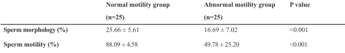
Table 1: Comparison of sperm morphology and motility after swim up method in the study subjects
P value Abnormal motility group
Normal motility group
(n=25) (n=25)
<0.001 16.69 ± 7.02
25.66 ± 5.61
Sperm morphology (%)
<0.001 49.78 ± 25.20
88.09 ± 4.58
Sperm motility (%)
Data are expressed as means ± SD. Comparison of mean values between both groups was performed with an independent t test.
Spermatozoa separation by swim-up procedure
After liquefaction, swim-up procedure was per-formed by adding 1 mL semen to the bottom of a Falcon tube (15 mL) containing 2 mL of fresh Ham’s F-10 medium (include 10% BSA; bovine serum albumin) using a sterile Pasteur pipette. The
tubes were then placed in a 45˚ angle and incubated at 37˚C in 5% CO2 for 30 minutes. After the incu-bation period, ~1.0 ml of the supernatant including motile spermatozoa was collected as a normal mo-tility sample and immobile spermatozoa under the tube were collected as an abnormal motility sam-ple. The samples were then centrifuged at 330×g for 7 minutes. The supernatants were aspirated and the pellets re-suspended in 0.5 mL of Ham’s F-10.
Preparation of human spermatozoa DNA
The total DNA of human spermatozoa was ex-tracted according to the method of Kao et al. (23)
with minor modiications. After centrifugation for
10 minutes at 2000×g at room temperature, the pellet of spermatozoa was washed twice with 0.9% NaCl solution and an aliquot of 2-3×10⁷
sperma-tozoa was incubated at 56˚C for 2 hours in a ly -sis buffer containing 2% sodium dodecyl sulphate
(SDS), 10 mM dithiothreitol (DTT), 100 µg/mL proteinase K and 50 mM Tris-Cl (pH=8.3). After digestion, supernatants were extracted with phe-nol, followed by phenol/chloroform (1:1, v/v), and chloroform. DNA was precipitated with isopro-panol (1:1, v/v) and one-tenth volume of 3 M
so-dium acetate (pH=5.6) and then incubated at -20˚C
overnight. After washing with 75% ethanol (v/v), the pellet was dried and re-suspended in
double-distilled water and stored at -20˚C until use.
Synthesis of oligonucleotide primers
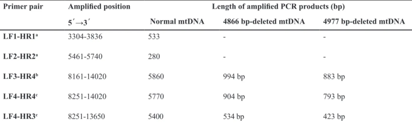
Oligonucleotide primers encompassing the target DNA sequence were chemically synthe-sized by Isogen Life Science (Demeen, Neth-erlands). The nucleotide sequences and sizes of the polymerase chain reaction (PCR) prod-ucts amplified from each of the primer pairs are shown in table 2. The LF1-HR1 and LF2-HR2 primer pairs were used for the amplification of 533 bp and 280 bp PCR products of total (de-leted and wild-type mtDNA), respectively. The primer pairs LF3-HR4, LF4-HR4 and LF4-HR3
were used for the detection of the ~ 5 kb deleted
mtDNA.
Table 2: Oligonucleotide primers used for PCR ampliication of the 4866 and 4977 bp deletions in the mtDNA of human
spermatozoa
Length of ampliied PCR products (bp) Ampliied position
Primer pair
4977 bp-deleted mtDNA 4866 bp-deleted mtDNA
Normal mtDNA 5´→3´
-533 3304-3836
LF1-HR1a
-280 5461-5740
LF2-HR2a
883 bp 994 bp
5860 8161-14020
LF3-HR4b
793 bp 904 bp
5770 8251-14020
LF4-HR4c
423 bp 534 bp
5400 8251-13650
LF4-HR3c
a; The primer sets used for the determination of the total mtDNA , b; The primer sets used for normal long-range PCR and c; The primer sets used for the determination of the 4866 bp and 4977 bp-deleted mtDNA.
LF1 (3304-3323) 5΄-AACATACCCATGGCCAACCT-3΄
LF2 (5461-5491) 5΄-CCCTTACCACGCTACTCCTA-3΄
LF3 (8161-8180) 5΄-CTACGGTCAATGCTCTGAAA-3΄
LF4 (8251-8270) 5΄-GCCCGTATTTACCCTATAGC-3΄
HR1 (3836-3817) 5΄-GGCAGGAGTAATCAGAGGTG-3΄
HR2 (5740-5721) 5΄-GGCGGGAGAAGTAGATTGAA-3΄
HR3 (13650-13631) 5΄-GGGGAAGCGAGGTTGACCTG-3΄
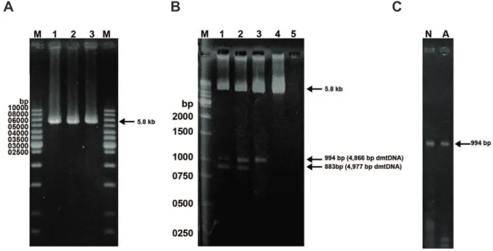
Long-range polymerase chain reaction
To detect the common mtDNA deletion (4977 bp),
a desired large segment of mtDNA (5.8 kb) was am
-pliied from 20 ng of DNA in a 50 µl reaction mixture
containing 200 µM of each dNTP, 0.5 µM of LF3 and HR4 primers (Fig 1, Table 2), 2 units of HLTaq DNA polymerase (Bioneer, Seoul, Korea), 40 mM KCl, 1.5 mM MgCl2 and 10 mM Tris-HCl, (pH=9.0) PCR was carried out for 35 cycles using the thermal
pro-ile of denaturation at 94˚C for 1 minute, annealing at 56˚C for 1 minute, and primer extension at 72˚C
for 5 minutes. The PCR products were separated on 1% agarose gel electrophoresis, stained with ethidium bromide (1 µg/ml) and visualized by transillumina-tion under UV light.
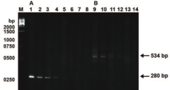
Primer-shift PCR
In order to ascertain that an ampliied DNA frag -ment was not due to mispriming in the presence of
the large-scale deletions in mtDNA, we identiied
mtDNA deletions by primer-shift PCR (28) and
us-ing primer pairs LF4-HR4 and LF4-HR3 (Table 2).
Semi-quantitative PCR
The proportion of the mtDNA with the 4866 bp deletion in each of the spermatozoa DNA samples was determined with a semi-quantitative PCR method (19). The total DNA of the spermatozoa was serially diluted twofold with distilled water. The primer pair LF2-HR2 was used for the
ampli-ication of a 280 bp DNA fragment from the total
mtDNA and the primer pair LF4-HR3 was used for
the ampliication of a 534 bp PCR product from
the mtDNA molecules with the 4866 bp deletion.
Ampliied DNA fragments were separated by elec -trophoresis on a 1.5% agarose gel. The proportion of mtDNA with the 4866 bp deletion was deter-mined as the ratio of the highest-fold dilution that allowed the 534 bp PCR product to be visible on the stained gel to the dilution that allowed the 280
bp PCR product to be visibly ampliied from the
total mtDNA under identical conditions.
Fig 1: Detection of large-scale deletions of mtDNA from human washed sperm by long-range PCR method. A: The 5860 bp band represents the PCR product of normal mtDNA with primer pair LF3-HR4. Lane M is the 1-kb DNA size. B: Using the primer sets LF3-HR4, the 5860 bp band was ampliied from the wild-type mtDNA, the 994 bp and 883 bands were ampliied from the 4866 bp and 4977 bp-deleted mtDNA, respectively. Spermatozoa in lanes 1-4 had the motility scores of 5.0, 20.0, 30.0, 40.0% respectively. Lane 5 is the blank, in which the sperm DNA was omitted from the reaction mixture. Lane M is the1-kb DNA size marker. C. The arrow indicates the band of 994 bp produced with primer pair LF3-HR4. Using a short extension time of 1 minute at 72˚C, the longer DNA product from wild-type mtDNA could not be produced and only mtDNA with 4866 bp-deletion was ampliied. Lanes N and A normal and abnormal groups, respectively.
DNA sequencing
The PCR product (534 bp mtDNA fragment)
ampli-ied from the 4866 bp deleted mtDNA using the LF4-HR3 primer pair was puriied with the PCR product recovery kit (Roche Applied Science, Mannheim, Germany). Direct sequencing of puriied PCR prod -uct was performed at Seq Lab (GOHingen, GmbH, Germany).
Statistical Analysis
The data were expressed as mean ± SD. The mean values were compared using the independent t test with SPSS 11 for Windows software (SPSS Inc., Chi-cago, IL, USA). McNemar’s test was used to compare the frequency of mtDNA deletions between the two groups.In all cases, p<0.05 was considered
statisti-cally signiicant.
Results
Based on standard semen analysis, motility and morphology of the spermatozoa after swim- up in
normal motility group were signiicantly higher
(p<0.001) in comparison with the abnormal motility group (Table 1). Using long-range PCR and primer-shift PCR techniques with the primer sets LF3-HR4, LF4-HR4 and LF4-HR3, we screened the existence of two large-scale deletions of mtDNA in human spermatozoa (Figs 1, 2). The results of long-range PCR with the primer set LF3–HR4 revealed three bands with lengths of 5860 bp from the wild-type mtDNA, 994 bp and 883 bp from deleted mtDNA. The primer-shift PCR results clearly demonstrated a novel 4866 bp deletion along with the common 4977 bp-deleted mtDNA in the spermatozoa with different motilities (Fig 2). By using the primer pairs LF4-HR4 and LF4-HR3, PCR products of 904, and 534 bp from the 4866 bp-deleted mtDNA and, 793, 423 bp from the 4977 bp-deleted mtDNA were obtained, respec-tively (Table 2).
Direct sequencing of the 534 bp PCR product
re-vealed that it was ampliied from the mtDNA with a
novel 4866 bp deletion. This deletion is located be-tween nucleotide position (np) 8270 and np 13136 and
lanked by a seven-nucleotide direct repeat of 5΄ -AC-CCCCT-3΄ within the deleted area, between np 8271-8277 and np 13127-13133 (Fig 3). DNA sequencing was also performed on a 432 bp PCR product from mtDNA. As expected, the analysis of the nucleotide
sequences lanking the break points of the 4977 bp
deletion revealed a 13 bp direct repeat (5΄ -ACCTC-CCTCACCA-3΄) associated with this common de-letion (data not shown). These two dede-letions were shown in both normal and abnormal motility groups.
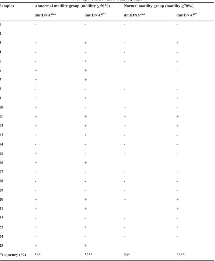
Also, 13 samples had both deletions of mtDNA (Ta-ble 3). The frequency of occurrence of mtDNA with the 4866 bp-deleted mtDNA (dmtDNA4866) and 4977
bp-deleted mtDNA (dmtDNA4977) was different in
both groups. The abundance of the these deletions in abnormal motility group was 56% for dmtDNA4866
and 52% for dmtDNA4977 in comparison with normal
motility group with 24% for dmtDNA4866 and 28%
for dmtDNA4977, respectively (Table 3). Overall, the
incidence of deleted mtDNA in the abnormal motility group was higher than the normal motility group.
Fig 2: Detection of the 4866 and 4977 bp-deleted mtDNA by
the primer shift PCR method in human spermatozoa. Lanes
1-2 represent the PCR products of 423 and 793 bp ampliied from the 4977 bp-deleted mtDNA with primer pair LF4-HR3 and LF4-HR4, respectively. Lanes 3-4 represent the PCR products of 534 and 904 bp ampliied from the 4866 bp-deleted mtDNA with primer pairs LF4-HR3 and LF4-HR4 respectively. Lane M indicates the 1-kb DNA size marker.
Fig 3: Semi-quantitative PCR analysis of mtDNA with the 4866
bp deletion using serial dilution method in human spermatozoa.
A. Lanes 1-8 represent the PCR products ampliied from total
mtDNA serially diluted 28, 29, 210, 211, 212, 213, 214, 215-fold, respec-tively with primer pair LF2-HR2. B. Lanes 9-14 represent the PCR products ampliied from 4866-bp deleted mtDNA serially
Table 3: The occurrence of the 4866 and 4977 bp deletions of mtDNA in the spermatozoa with different motility after
swim-up method in the two study groups
Normal motility group (motility ≥70%) Abnormal motility group (motility ≤ 50%)
Samples dmtDNA4977 dmtDNA4866 dmtDNA4977 dmtDNA4866 -1 -2 + + + + 3 -4 -+ -5 -+ + 6 -+ + 7 -8 + + + + 9 -+ -+ 10 + + + + 11 + + + + 12 -+ + 13 -14 -+ 15 -+ + 16 -17 -18 -19 + + + + 20 + -+ + 21 -22 + -+ + 23 -24 -+ + 25 28** 24* 52** 56* Frequency (%)
The spermatozoa with different motility were separated by swim-up method and divided to two groups. dmtDNA4866: 4866 bp deleted mtDNA, dmtDNA4977: 4977 bp deleted mtDNA. +; Presence of the indicated mtDNA deletion; -; Absence of the indicated
Discussion
Sperm motility is one of the most important factors of fertility in men (1, 2). Several studies have shown that an increase in the concentra-tion of individual mitochondrial OXPHOS in-hibitors results in a decline in sperm motility (29, 30). A correlation has been found between semen quality and the respiratory chain func-tion in sperm mitochondria (30, 31). This ap-peared to be a relationship between mitochon-drial DNA T-haplotype and poor sperm motility (30). Spiropoulos et al (13). reported that the high frequency of the A3243G mtDNA muta-tion strongly correlates with low sperm motil-ity. Thangaraj et al. (32) identified two nucle-otide deletions in the COII genes (at np 8195 and 8196) of sperm mtDNA, introducing a stop codon (AGA), which might be responsible for low sperm motility.
Kumar et al. (14) showed high frequency of some nucleotide changes in the mitochondrial genes including ATPase (6 and 8), ND (2, 3, 4 and 5) in the semen of the oligoasthenozoo-spermic (OA) infertile men. Pereira et al. (33) did not find any correlation between mutation C11994T in ND4 gene and low sperm motil-ity in OA infertile men. It is believed that any defects or abnormal changes in the arrange-ment of mitochondrial DNA may affect sperm motility in idiopathic asthenozoospermic and OA patients (34). So far, multiple large-scale mtDNA deletions have been identified in the sperm of infertile men, especially in men with low sperm motility (1, 21-25). Some studies observed a negative relationship between the common 4977 bp mtDNA deletion and sperm motility (21, 25). Kao et al. (1) first observed a higher incidence of the common 4977-bp dele-tion in the mtDNA of lower Percoll-fracdele-tionated spermatozoa of patients with infertility or sub-fertility. They also identified presence of two novel deletions, of 7345 and 7599 bp in length in mtDNA of poor motile sperm (23).
In one study from a Northern Iranian popu-lation, the occurrence of the 4977 bp deletion of spermatozoa in infertile men with varicocele was significantly higher than in control healthy men (22). However, some studies have not
found a direct correlation between the 4977 bp
and 7.4-7.6 kb deletions, and low sperm motil -ity (25) or for the 4977 bp deletion and semen quality (35). Although, they showed the persis-tence of multiple mtDNA deletions in both nor-mozoospermic and oligoasthenoteratozoosper-mic men. Lestienne et al. revealed the presence of multiple mtDNA deletions in both
spermato-zoa and skeletal muscle in a patient with OA.
They suggested that the multiple human mtD-NA deletions might be of nuclear origin since at least three nuclear loci have been ascribed to multiple mtDNA deletions: 10 q 23.3-24.3, 3 p 14.1-21.2 and 4 p 16 (36).
In the present study, we investigated cor-relation between large-scale deletions of the mtDNA with sperm motility. Our PCR analysis demonstrated a novel 4866 bp (Fig 4) and the common 4977 bp deletions from the mtDNA of spermatozoa (Fig 1). We also confirmed the persistence of these mtDNA deletions in both groups (Fig 2). Our results showed a higher in-cidence of mtDNA with the 4977 bp and 4866 bp deletions in abnormal motility group than in normal motility group. While we found the 4977 bp and a novel 4866 bp deleted mtDNA with primer pair LF4-HR3 (8251-13650), Kao et al. (1) only identified the 4977 bp mtDNA deletion in the human spermatozoa with this primer pair. Furthermore, Fahn et al. (37) also observed the 4977 bp deletion along with a novel 4839 bp deletion with the same primer set in lung tissue. It appears that one of the causes of differences in such mutations might be a reflection of the differences in tissues or populations. Therefore, it is important to note that some other mtDNA deletions may exist that have been undetected. Reynier et al. found that about 85% of sperm samples contained large-scale mtDNA dele-tions of variable sizes, and that most subjects had 2 to 7 deletions of mtDNA. They suggested that these mtDNA deletions are similar to those
observed in skeletal muscle, myocardium, and
other tissues of aged individuals (24). In our study, 13 of the samples had 2 large-scale
mtD-NA deletions. Ieremiadou and Rodakis showed
dele-tions result in complete removal or truncation of some structural genes and tRNA genes of mtDNA. The defective protein subunits en-coded by the deleted mtDNA are assembled with nDNA encoded subunits to yield impaired respiratory enzymes that may further enhance ROS or free radical production and result in a progressive decline in the bioenergetic function of mitochondria and hence low sperm motility.
Thus, random attacks on the naked mtDNA by
ROS or free radicals may cause mutations in the mtDNA with pathological consequences (2, 23). It is suggested that ROS elicited oxidative damage to DNA might be fixed as large-scale deletions of mtDNA in spermatozoa (2).
Furthermore, spermatozoa are especially sus-ceptible to oxidative stress because their plasma membranes are rich in unsaturated fatty acids (38). One of the common oxidative byprod-ucts of DNA, 8-hydroxy-2-deoxy guanosine (8-OHdG) was identified in the human sper-matozoa. Also, the level of sperm 8-OHdG in infertile patients was significantly higher than the healthy subjects (39). However, the mecha-nisms on how these deletions are generated
re-main unclear, but two major hypotheses have been considered to generate these deletions: i. replication through slipped mispairing between two repeats and ii. repair mediated by mtDNA
double-strand breaks (11). A predominance of
rearranged molecules over wild-type, (hetero-plasmy), or the persistence of mutated or delet-ed molecules only (homoplasmy), can result in the onset of mtDNA disease (4). Though energy
requiring organs like brain, muscle and heart
are mostly affected by heteroplsamy (10), such effect on mtDNA of the spermatozoa is not well studied. More studies are needed to understand the role of heteroplasmy in sperm mtDNA of in-fertile men, since homoplasmy mutant mtDNA has been found in OA infertile patients (40). According to our results, the frequency of the 4977 bp and a novel 4866 bp- deleted mtDNA in the abnormal motility group was higher than the normal motility group (p<0.05). Our results indicated that the difference of frequency be-tween the two groups is nonrandom and suggest an association between mtDNA deletions and poor sperm motility, similar to the findings of Kao et al. (1, 23).
Conclusion
We conclude that there is a direct correla-tion between large-scale mtDNA delecorrela-tions and low sperm motility. These deletions might be an important factor for poor sperm motility es-pecially in asthenoteratozoospermic patients, but we can not say certainly that declined fer-tility in men is associated with these deletions. Therefore, more studies are required with larger samples of diagnostically categorized infertile males. Furthermore, the identification of large-scale deletions of mtDNA could be important to better understand the etiology of idiopathic infertility and treatment/ assisted reproductive techniques.
Acknowledgements
We thank Dr. Evangeline Foronda for the
English editing of this article. This research was supported by University of Mazandaran of the Babolsar and Razi University, Kermanshah, in Iran. There is no conflict of interest in this study.
References
1. Kao SH, Chao HT, Wei YH. Mitochondrial deoxyribonu-cleic acid 4977-bp deletion is associated with diminished fertility and motility of human sperm. Biol Reprod. 1995; 52(4): 729-736.
2. Wei YH, Kao SH. Mitochonderial DNA mutation and dele-tion are associated with decline of fertility and motility of human sperm. Zool Stud. 2000; 39(1): 1-12.
3. St John JC, Sakkas D, Barratt CL. A role for mitochondrial DNA and sperm survival. J Androl. 2000; 21(2): 189-199. 4. St John JC, Jokhi RP, Barratt CL. The impact of
mitochon-drial genetics on male infertility. Int J Androl. 2005; 28(2): 65-73.
5. Anderson S, Bankier AT, Barrell BG, de Bruijn MH, Coul-son AR, Drouin J, et al. Sequence and organization of the human mitochondrial genome. Nature. 1981; 290(5806): 457-465.
6. Moore FL, Reijo-Pera RA. Male sperm motility dictated by mother’s mtDNA. Am J Hum Genet. 2000; 67(3): 543-548. 7. Clayton DA, Doda JN, Friedberg EC. The absence of a py-rimidine dimer repair mechanism in mammalian mitochon-dria. Proc Natl Acad Sci USA. 1974; 71(7): 2777-2781. 8. Beckman KB, Ames BN. The free radical theory of aging
matures. Physiol Rev. 1998; 78(2): 547-581.
9. Venkatesh S, Deecaraman M, Kumar R, Shamsi MB, Dada R. Role of reactive oxygen species in the patho-genesis of mitochondrial DNA (mtDNA) mutations in male infertility. Indian J Med Res. 2009; 129(2): 127-137. 10. Wallace DC. Mitochondrial DNA sequence variation in
human evolution and disease. Proc Natl Acad Sci USA. 1994; 91(19): 8739-8746.
11. Chen T, He J, Huang Y, Zhao W. The generation of mi-tochondrial DNA large-scale deletions in human cells. J Hum Genet. 2011; 56(10): 689-694.
12. Holyoake AJ, McHugh P, Wu M, O'Carroll S, Benny P, Sin IL, et al. High incidence of single nucleotide substitutions in the mitochondrial genome is associated with poor se-men parameters in se-men. Int J Androl. 2001; 24(3): 175-182.
13. Spiropoulos J, Turnbull MD, Chinnery PF. Can mitochon-derial DNA mutations cause sperm dysfuunction?. Mol Hum Reprod. 2002; 8(8): 719-721.
14. Kumar R, Venkatesh S, Kumar M, Tanwar M, Shamsi MB, Kumar R, et al. Oxidative stress and sperm mitochondrial DNA mutation in idiopathic oligoasthenozoospermic men. Indian J Biochem Biophys. 2009; 46(2): 172-177. 15. Zhao XT, Feng JB, Li YW, Luo Q, Yang XC, Lu X, et al.
Identiication of two novel mitochondrial DNA deletions
induced by ionizing radiation. Biomed Environ Sci. 2012; 25(5): 533-541.
16. Holt IJ, Harding AE, Morgan-Hughes JA. Deletions of muscle mitochondrial DNA in patients with mitochondrial myopathies. Nature. 1988; 331(6158): 717-719.
17. Shoffner JM, Lott MT, Voljavec AS, Soueidan SA, Costi-gan DA, Wallace DC. Spontaneous Kearns-Sayre/chronic progressive external ophthalmoplegia plus syndrome as-sociated with a mitochondrial DNA deletion: a slip-repli-cation model and metabolic therapy. Proc Natl Acad Sci USA. 1989; 86(20): 7952-7956.
18. Wallace DC. Disease of the mitochondrial DNA. Annu Rev Biochem. 1992; 61: 1175-1212.
19. Fahn HJ, Wang LS, Hsieh RH, Chang SC, Kao SH, Huang MH, et al. Age-related 4977 bp deletion in human lung mi-tochondrial DNA. Am J Respir Crit Care Med. 1996; 154 (4 Pt 1): 1141-1145.
20. Arai T, Nakahara K, Matsuoka H, Sawabe M, Chida K, Matsushita S, et al. Age-related mitochondrial DNA dele-tion in human heart: its reladele-tionship with cardiovascular diseases. Aging Clin Exp Res. 2003; 15(1): 1-5.
21. Ieremiadou F, Rodakis GC. Correlation of the 4977 bp mi-tochondrial DNA deletion with human sperm dysfunction. BMC Res Notes. 2009; 2: 18.
22. Gashti NG, Salehi Z, Madani AH, Dalivandan ST. 4977-bp mitochondrial DNA deletion in infertile patients with varico-cele. Andrologia. 2014; 46(3): 258-262.
23. Kao SH, Chao HT, Wei YH. Multiple deletion of mitochon-drial DNA are associated with the decline of motility and fertility of human spermatozoa. Mol Hum Reprod. 1998; 4(7): 657-666.
24. Reynier P, Chretien MF, Savagner F, Larcher G, Rohmer V, Barriere P, et al. Long PCR analysis of human gamete mtDNA suggests defective mitochondrial maintenance in spermatozoa and supports the bottleneck theory for oocytes. Biochem Biophys Res Commun. 1998; 252(2): 373-377.
25. St John JC, Jokhi RP, Barratt CL. Men with oligoasthe-noteratozoospermia harbour higher numbers of multiple mitochondrial DNA deletions in their spermatozoa, but in-dividual deletions are not indicative of overall aetiology. Mol Hum Reprod. 2001; 7(1): 103-111.
26. World Health Organization.WHO Laboratory manual for the examination of human semen and semen- cervical mucus interaction, 4th ed. New York: Cambridge University
Press; 1999; 4-23.
27. Kruger TF, Acosta AA, Simmons KF, Swanson RJ, Matta JF, Veeck LL, et al. New method of evaluating sperm mor-phology with predictive value for human in vitro fertiliza-tion. Urology. 1987; 30(3): 248-251.
29. St John JC, Cooke ID, Barratt CL. Mitochondrial muta-tions and male infertility. Nat Med. 1997; 3(2): 124-125. 30. Ruiz-Pesini E, Lapena AC, Diez C, Alvarez E, Enriquez
JA, Lopez-Perez MJ. Seminal quality correlates with mi-tochondrial functionality. Clin Chim Acta. 2000; 300(1-2): 97-105.
31. Kasai T, Ogawa K, Mizuno K, Nagai S, Uchida Y, Ohta S, et al. Relationship between sperm mitochondrial mem-brane potential, sperm motility, and fertility potential. Asian J Androl. 2002; 4(2): 97-103.
32. Thangaraj K, Joshi M, Reddy AG, Rasalkar AA, Singh L. Sperm mitochondrial mutations as a mause of low sperm motility. J Androl. 2003; 24(3): 388-392.
33. Pereira L, Gonçalves J, Bandelt HJ. Mutation C11994T in the mitochondrial ND4 gene is not a cause of low sperm motility in Portugal. Fertil Steril. 2008; 89(3): 738-741. 34. Sun ZM, Ding CF, Yan ZZ, Bao YZ. Ultrastructure and
function of mitochondria in idiopathic asthenospermia: study of 151 cases. Zhonghua Yi Xue Za Zhi. 2007; 87(18): 1263-1265.
35. Cummins JM, Jequier AM, Martin R, Mehmet D, Goldblatt J. Semen levels of mitochondrial DNA deletions in men
at-tending an infertility clinic do not correlate with phenotype. Int J Androl. 1998; 21(1): 47-52.
36. Lestienne P, Reynier P, Chretien MF, Penisson-Besnier I, Malthiery Y, Rohmer V. Oligoasthenospermia associated with multiple mitochondrial DNA rearrangements. Mol Hum Reprod. 1997; 3(9): 811-814.
37. Fahn HJ, Wang LS, Kao SH, Chang SC, Huang MH, Wei YH. Smoking associated mitochondrial DNA mutations and lipid peroxidation in human lung tissues. Am J Respir Cell Mol Biol. 1998; 19(6): 901-909.
38. Aitken RJ, Clarkson JS, Fishel S. Generation of reac-tive oxygen species, lipid peroxidation and human sperm function. Biol Reprod. 1989; 41(1): 183-197.
39. Shen HM, Chia SE, Ong CN. Evaluation of oxidative DNA damage in human sperm and its association with male infertility. J Androl. 1999; 20(6): 718-723.