Research Article Open Access
Benzotropolone moiety in theaflavins is responsible for inhibiting
peptide-transport and activating AMP-activated protein kinase in Caco-2 cells
Ha-Young Park, Yuri Kunitake, and Toshiro Matsui*
Department of Bioscience and Biotechnology, Division of Bioresources and Biosciences, Faculty of Agriculture, Graduate School of Kyushu University, 6-10-1 Hakozaki, Fukuoka, 812-8581, Japan
*Corresponding Author: Toshiro Matsui, PhD, Professor, Department of Bioscience and Biotechnology, Division of Bioresources and Biosciences, Faculty of Agriculture, Graduate School of Kyushu University, 6-10-1 Hakozaki, Fukuoka, 812-8581, Japan
Submission date: April 23, 2013; Acceptance date: May 23, 2013; Publication date: May 24, 2013
ABSTRACT
Objective: In the small intestine, peptide transporter 1 (PEPT1) plays a role in the transport of di- and tri-peptides. Recently, we found that theaflavins (TFs), dimeric catechins, inhibited the transport of di-peptides across Caco-2 monolayers by suppressing the expression of PEPT1 through AMP-activated protein kinase (AMPK) activation. In this study, we investigated the structural requirement of theaflavins for the effect, and the mechanism(s) underling theaflavin-induced AMPK activation.
Methods: Theaflavin-3’-O-gallate (TF3’G) was used for this study, since it possessed the most potent inhibition power for peptide-transport among theaflavins. Absorption ability was measured with Caco-2 cell monolayers treated with or without 20 M sample (TF3’G or its related compounds) in an Ussing Chamber. The amount of Gly-Sar (a model of PEPT1-transporing peptide) transport at fixed time-points to 60 min was determined by fluorescent naphthalene-2,3-dicarboxaldehyde-derivatized assay(Ex/Em: 405 nm/460 nm). The apparent permeability coefficient (Papp) was used to evaluate the permeability. Expression of PEPT1
protein in Caco-2 cells treated with or without 20 M TF3’G in the presence or absence of
inhibitor (10 μM compound C as AMPK inhibitor or 25 μM STO-609 as CaMKK inhibitor) was evaluated by Western blot.
Results: The Papp value of Gly-Sar significantly (P < 0.05) decreased in 20 μM
purprogallin-treated Caco-2 cells as well as in TF3’G-treated cells, together with the reduction of PEPT1 expression, while their monomeric catechins did not show any Papp reduction. In TF3’G
-treated Caco-2 cells, the recovery of the reduced PEPT1 expression was found by 10 μM compound C, but not STO-609.
crucial structural requirement for exerting the inhibition of intestinal peptide-transport, and the suppression of PEPT1 expression by theaflavins would be caused by activating LKB1/AMPK pathway, but not CaMKK/AMPK pathway.
Keywords: Theaflavin-3’--gallate, Peptide transport, PEPT1, Benzotropolone, AMP-activated protein kinase, Calmodulin-dependent protein kinase kinase
BACKGROUND:
To date, many food scientists have paid attention to physiological functions of small peptides in the body, some of which have been proven to be effective for the prevention of life-style related diseases such as hypertension [1, 2], hypercholesterolemia [3] and inflammation [4]. Previous researches provided the health-benefit of peptide intake that can be absorbed via intestinal peptide transporter 1 (PEPT1) [5].
PEPT1 that is expressed as peptide transporter in the intestinal membrane can recognize di-and tri-peptide lengths [5, 6]. In intestinal membranes it has been reported that the expression of PEPT1 was closely associated with AMP-activated protein kinase (AMPK, a serine/threonine kinase) activation [7, 8]. Pieri et al. demonstrated that the activated AMPK resulted in a decrease of peptide absorption, along with the suppression of PEPT1 proteins in Caco-2 cells [7]. In our previous report, we also found that the transport of di-peptides across Caco-2 monolayers was markedly inhibited by theaflavin-induced AMPK activation [9]. So far, although it has been reported that catechins [10] and epigallocatechin-3-gallate [11] could stimulate AMPK activity in the liver, no study was found for AMPK activation by condensed catechins or theaflavins in the intestine. In addition, involvement(s) of up-streamed kinases including liver kinase B1 (LKB1) and Ca2+/calmodulin-dependent protein kinase kinase (CaMKK) on the theaflavin-induced AMPK activation still remains unclear.
In this study, we investigated the structural requirement of theaflavins for the inhibition of intestinal peptide-transport in Caco-2 cells, and clarified whether LKB1/ or CaMKK/AMPK was involved in theaflavin-induced AMPK activation. We used theaflavin-3’-O-gallate (TF3’G) throughout this study, since it possessed the most potent inhibition power for peptide-transport among theaflavins [9].
MATERIALS AND METHODS:
Reagents: Dulbecco’s modified Eagle’s medium (DMEM) and fetal bovine serum (FBS) were purchased from GIBCO Life Technologies (Grand Island, NY, USA). Dimethylsulfoxide (DMSO) was obtained from Sigma-Aldrich (St. Louis, MO, USA).
Caco-2 cell culture: Caco-2 cells were cultured under standard cell culture conditions as described previously [9]. Caco-2 cells used in this study were between passages 50 and 60. For transport studies, cells were grown on a BD Falcon™ cell culture insert (PET membrane, 0.9 cm2, 1.0 µm pore size; BD Biosciences, Bedford, MA, USA) coated with type I collagen (collagen gel culturing kit, Cellmatrix type I-A, Nitta Gelatin, Osaka, Japan). The cells were seeded at a density of 4.0 × 10⁵ cells/mL and cultured in a seeding basal medium
containing MITO+™ serum extender for 48 h. After the medium was replaced with an
enterocyte differentiation medium containing MITO+™ serum extender, cells were incubated for another 72 hours to form monolayers. The medium was freshly changed every 24 hours. The integrity of Caco-2 monolayers was evaluated by measuring the transepithelial electrical resistance (TEER) with a multi-channel EVC-4000 system (World Precision Instruments, FL, USA). Monolayers with TEER values of >100 cm2 were used for peptide-transport experiments.
Cell treatment: Treatment of Caco-2 cell monolayers with sample (TF3’G or its related compounds) were performed by replacing 10% FBS in DMEM with the medium containing
20 μM sample and allowing them to incubate for 24 hours at 37 ˚C at O2:CO2 (95:5). Cells
incubated in sample-free medium were used as a control. The medium containing the sample was prepared by diluting a stock solution of each sample, dissolved in DMSO, with 10% FBS in DMEM. The final concentration of DMSO was 0.5% throughout the experiments to avoid any cell damage by DMSO [13].
Transport study: Transport experiments in Caco-2 cell monolayers were performed using an Ussing Chamber system (Model U-2500; Warner Instrument Co., Hamden, CT, USA). Caco-2 cell monolayers grown in transwell inserts were gently rinsed with Hanks’ balanced salt solution (HBSS) buffer (pH 6.0) to wash or remove any free samples in the medium, and carefully mounted in the Ussing Chamber using blunt forceps. An aliquot (7.5 mL) of HBSS buffer (adjusted to pH 6.0 with 10 mM MES [2-(N-morpholino) ethanesulfonic acid]) or HBSS buffer (adjusted to pH 7.4 with 10 mM HEPES [4-(2-hydroxyethyl)-1-piperazine ethanesulfonic acid]) was added to the apical or basolateral side, respectively. After equilibration for 15 min at 37 ˚C, the transport experiments were started by replacing the apical buffer with fresh HBSS buffer (pH 6.0) containing 10 mM Gly-Sar for Caco-2 cells treated with or without 20 M sample. During the experiments, solutions on both sides were bubbled continuously with a mixture of O2:CO2 (95:5). Aliquots (0.2 mL each) of solution
were drawn from the basolateral side at time intervals of 15, 30, 45 and 60 minutes to determine the amount of transported Gly-Sar. The same volume of fresh buffer was added to the basolateral side at each time point. The membrane barrier and transport ability of cells were maintained during the 60 min-transport experiments without any reduction of TEER value (>100 cm2) by Gly-Sar. The amount of Gly-Sar transported to the basolateral side was determined by a fluorescent NDA-derivatization method [14]. Briefly, 10 μL of 20 mM sodium cyanide in borate buffer (pH 9.5) was added to the basolateral solution taken at each
time point (100 μL) in a 96-well microplate, followed by the addition of 50 μL of 0.1 mM
NDA solution in methanol. After incubating at 25˚C for 60 min, the fluorescence intensity of
intensity of basolateral solution taken at each time point for blank (no Gly-Sar) transport experiments was used to cancel an increase in intensity by any contaminants from Caco-2 cells.
The apparent permeability coefficient (Papp) was calculated from the following equation:
Papp (cm/s) =
V
AC0 dC
dt
dC/dt is the change in concentration at the basolateral side over time (mmol/s), V is the volume of solution in the basolateral compartment (7.5 mL), A is the surface area of the membrane (0.2826 cm2) and C0 is the initial concentration at the apical side (mmol). The
effect of sample (TF3’G or its related compounds) on Papp was expressed relative to control
(%).
Protein extraction and Western blotting: Expression of PEPT1 protein in Caco-2 cells
treated with or without TF3’G (20 μM) in the presence or absence of inhibitor (10 μM
compound C as AMPK inhibitor or 25 μM STO-609 as CaMKK inhibitor) for 24 h at 37 ˚C was evaluated by Western blot analysis. Briefly, protein (1.3 mg/mL) extracted from Caco-2 cells using radioimmunoprecipitation assay buffer was mixed with an equal volume of sample buffer (20% glycerol, 4% sodium dodecylsulfate, 3% dithiothreitol, 0.002% bromophenol blue and 0.125 M Tris-HCl, pH 6.8) and boiling in the water for 5 min. An aliquot (9.75 µg) of the prepared sample was applied to 10% SDS polyacrylamide gel electrophoresis for 2 h at 20 mA and transferred onto PVDF membrane (Hybond-P, GE Healthcare, Piscataway, NJ, USA) for 1.5 h at 40 mA. The membrane was blocked with 5% non-fat dried milk in TBS-T (Tris-buffered saline containing 0.05% Tween 20) for 1 h at
room temperature and then probed with the primary antibody for PEPT1 or β-actin, followed
by rabbit anti PEPT1 antibody (1:1000, Abbiotec, San Diego, CA, USA) or mouse anti β -actin antibody (1:1000, Applied biological materials inc, Richmond, BC, Canada) overnight
at 4˚C. The membrane was then re-probed with HRP-conjugated donkey anti-rabbit IgG antibody or HRP-conjugated sheep anti-mouse IgG antibody (1:1000, GE Healthcare, Buckinghamshire, UK) for 1 h at room temperature. The membrane was detected with ECL plus detection reagent using an ImageQuant LAS 4000 (GE Healthcare, Piscataway, NY, USA)). Quantitation of PEPT1 and β-actin were performed with ImageQuant TL 7.0 software (GE Healthcare), and the PEPT1 protein levels in Caco-2 cells treated with TF3’G or TF3’G in the presence or absence of inhibitor were expressed relative to the control (or TF3’G -untreated cells).
Statistical analysis: Results are expressed as the mean ± SEM. The statistical significance between groups was analyzed using one-way ANOVA followed by Tukey-Kramer’s t-test for
post hoc analysis. P < 0.05 was considered to be significant. Statistical analyses were conducted with StatView J5.0 (SAS Institute Inc., Cary, NC, USA).
(NO) in aorta [19]. However, as we have clearly demonstrated in our previous report [9], theaflavins themselves were not a penetrant through intestinal membrane, irrespective of the above-mentioned health-benefits. In addition, we also demonstrated that non-absorbable theaflavins had potency in inhibiting intestinal peptide-transport [9], whereas no further useful structural information and underlying mechanism(s) of theaflavin-induced inhibition of peptide-transport was obtained.
Fig. 1. Chemical structures of theaflavin-3’-O-gallate (TF3’G), catechins and purpurogallin used in this study
Effect of TF3’G and its related compounds on Gly-Sar transport across Caco-2 cell monolayers: In order to get information on structural requirement of TFs exerting
peptide-transport inhibition, compounds related to TF3’G skeleton as shown in Fig. 1 were used for Gly-Sar transport experiments across Caco-2 monolayers. As shown in Fig 2, four catechin monomers did not show any significant Papp reduction in Caco-2 cells, while the Papp of
Gly-Sar was significantly (P < 0.05) reduced in 20 M purprogallin-treated cells as similar to TF3’G treated cells (TF3’G, 41%; purprogallin, 30% of control in Papp). This clearly
demonstrated that the benzotropolone moiety in theaflavins, which has a characteristic 7-ring structure [20], was a crucial structural requirement for exerting the inhibition of intestinal peptide-transport in Caco-2 cells.
Effect of AMPK inhibition on PEPT1 protein expression in TF3’G-treated Caco-2 cells:
Fig. 2. Effect of theaflavin-3’-O-gallate and its related compounds on Gly-Sar transport.
Caco-2 cells were treated with 20 μM sample for 24 h. Theaflavin-3’-O-gallate, TF3’G; (
-)-epigallocatechin, EGC; epicatechin gallate, ECG; epigallocatechin gallate, EGCG;
(-)-epicatechin, EC. Control: transport of Gly-Sar in Caco-2 cells without TF3’G treatment. The values
(relative Papp value vs. control, %) are expressed as the mean ± SEM (n=3-5). Means without a
common letter are significantly different; P < 0.05. Significant difference between groups was
evaluated by Tukey–Kramer’s t-test.
Fig. 3. Effect of AMPK inhibition on PEPT1 protein expression in Caco-2 cells treated with theaflavin-3’-O-gallate. Caco-2 cells were treated with 20 μM theaflavin-3’-O-gallate
(TF3’G) in the presence or absence of 10 μM compound C. Values are expressed as the mean ± SEM
(n = 3). Means without a common letter are significantly different; P < 0.05. Significant difference
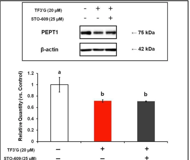
Effect of CaMKK inhibition on PEPT1 protein expression in TF3’G-treated Caco-2 cells: Effect of STO-609, a selective inhibitor of CaMKK, on PEPT1 protein expression in Caco-2 cells treated with 20 μM TF3’G was evaluated by Western blot analysis. As shown in Fig. 4, no significant recovery of the reduced PEPT1 expression by STO-609 was obtained in
the TF3’G-treated Caco-2 cells. This result clearly suggested that the CaMKK-activated AMPK pathway [11, 21-22] could be excluded from the mechanism(s) underlying TF3’G -induced PEPT1 suppression.
Fig. 4. Effect of CaMKK inhibition on PEPT1 protein expression in Caco-2 cells treated with theaflavin-3’-O-gallate. Caco-2 cells were treated with 20 μM theaflavin-3’-O-gallate
(TF3’G) in the presence or absence of 25 μM STO-609. Values are expressed as the mean ± SEM
(n=3). Means without a common letter are significantly different; P < 0.05. Significant difference
between groups was evaluated by Tukey–Kramer’s t-test.
There have been some reports on the potential of polyphenols in the activation of CaMKK/AMPK signaling pathway [8, 11, 20]. Collins et al. reported that EGCG reduced hepatic gluconeogenesis by activating CaMKK/AMPK pathway [11]. However, in this study,
Fig. 5. Proposed upstream mechanism(s) of theaflavins on signaling pathway in Caco-2 cells.
As summarized in Fig. 5, the present findings suggested that theaflavins may stimulate LKB1/AMPK signaling so as to suppress PEPT1 expression in Caco-2 cells, although mechanisms underlying LKB1 activation by either transforming growth factor-β-activated kinase (TAK-1) [23-25] or Sirtuin 1 (SIRT1) [26, 27] in Caco-2 cells remain unclear. So far, Lin et al. [28] have reported that in HepG2 cell experiments theaflavins could accumulate hepatic lipids by activating LKB1/AMPK pathway. Considering the report and the expression of LKB1 in Caco-2 cells [29], we can speculate the possible role of theaflavins in the LKB1/AMPK signaling pathway to suppress PEPT1 expression in intestinal membrane. In addition, there might be specific receptor(s) that recognizes theaflavins or benzotropolone ring in Caco-2 cells, and experiments for binding of theaflavins to intestinal membrane are now in progress.
CONCLUSIONS:
We demonstrated that the benzotropolone moiety in theaflavins, which has a characteristic 7-ring structure, was a crucial structural requirement for exerting the inhibition of intestinal peptide-transport in Caco-2 cells. It seems likely that theaflavins may act as an activator for LKB1/AMPK pathway, but not CaMKK/AMPK pathway, to suppress PEPT1 expression.
Abbreviations: PEPT1, peptide transporter 1; AMPK, AMP-activated protein kinase; LKB1, liver kinase B1; CaMKK, calmodulin-dependent protein kinase kinase; TF3’G, theaflavin-3’
-O-gallate; NDA, naphthalene-2,3-dialdehyde; TEER, transepithelial electrical resistance; Papp,
kinase; SIRT1, sirtuin 1
Competing interests: There were no competing interests.
Authors’ contributions: All authors have been contributed
Acknowledgements and funding: This study was in part supported by a Grant-in-aid for the Ministry of Education, Science, Sports and Culture of Japan (No. 22248014) to TM. The authors thank Junko Takeda in Kyushu University for her kind technical supports.
REFERENCES:
1. Matsui T, Matsumoto K: Antihypertensive peptides from natural resources. Advances in Phytomedicine 2006, 2:255-271.
2. Kawasaki T, Seki E, Osajima K, Yoshida M, Asada K, Matsui T, Osajima Y:
Antihypertensive effect of valyl-tyrosine, a short chain peptide derived from sardine muscle hydrolyzate, on mild hypertensive subjects. J Hum Hypertens 2000, 14: 519-523.
3. Nagaoka S, Futamura Y, Miwa K, Awano T, Yamauchi K, Kanamaru Y, Tadashi K, Kuwata T: Identification of novel hypocholesterolemic peptides derived from bovine. Biochem Biophys Res Commun 2001, 281:11-17.
4. Wang YF, Xu X, Fan X, Zhang C, Wei Q, Wang X, Guo W, Xing W, Yu J, Yan JL, Liang HP: A Cell-penetrating peptide suppresses inflammation by inhibiting NF-κB signaling. Mol Ther 2011, 12:1849-1857.
5. Shiraga T, Miyamoto K, Tanaka H, Yamamoto H, Taketani Y, Morita K, Tamai I, Tsuji A, Takeda E: Cellular and molecular mechanisms of dietary regulation on rat intestinal H+/Peptide transporter PepT1. Gastroenterology 1999, 116:354-62.
6. Fei YJ, Kanai Y, Nussberger S, Ganapathy V, Leibach FH, Romero MF, Singh SK, Boron WF, Hediger MA: Expression cloning of a mammalian proton-coupled oligopeptide transporter. Nature 1994, 368:563-566.
7. Pieri M, Christian HC, Wilkins RJ, Boyd CA, Meredith D: The apical (hPepT1) and basolateral peptide transport systems of Caco-2 cells are regulated by AMP-activated protein kinase. Am J Physiol Gastrointest Liver Physiol 2010, 299:G136-143.
8. Hallows KR: Emerging role of AMP-activated protein kinase in coupling membrane transport to cellular metabolism. Curr Opin Nephrol Hypertens 2005, 14:464-471. 9. Takeda J, Park HY, Kunitake Y, Yoshiura K, Matsui T: Theaflavins, dimeric
catechins, inhibit peptide transport across Caco-2 cell monolayers via down-regulation of AMP-activated protein kinase-mediated peptide transporter PEPT1. Food Chem 2013, 138: 2140-2145.
10.Murase T, Misawa K, Haramizu S, Hase T: Catechin-induced activation of the LKB1/AMP-activated protein kinase pathway. Biochem Pharmacol 2009, 78:78-84. 11.Collins QF, Liu HY, Pi J, Liu Z, Quon MJ, Cao W: Epigallocatechin-3-gallate
(EGCG), a green tea polyphenol, suppresses hepatic gluconeogenesis through 5'-AMP-activated protein kinase. J Biol Chem 2007, 282:30143-30149.
13.Da Violante G, Zerrouk N, Richard I, Provot G, Chaumeil JC, Arnaud P: Evaluation of the cytotoxicity effect of dimethyl sulfoxide (DMSO) on Caco2/TC7 colon tumor cell cultures. Biol Pham Bull 2002, 25:1600-1603.
14.Ueno T, Tanaka M, Matsui T, Matsumoto K: Determination of antihypertensive small peptides, Val-Tyr and Ile-Val-Tyr, by fluorometric high-performance liquid chromatography combined with a double heart-cut column-switching technique. Anal Sci 2005, 21:997-1000.
15.Krishnan R, Maru GB: Inhibitory effect(s) of polymeric black tea polyphenols on the formation of B(a)P-derived DNA adducts in mouse skin. J Environ Pathol Toxicol Oncol 2005, 24:79-90.
16.Sang S, Lambert JD, Tian S, Hong J, Hou Z, Ryu JH, Stark RE, Rosen RT, Huang MT, Yang CS, Ho CT: Enzymatic synthesis of tea theaflavin derivatives and their anti-inflammatory and cytotoxic activities. Bioorg Med Chem 2004, 12:459-467. 17.Leung LK, Su Y, Chen R, Zhang Z, Huang Y, Chen ZY: Theaflavins in black tea and
catechins in green tea are equally effective antioxidants. J Nutr 2001, 131:2248-2251. 18.Tanaka T, Inoue K, Betsumiya Y, Mine C, Kouno I: Two types of oxidative
dimerization of the black tea polyphenol theaflavin. J Agric Food Chem 2001, 49:5785-5789.
19.Huang Y, Zhang A, Lau CW, Chen ZY: Vasorelaxant effects of purified green tea epicatechin derivatives in rat mesenteric artery. Life Sci 1998, 63:275-283.
20.Wang C. and Li Y: Research progress on property and application of theaflavins. Am J Biotechnol 2006, 5:213-218.
21.Gormand A, Henriksson E, Ström K, Jensen TE, Sakamoto K, Göransson O: Regulation of AMP-activated protein kinase by LKB1 and CaMKK in adipocytes. J Cell Biochem 2011, 112:1364-1375.
22.Towler MC, Hardie DG: AMP-activated protein kinase in metabolic control and insulin signaling. Circ Res 2007, 100:328-341.
23.Momcilovic M, Hong SP, Carlson M: Mammalian TAK1 activates Snf1 protein kinase in yeast and phosphorylates AMP-activated protein kinase in vitro. J Biol Chem 2006, 281:25336-25343.
24.Chen A, Davis BH, Sitrin MD, Brasitus TA, Bissonnette M: Transforming growth factor-beta 1 signaling contributes to Caco-2 cell growth inhibition induced by 1,25(OH)2D3. Am J Physiol Gastrointest Liver Physiol 2002, 283: G864-874.
25.Xie M, Zhang D, Dyck JR, Li Y, Zhang H, Morishima M, Mann DL, Taffet GE, Baldini A, Khoury DS, Schneider MD: A pivotal role for endogenous TGF-beta-activated kinase-1 in the LKB1/AMP-TGF-beta-activated protein kinase energy-sensor pathway. Pro Natl Acad Sci USA 2006, 103:17378-17383.
26.Chung S, Yao H, Caito S, Hwang JW, Arunachalam G, Rahman I: Regulation of SIRT1 in cellular functions: role of polyphenols. Arch Biochem Biophys 2010, 501:79-90.
27.Hou X, Xu S, Maitland-Toolan KA, Sato K, Jiang B, Ido Y, Lan F, Walsh K, Wierzbicki M, Verbeuren TJ, Cohen RA, Zang M: SIRT1 regulates hepatocyte lipid metabolism through activating AMP-activated protein kinase. J Biol Chem 2008, 283:20015-20026.