necrosis stem pitting-associated virus
from China
Linning Qu1,2, Hongguang Cui1, Guanwei Wu1, Jufang Zhou1, Jiaming Su3, Guoping Wang1,2*, Ni Hong1,2*
1College of Plant Science and Technology, Huazhong Agricultural University, Wuhan, Hubei, China, 2National Key Laboratory of Agromicrobiology, Huazhong Agricultural University, Wuhan, Hubei, China,3Yantai Agricultural Science and Technology Institute, Yantai Academy of Agricultural Science, Yantai, Shandong, China
Abstract
Plum bark necrosis stem pitting-associated virus(PBNSPaV), a member of the genusAmpelovirusin the familyClosteroviridae,
infects differentPrunusspecies and has a worldwide distribution. Yet the population structure and genetic diversity of the
virus is still unclear. In this study, sequence analyses of a partial heat shock protein 70 homolog (HSP70h) gene and coat
protein (CP) gene of PBNSPaV isolates from sevenPrunus species grown in China revealed a highly divergent Chinese
PBNSPaV population, sharing nucleotide similarities of 73.1–100% with HSP70h gene, and 83.9–98.6% with CP gene. Phylogenetic analysis of HSP70h and CP sequences revealed segregation of global PBNSPaV isolates into four phylo-groups (I–IV), of which two newly identified groups, II and IV, solely comprised Chinese isolates. Complete genome sequences of three PBNSPaV isolates, Pch-WH-1 and Pch-GS-3 from peaches, and Plm-WH-3 from a plum tree, were determined. The three isolates showed overall nucleotide identities of 90.0% (Pch-GS-3) and 96.4% (Pch-WH-1) with the type isolate PL186, and the lowest identity of 70.2–71.2% with isolate Nanjing. For the first time, to the best of our knowledge, we report evidence of significant recombination in the HSP70h gene of PBNSPaV variant Pch2 by using five programs implemented in RDP3; in addition, five codon positions in its CP gene (3, 8, 44, 57, and 88) were identified that appeared to be under positive selection. Collectively, these results indicate a divergent Chinese PBNSPaV population. In addition, our findings provide a foundation for elucidating the epidemiological characteristics of virus population.
Citation:Qu L, Cui H, Wu G, Zhou J, Su J, et al. (2014) Genetic Diversity and Molecular Evolution ofPlum bark necrosis stem pitting-associated virusfrom China. PLoS ONE 9(8): e105443. doi:10.1371/journal.pone.0105443
Editor:Darren P. Martin, Institute of Infectious Disease and Molecular Medicine, South Africa
ReceivedJune 24, 2014;AcceptedJuly 21, 2014;PublishedAugust 21, 2014
Copyright:ß2014 Qu et al. This is an open-access article distributed under the terms of the Creative Commons Attribution License, which permits unrestricted use, distribution, and reproduction in any medium, provided the original author and source are credited.
Data Availability:The authors confirm that all data underlying the findings are fully available without restriction. The GenBank accession numbers for the sequences are KJ792814 to KJ792828 for the CP gene, KJ792829 to KJ792851 for the HSP70h gene, and KJ792852 to KJ792854 for the complete genome.
Funding:This work was financially supported by the agricultural projects (nyhyzx-201203076-03 and nyhyzx-200903004) administered by the Chinese Ministry of Agriculture. The funders had no role in study design, data collection and analysis, decision to publish, or preparation of the manuscript.
Competing Interests:The authors have declared that no competing interests exist.
* Email: [email protected] (GPW); [email protected] (NH)
Introduction
Plum bark necrosis stem pitting-associated virus(PBNSPaV) is a
member of the genus Ampelovirus in the family Closteroviridae
[1]. The stem-pitting disease of sweet cherry trees was described in the 1960s in North America [2,3]. Subsequently, the disease has
been observed in many cultivated and ornamentalPrunusspecies,
including Japanese plum (Prunus salicina), apricot (P. armeniaca),
peach (P. persica), cherry (P. avium), and almond (P. dulcis) [4–9].
To date, the disease has been reported in Italy [4], Morocco [10], Serbia [11,12], Jordan [13], Egypt [14], Turkey [15], France [16], and China [17]. Some diseased trees show decline, gummosis, flattening of scaffold branches, severe necrosis of bark tissues, and necrotic pitting on the woody cylinders [18,19]. The plum bark necrosis-stem pitting disease was first confirmed to be graft-transmissible on a Japanese plum cv. Black Beaut grown in the United States [20]. High-molecular-weight double-stranded RNAs (dsRNA) were first recovered from symptomatic cherry trees in California [21]. The first successful amplification of cDNA fragments, using HSP70h (a homolog of the heat shock protein
70 gene)-degenerate primers for viruses in the family
Closterovir-idae, identified the virus as a closterovirus, and it was namedPlum
bark necrosis stem pitting-associated virus (PBNSPaV) [7]. The association of the virus with the PBNSP disease of stone fruit trees was further confirmed by using degenerate primers, and primers designed within the fragment amplified by the degenerate primers [4,22]. The first complete genome sequence of a PBNSPaV
isolate, PL186, was determined by Al Rwahnihet al.[23]. Its plus
and single-stranded RNA genome of 14,214 nucleotides (nts) long consists of seven open reading frames (ORFs), and two
untrans-lated regions (UTRs) at the 59 and 39termini. ORFs 1a and 1b
encode a large polyprotein with a molecular mass (Mr) of 259.6 kDa, containing conserved domains characteristic of a papain-like protease, a methyltransferase, and a helicase (ORF1a), and a 64.1-kDa RNA-dependent RNA polymerase (RdRp) (ORF1b), respectively. ORF2 and ORF4 encode a 6.3-kDa hydrophobic protein and a 61.6-kDa protein of unknown function, respectively. ORF3 encodes a 57.4-kDa heat shock protein homolog (HSP70h). ORF5 and ORF6 encode a 35.9-kDa capsid protein (CP) and a 25.2-kDa minor capsid protein (CPm), respectively. The molecular characterization of the virus
support-ed its classification in the genusAmpelovirus[23]. Recently, the
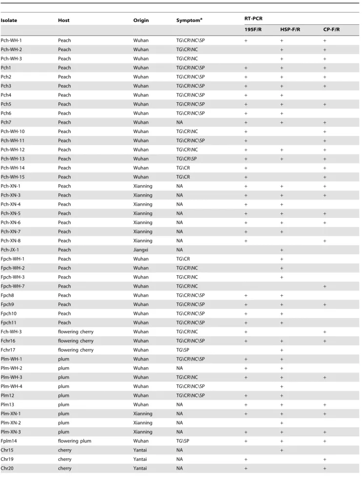
Table 1.Isolates of PBNSPaV collected from China and sequenced for their HSP70 and CP genes.
Isolate Host Origin Symptoma RT-PCR
195F/R HSP-F/R CP-F/R
Pch-WH-1 Peach Wuhan TG\CR\NC\SP + + +
Pch-WH-2 Peach Wuhan TG\CR\NC + +
Pch-WH-3 Peach Wuhan TG\CR\NC + +
Pch1 Peach Wuhan TG\CR\NC\SP + + +
Pch2 Peach Wuhan TG\CR\NC\SP + + +
Pch3 Peach Wuhan TG\CR\NC\SP + + +
Pch4 Peach Wuhan TG\CR\NC\SP + +
Pch5 Peach Wuhan TG\CR\NC\SP + + +
Pch6 Peach Wuhan TG\CR\NC\SP + +
Pch7 Peach Wuhan NA + + +
Pch-WH-10 Peach Wuhan TG\CR\NC + +
Pch-WH-11 Peach Wuhan TG\CR\NC\SP + +
Pch-WH-12 Peach Wuhan TG\CR\NC + + +
Pch-WH-13 Peach Wuhan TG\CR\SP + + +
Pch-WH-14 Peach Wuhan TG\CR + +
Pch-WH-15 Peach Wuhan TG\CR + +
Pch-XN-1 Peach Xianning NA + + +
Pch-XN-3 Peach Xianning NA + + +
Pch-XN-4 Peach Xianning NA + +
Pch-XN-5 Peach Xianning NA + + +
Pch-XN-6 Peach Xianning NA + + +
Pch-XN-7 Peach Xianning NA + +
Pch-XN-8 Peach Xianning NA + +
Pch-JX-1 Peach Jiangxi NA +
Fpch-WH-1 Peach Wuhan TG\CR +
Fpch-WH-2 Peach Wuhan TG\CR\NC +
Fpch-WH-3 Peach Wuhan TG\CR\NC +
Fpch-WH-7 Peach Wuhan TG\CR\NC +
Fpch8 Peach Wuhan TG\CR\NC\SP + +
Fpch9 Peach Wuhan TG\CR\NC\SP + + +
Fpch10 Peach Wuhan TG\CR\NC\SP + +
Fpch11 Peach Wuhan TG\CR\NC\SP + +
Fch-WH-3 flowering cherry Wuhan TG\CR\NC + +
Fchr16 flowering cherry Wuhan TG\CR\NC\SP + + +
Fchr17 flowering cherry Wuhan TG\SP +
Plm-WH-1 plum Wuhan TG\CR\NC\SP + +
Plm-WH-2 plum Wuhan NA + +
Plm-WH-3 plum Wuhan TG\CR\NC + + +
Plm-WH-4 plum Wuhan TG\CR\NC\SP +
Plm12 plum Wuhan TG\CR\NC\SP + +
Plm13 plum Wuhan NA + + +
Plm-XN-1 plum Xianning NA + + +
Plm-XN-2 plum Xianning NA +
Plm-XN-3 plum Xianning NA + + +
Fplm14 flowering plum Wuhan TG\SP + + +
Chr15 cherry Yantai NA +
Chr19 cherry Yantai NA + +
showed that the PBNSPaV population is diverse; a PBNSPaV isolate named ‘Nanjing,’ from a Chinese peach sample, was found to be highly divergent, showing only 74.4–76.9% identity to other PBNSPaV isolates [16].
China is an important country for fruit production, and stone
fruit trees and ornamental trees in the genusPrunusare grown in
almost all regions of China. However, the incidence and
distribution of PBNSPaV in the variousPrunusspecies grown in
China is unknown. Recently, during field investigations for the incidence of viral diseases in stone fruit trees in China, some
cultivated and ornamental Prunus species were found to show
trunk gummosis, stem pitting or grooving and some plum trees were found to have died from the disease. The presence and divergence of PBNSPaV isolates in China was first confirmed by reverse transcription (RT)-PCR and sequencing of a 590-nt fragment of HSP70h gene [17]. The origins and evolutionary status of PBNSPaV isolates are poorly understood. Given the apparent effect of the disease associated with PBNSPaV on fruit production, it is imperative to better understand the genetic variation and population structure of PBNSPaV. In this study,
PBNSPaV isolates from differentPrunusspecies grown in China
were characterized by sequencing their CP and HSP70h genes; the complete genomes of three representative isolates were also sequenced. The objectives of this study were (i) to determine the
incidence of PBNSPaV inPrunustrees grown in China, and (ii) to
characterize the genetic variability of the PBNSPaV isolates
obtained from differentPrunusspecies and from various regions
of China. This study provides useful information for enhanced understanding of molecular evolution within the global PBNSPaV population, which will be helpful for epidemiological investigations and for developing more efficient molecular detection methods for the diagnosis of the viral disease.
Results
RT-PCR analysis shows wide PBNSPaV infections in stone fruit trees
RT-PCR using the primer pair PBN-HSP-P1/PBN-HSP-P2 showed that of the 256 samples analyzed, 52 samples (20.3%), including 25 from peach, 5 from flowering peach, 1 from
nectarine, 10 from plum, 1 from apricot, 7 from cherry, and 3 from flowering cherry, were positive for PBNSPaV. In peach, flowering peach, and plum trees, PBNSPaV was usually associated with symptoms such as stem pitting and bark cracks on the trunks (Table 1, Fig. 1), whereas other trees were symptomless or showed only dark-colored gummosis.
Phylogenetic analysis for HSP70h and CP sequences of PBNSPaV isolates reveals existence of divergent variants
The sequences of 87 HSP70h clones from 31 isolates and 61 CP clones from 38 isolates were determined (Table 1). The product sizes for the HSP70h and CP genes were 590 bp and 1041 bp
(including 978 bp of the complete CP gene and 48,63 bp of the
intergenic region preceding the CP gene), respectively. The GenBank accession numbers for the determined sequences are KJ792814 to KJ792828 for the CP gene, KJ792829 to KJ792851 for the HSP70h gene. Sequence comparison showed that PBNSPaV isolates from China were highly divergent and the Table 1.Cont.
Isolate Host Origin Symptoma RT-PCR
195F/R HSP-F/R CP-F/R
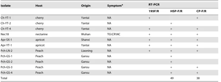
Ch-YT-1 cherry Yantai NA + +
Ch-YT-2 cherry Yantai NA +
Ch-YT-4 cherry Yantai NA + + +
Nec18 nectarine Wuhan TG\CR\NC + + +
Apr-SX-1 apricot Shanxi NA + + +
Apr-YT-1 apricot Yantai NA + + +
Pch-LN-2 Peach Liaoning NA + + +
Pch-GS-1 Peach Gansu NA +
Pch-GS-2 Peach Gansu NA +
Pch-GS-3 Peach Gansu NA + +
Pch-GS-4 Peach Gansu NA + +
Total 49 38
aTG: trunk gummosis, CR: cracking, NC: necrosis, SP: stem pitting, NA: not observed.
doi:10.1371/journal.pone.0105443.t001
Figure 1. Stem grooving (A) and brown necrotic lines (B) on the trunks of plum trees, and stem pitting on the trunk of a plum tree.
HSP70h sequences (including the 18 sequences reported previ-ously) shared 73.1–100% nt and 86.8–100% amino acid (aa) similarities, respectively. The CP gene sequences of the 38 Chinese isolates shared 74.3–98.6% nt and 87.0%–99.1% aa similarities, respectively. Intra-isolate sequence diversity was found in isolates Pch-GS-3 and Pch-XN-3. The level of nucleotide sequence identity ranged from 73.6–99.3% among distinct HSP70h variants of Pch-GS-3, and from 76.7–86.2% among distinct CP variants of Pch-XN-3.
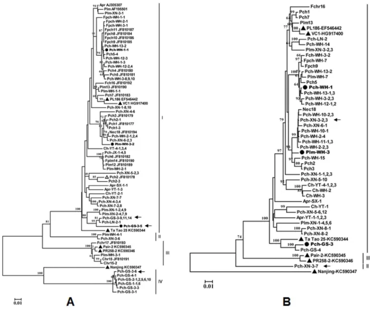
Phylogenetic analysis of the HSP70h nucleotide sequences from Chinese isolates determined in the present study, those previously reported by our group, and eight isolates available in GenBank (Table S1 in File S1) using the neighbor-joining method revealed four major groups (designated as groups I–IV) with strong (100%)
bootstrap support values (Fig. 2A). Among those groups, group I and group II are separated by a bootstrap value of 81%, and group IV is separated from the isolate Nanjing KC5990347 by a bootstrap value of 100%. Group I was the largest and contained isolates or variants from all species sampled (81.3% of all obtained sequences) and four sequences (AJ305307, AF159901, EF546442, and KC590344) referred from GenBank. Group II contained only two Chinese isolates from a plum and a peach samples. Group III contained six sequences, including one sequence (Plm-WH-3-1) determined in the present study, three sequences (Fch-WH-2-1, Ch-WH-1-1, Ch-WH-1-2) previously reported by our group (Cui
et al., 2011), and two sequences (KC590345 and KC590346) of isolates, Pair-2 and PR258-2, from France, recently reported by
Maraiset al.[16]. Therefore, group III appears to correspond to
Figure 2. Phylogenetic analysis of isolates of Plum bark necrosis stem pitting-associated virus (PBNSPaV) based on the nucleotide
sequences of their partial HSP70h gene (590 nt) (A) and complete coat protein gene (B).The CP sequences of 38 isolates obtained in this
study, the partial HSP70 sequences of 41 isolates obtained in this study, and 18 Chinese isolates previously reported were included in the analysis. The six isolates for which the complete genome sequence are available from GenBank are shown with ‘‘m’’. The three isolates for which the complete genomes were sequenced in the present study are indicated by ‘‘
N
’’. Sequences previously reported by our group and the corresponding sequences available from GenBank are identified by their GenBank accession number following their isolate names. The divergent HSP70h variants within isolate Pch-GS-3 and the CP variants within isolate Pch-XN-3 are indicated by black arrows. The trees were constructed using the neighbor-joining method. The numbers at the nodes indicate bootstrap support (1,000 replicates). Values below 60% are not shown. The scale bar represents 0.01 substitutions per site.Group 2 identified by Marais et al. [16], based on nucleotide sequences of PBNSPa-3f/2r (HSP70h). Four isolates from peach grown in Gansu province formed a separate group, group IV, which was most closely related to isolate Nanjing (KC590347), sharing 81.6–82.4% nt and 92.3–92.9% aa similarities.
The phylogenetic tree inferred from the nucleotide sequences of the complete CP gene sequences showed a similar topology to the tree inferred from the HSP70h sequences (Fig. 2B); most sequences clustered in a large group with sequence EF546442, obtained from the plum PBNSPaV type isolate PL186. However, no group corresponding to the HSP70h group IV was identified in the analysis of the viral CP sequences.
Sequence alignment of the intergenic spacer (IS) region immediately preceding the start codon of the CP gene showed that the region was highly variable, rich in the bases A and T, and that its size varies from 48 nts to 63 nts (Fig. S1 in File S2). In the IS-based phylogenetic tree, most isolates occupied similar positions to those they occupied in the CP-based phylogenetic tree, indicating that the IS sequence diversity of PBNSPaV isolates may reflect the phylogenetic relationships between viral variants.
Genome sequences of three PBNSPaV isolates
The complete genome sequences of Pch-WH-1, Plm-WH-3, and Pch-GS-3 consisted of 14,208, 14,211, and 14,211 nts, respectively (Table 2). The sequences have been deposited in the
GenBank database with accession numbers KJ792852,
KJ7928523, and KJ792854. The Pch-WH-1 and Plm-WH-3 isolates had highly similar genome sequences, with 94.8% identity at the nt level, and over 98% identity at the aa level, except for the polymerase encoded by ORF1. The isolate Pch-GS-3 shared only 88.9–89.7% nt identity with isolates Plm-WH-3 and Pch-WH-1. The genome-wide nt sequence identities between these three isolates and the type PBNSPaV isolate PL186 (EF546442) were
90.0% (Pch-GS-3) and,96% (Pch-WH-1 and Plm-WH-2), and
these three isolates showed low identity (70.2–71.2%) to an isolate Nanjing (KC590347). The genome structure of these three isolates was identical to that of the type PBNSPaV isolate (EF546442), and consisted of seven ORFs. Each ORF was the same size as the corresponding ORF in EF546442, except ORF1b, which was three nts smaller, and the gene encoding the minor coat protein (CPm), which was 16 nts longer than the corresponding genes of isolates Pair-2 and Nanjing. In general, the 39-UTRs were highly conserved and were 99.0% identical to those of EF546442. ORF1 showed the highest variability, and the three isolates showed 88.2– 95.8% nt and 86.5–96.4% aa sequence identities overall to EF546442, and only 66.3–67.6% nt and 67.3–70.6% aa sequence identities to the isolate from Nanjing. The CP and p6 genes appeared to be the most highly conserved proteins, sharing over 93% and 91.2% aa identities among these three isolates and the four reference PBNSPaV isolates. The Pch-GS-3 isolate was the most divergent of the three isolates sequenced here. The 59-UTR of the Pch-GS-3 isolate was only 87.8% identical to that of EF546442, and its seven ORFs showed overall 88.2–94.3% nt identities to those of EF546442.
Phylogenetic analysis of the complete genomic sequences of the three isolates examined in the present study and the five isolates available in GenBank revealed four groups with 100% bootstrap support values (Fig. 3). Pch-WH-1 and Plm-WH-3 clustered with the type isolate, forming a group I, and Pch-GS-3 clustered with a Ta Tao 25 isolate from a Chinese Ta Tao sample in group II, two isolates (Pair-2 and PR258-2) from France form a group III, and one isolate from Nanjing, China [16], forming a group IV.
Genetic parameters confirms the genetic diversity of PBNSPaV population
The genetic distance within and between HSP70h groups was calculated using MEGA 5 [24]. The within-group genetic distance was highest in group I (0.052) and lowest in group III (0.009). The between-group genetic distance ranged from 0.167 to 0.342 (Table 3). Group IV, consisting of peach isolates from Gansu province, was found to have the greatest genetic distance (0.332– 0.342) from the other three groups. The Nanjing isolate, which was not included in any of the groups, showed the greatest genetic distance (0.335) from group I and the lowest distance (0.211) from group VI. These data strongly supported the topology of the tree inferred from the HSP70h sequences, with four major groups.
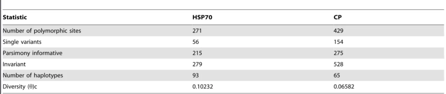
HSP70h and CP differed in diversity, polymorphic sites, and number of haplotypes (Table 4). An analysis of all available data showed that 47.8% of the nucleotides in HSP70h were polymor-phic and 39.1% were parsimony-informative, with a diversity statistic of 0.10521. In the CP gene, 44.8% of the nucleotides were polymorphic and 28.7% were parsimony-informative, with a diversity statistic of 0.06582. These results confirmed that HSP70h was more variable than CP.
The population genetic parameters of the partial HSP70 (590 nt) and CP gene sequences obtained in this study and those available in GenBank were calculated using Dnasp5 (Table 5).
The overall nucleotide diversity (p) values for HSP70h and CP
were found to be less than 1 and the haplotype diversity (Hd) values were found to be 0.996 and 1.000, indicating low genetic diversity. Negative values were obtained for the Fu and Li’s D statistical tests, suggesting that the viral population may be in expansion; however, these values were not statistically significant. The ratios (dN/dS) of nucleotide substitutions at non-synonymous (dN) to synonymous positions (dS) for HSP70h and CP were 0.083 and 0.099, much less than 1, suggesting that the two proteins have been under strong purifying selection.
Selective pressure analyses identified positively selected codons in the PBNSPaV CP gene
Furthermore, the strength of the selective forces acting on codons of the two genes were analyzed using three complementary methods, namely, SLAC, FEL, and IFEL [25]. The results showed that the distribution of codon positions under purifying, neutral, and positive selection differed between the two genes, indicating
that the two genes had been exposed to different selective pressures (Table 6). The CP gene contained more neutral codons than HSP70h. Most of the codons in the CP gene were found to be under purifying selection. Five codon positions (3, 8, 44, 57, and 88) of the CP gene were detected to be under positive selection; three of these (3, 8, and 88) were identified by all three methods. However, no codons in the HSP70h gene were found to be under positive selection.
The HSP70h gene of PBNSPaV exhibits significant recombination
One HSP70h variant sequence of a previously reported peach isolate, Pch2 (JF810178), showed significant evidence of
recombi-nation based on analysis using RDP3 (MaxChi61.356 10203,
SiScan 7.128610206, LARD 1.313610201, 3Seq 3.023610205,
Chimaera 8.270610204). Recco analysis showed that the
recom-bination junction was between nt 313–331 (Fig. S2 in File S2), with Pch3 (JF810179) and Pch1 (JF810177) identified as the major and minor parents, sharing 98.73% and 93.82% nt, and 98.91% and 97.81% aa similarities, respectively. Moreover, two other HSP70h variant sequences, Apr-YT-1-3 from apricot and Pch2-3 from peach, showed potential evidence of recombination based on Recco analysis (Fig. S2 in File S2). Of those recombinants, variant Pch2-3 and Pch2 (JF810178) were obtained from the same peach plant. However, the variant Pch2-3 showed evidence of a different recombination junction at nts 76–94, with Pch2-1 and Pch6-3 as its parents. The recombination junction of Apr-YT-1-3 was found to be between nts 233 and 245; Ch-YT-2-1 was identified as one of its parents, and the other parent was unknown.
Discussion
Of the 256Prunussamples examined in the present study, 56
were found to be infected by PBNSPaV, representing an incidence of 20.3% in stone fruit trees, similar to results obtained for samples collected in Italy [26], but much higher than the incidence identified in other studies [16]. The wide species of stone fruit hosts identified and the high incidence of PBNSPaV suggest that the virus may be efficiently transmitted in nature, similar to other closteroviruses.
To date, only a few partial sequences of the HSP70h gene of PBNSPaV are available, and no studies have been conducted on the phylogenetic characteristics of the CP gene, with the exception
Figure 3. Phylogenetic tree of the complete genome sequences of the three Chinese PBNSPaV isolates identified in this study and six isolates from GenBank.The tree was constructed using neighbor-joining analysis. The reference isolates are identified using their GenBank accession numbers. Bootstrap analysis with 1000 replicates was performed. Only.60% bootstrap values are shown.
of the six fully sequenced isolates. In this study, sequence analyze of a 590-bp fragment of HSP70h from 31 isolates and the complete CP gene from 38 isolates shows that the Chinese PBNSPaV population consists of highly divergent isolates, which can be divided into four distinct HSP70h clusters. Two HSP70h groups, II and IV, consist solely of Chinese PBNSPaV isolates sequenced in the present study and are likely to represent two novel phylo-groups. Especially, group IV consisting of molecular variants of four peach isolates from Gansu province were the most divergent from the other groups, with genetic distances of 0.332– 0.342, and a distance of 0.207 from the Nanjing isolate. Unfortunately, CP gene sequences of the isolates in HSP70h group IV could not be obtained, although several primer pairs were designed in attempts to amplify their CP gene. The failure of primers that successfully amplified most isolates to amplify CP gene of isolates Pch-GS-1, 2, 3 and 4 suggests a high level of sequence divergence in the genome of these isolates as compared
to the other isolates. Maraiset al.experienced a similar problem
[16]. The similar topologies of the phylogenetic trees constructed using all available CP and HSP70h sequences from PBNSPaV isolates indicate a co-evolutionary tendency between the two genes [27]. However, phylogenetic analyses did not show a clear relationship between genetic variability of PBNSPaV isolates and geographical or host origin excerpt that a few sequences (Pch-GS-1, 2, 3 and 4) from peach grown in Gansu formed a separated group. Furthermore, to the best of our knowledge, our study is the first to show the occurrence of mixed infections of highly divergent variants of PBNSPaV in individual host plant. The Pch-GS-3 and Pch-XN-3 isolates contained HSP70h variants in groups I and IV and CP variants in groups I and II, respectively. The presence of divergent variants in a single host plant may be a result of different infection events during horticultural operations or of vector (if it presents) transmission. Mixed infections with divergent variants could increase viral genotypic complexity, with implications for phylogenetic analysis and the evolutionary history of the virus.
Analysis of the complete genome sequences of three Chinese PBNSPaV isolates determined in this study, combined with two Chinese PBNSPaV isolates (Ta Tao 25 and Nanjing) sequenced by other researchers [16] also showed the genetic complexity of the Chinese PBNSPaV population. The genomic sequences of Pch-WH-1 from a peach and Plm-WH-3 from a plum were closely related to the type isolate PL186, sharing 96% overall nucleotide identity. However, Pch-GS-3 was found to be closely related to Ta Tao 25 (92.7% overall nucleotide identity), but less closely related to Plm-WH-3 and WH-1. Although Pch-WH-1 and Plm-WH-3 recovered from the same region (Wuhan) share high similarity of 94.8%, we cannot postulate that the sequence similarity is related to their same geographic origin. This similarity could be related to origin of their foremost isolate, from which variants were separated during dissemina-tion. In addition, although it has been observed that the infection of PBNSPaV can induce severe symptoms or be latent, more extensive studies will be done to reveal the relationships between host phenotypes and viral genotypes. Moreover, the clustering pattern of the complete genome sequences was substantially concordant with the topology of the HSP70h-derived trees, indicating that the evolutionary relationship of global PBNSPaV isolates can be reliably inferred using HSP70h sequences.
Recombination is a powerful driving force for generating new variants in RNA viruses [28–30]. Recombination events have been reported to be evolutionarily important in shaping the
genomes of some viruses in the family Closteroviridae [27].
Multiple recombination events have been identified between
Table
3.
Genetic
distances
between
and
within
variant
groups
of
PBNSPaV
partial
HSP70
gene
and
complete
CP
gene.
Group
a
HSP70h
CP
Between
variant
groups
Within
variant
groups
Between
variant
groups
Within
variant
groups
Gp-I
0.05244
0.05427
Gp-II
0.16650
0.00915
0.16757
–
Gp-III
0.18705
0.17264
0.01872
0.16369
0.17544
0.01266
Gp-IV
0.33714
0.34149
0.33186
0.04051
–
–
–
–
aThe
isolate
Nanjing
(KC590347)
is
not
included
in
this
analysis.
doi:10.1371/journal.pone.
sequence variants of the Citrus tristeza virus [31–33]. As more sequences of PBNSPaV variants become available, we may be able to identify more potential recombination events between different variants. The presence of divergent PBNSPaV variants and co-infection with different variants enable the occurrence of recombination events. For the first time, we report evidence of significant recombination in the HSP70h gene of PBNSPaV variant Pch2. Another HSP70h variant, Pch2-3, also from the Pch2 isolate, and two other variants, Apr-YT-3-1 and Pch-XN-4-6, also showed clear crossover sites at regions having high sequence similarity with their parental variants [34], although these were not detected by the programs implemented in RDP3. However, no recombination events were detected in the CP gene. The results indicate that the HSP70h gene could be a hotspot of recombination in the PBNSPaV genome; our results contrast with those from GLRaV-3, which has been shown to contain recombination hotspots in its CP gene [35], although both viruses are in the same genus.
The HSP70h genes of plant viruses in the familyClosteroviridae
have multiple biological functions and are used for phylogenetic classification of viruses in this family [1]. Most plant closteroviruses are insect-vector transmissible [27]. Thus, their capsid proteins play important roles in their ability to survive in both plant and insect environments. To date, no insect vector has been identified for PBNSPaV. Although mealybugs were frequently observed on PBNSPaV-infected fruit trees during this investigation, no insects were found to be PBNSPaV-positive in RT-PCR tests (data not shown). Some studies have indicated that vector-borne plant viruses are subjected to greater purifying selection on their capsid proteins than non-vectored viruses [36]. The population genetic parameters of both the HSP70h and CP genes suggest the possibility of a PBNSPaV population in expansion. Given the low dN/dS ratios found for HSP70h and CP, combined with the large portion of negatively selected sites identified in both genes, it appears that the virus may have undergone purifying selection, which is the primary evolutionary force acting on many plant viruses [37]. In addition, although HSP70h is more variable than CP as it is showed in Table 4, the higher dNs/dS value and the presence of positively selected sites in the CP gene indicate that the CP gene is under stronger selection pressure than that acting on the HSP70 gene. In accordance with the expectation that most mutations are deleterious or lethal to plant RNA viruses due to the compactness of viral genomes [38], the large portion of negatively selected sites existing in those two gene. The presence of a small number of positively selected sites in the CP gene might be necessary for adaption to different biological conditions. Since the successful infection of a virus in hosts depends on multiplex interactions between the virus and its host, beneficial mutations (positively selected sites) may affect interactions with host receptors and other host-specific molecules [39]. Extensive investigations
will be necessary to generate detailed information about the viral population structure and the biological and epidemiological implications of its genetic diversity and possible transmission vectors.
Because the genetic diversity within global PBNSPaV
populations has not been studied extensively, the results of this study provide important information on the genetic diversity of the Chinese PBNSPaV population and will advance the improved understanding of the epidemiology of the virus on a global scale and give insights into viral disease management [40]. From a practical perspective, the genetic diversity data generated in this study show that the HSP70h-specific primer pair is more robust and better, and that it is able to detect more PBNSPaV isolates than the primers used to amplify the CP gene. Considering the economic impact of the virus on many stone fruits [41], it is necessary and urgent to establish an efficient scheme for reducing the global transmission of the virus via propagation materials.
Materials and Methods Sample collection
In total, 256 samples from 7 species ofPrunus, including 98
from peach (P. persica), 49 from plum (P. domestica), 50 from
sweet cherry (P. avium), 10 from flowering cherry (P. serrulata),
24 from flowering peach (P. persica), 18 from apricot (P.
armeniaca), and 7 from nectarine (P. persica var. nucipersica), were collected in China between 2009 and 2013. All sample collections were conducted with approval from local institutes, and no specific permissions were required for these locations/activities. The study did not involve endangered or protected species. Symptoms of sampled trees consisting of stem pitting on tree trunks with thick corky bark, and dark-colored gummosis or spongy texture and severe cracks in the bark were observed on the trunks of some peach and plum trees. Virus-free seedlings of peach GF305 were used as negative controls in all tests for the detection of the virus.
The complete genome sequences of three PBNSPaV isolates, namely, Pch-WH-1, Plm-WH-3, and Pch-GS-3, were determined. Isolates Pch-WH-1 and Plm-WH-3 were collected from a peach tree and a plum tree grown in Wuhan city in central China, and the isolate Pch-GS-3 was taken from a peach tree grown in the Gansu province in western China. Based on our primary results of HSP70h sequencing, the three isolates distributed into three phylogenetic clades or sub-clades. Both peach tree infected by Pch-WH-1 and plum tree infected by Plm-WH-3 showed visible symptoms (Table 1) and represent two important host species of the virus.
Table 4.Phylogenetically informative statistics for HSP70h and CP genes.
Statistic HSP70 CP
Number of polymorphic sites 271 429
Single variants 56 154
Parsimony informative 215 275
Invariant 279 528
Number of haplotypes 93 65
Diversity (h)c 0.10232 0.06582
n Hd p Fu & Li’sD Fu & Li’sF dNs dS dNs/dS
HSP70 113 0.996 0.10232 20.95705 21.09359 0.02739 0.32974 0.083
CP 67 1.000 0.06582 22.26314 22.43963* 0.02107 0.21313 0.099
*Statistical significance: P,0.05. doi:10.1371/journal.pone.0105443.t005
Table 6.Estimates of selection pressures acting on the HSP70h and coat protein (CP) genes of global PBNSPaV isolates.
Coding regiona Selection pressureb
Normalized dN-dSc
Detection methods Positive Negative Neutral
Log (L) Mean No.d % No. % No. %
HSP70h 24306.36 0.0811 SLAC 0 0 99 54.1 84 45.9
FEL 0 0 136 74.3 47 25.7
IFEL 0 0 133 72.7 50 27.3
CP 26404.63 0.1199 SLAC 3 (3, 8, 88) 0.9 133 41.7 183 57.4
FEL 5 (3, 8, 44, 57, 88) 1.6 201 63.6 113 35.4
IFEL 4 (3, 8, 44, 88) 1.3 179 56.1 136 42.6
aData sets are obtained by the analysis of 183 codons for HSP70h region and 319 codons for CP region, respectively.
bPositively or negatively selected sites are identified by four selection pressure detection methods: single-likelihood ancestor counting (SLAC), fixed-effects likelihood (FEL), internal fixed-effects likelihood (IFEL), and random-effects
likelihood (REL).
cNormalized values of the ratio of nonsynonymous substitutions per nonsynonymous site (dN) to synonymous substitutions per synonymous site (dS) (dN–dS). Normalized dN–dS values,1 indicates negative or purifying,
normalized dN–dS values = 1 suggests neutral selection, and normalized dN-dS values.1 indicates positive selection.
dThe positively selected amino acid sites are indicated in brackets.
doi:10.1371/journal.pone.0105443.t006
Molecular
Character
ization
of
PBNSPaV
ONE
|
www.ploson
e.org
9
August
2014
|
Volume
9
|
Issue
8
|
RT-PCR and Sequencing of HSP70h and CP genes Total RNA was extracted from leaves using a CTAB protocol,
as described by Li et al. [42]. The presence of PBNSPaV was
tested by reverse transcription (RT)-PCR using the primer set PBN-195-F/PBN-195-R
(59-CTGGTCTTCCTGCTACTCCT-T-39/59-CGCTCTGAGATTGTGGGCTT -39), designed for
the detection of the coat protein (CP) gene of the virus [17]. The primer pairs PBN-HSP-F/PBN-HSP-R (59-GGAATT-
GACTTCGGTACAAC-39/59-TTCGGTGGTGGTACTTTCG-A-39) and PBN-CP-F/PBN-CP-R (59
-TCTTGTTGGATCGGG-GAATA-39/59-CATCTTCCACCGGACTGATT-39), designed
based on the corresponding sequences of the American PBNSPaV isolate PL186, were used for the amplification of partial HSP70h gene sequences and complete CP gene sequences, respectively.
Reverse transcription was performed at 37uC for 1.5 h using 3mL of
total RNA and 1mL of random primer in a 20-mL reaction volume
with Maloney murine leukemia virus (M-MLV) reverse transcrip-tase (Promega, Madison, WI, USA), according to the manufactur-er9s protocols. PCR was performed in a 25-mL volume of reaction
mixture containing 2.5mL of 106PCR buffer, 0.5 mM dNTPs,
0.5 mM of each primer, one unit of Taq DNA polymerase
(TaKaRa, Dalian, China), and 3mL cDNA. PCR was performed
using an iCycler Thermocycler (Bio-Rad, Hercules, CA). The PCR profile employed for all primer sets consisted of an initial denaturation at 95uC for 30 s followed by 35 cycles of 95uC for
3 min, 52uC for 30 s, 72uC for 1 min, and a final extension for
10 min at 72uC. The PCR products were separated by
electropho-resis on a 1.2% agarose gel, stained with ethidium bromide, and visualized under UV light.
PCR products were gel purified and ligated into the pMD18-T vector (Takara, Dalian, China), following the manufacturer’s instructions. The recombinant plasmids were identified after
transformation intoEscherichia coliDH5a. In order to obtain a
view of molecular composition intra each isolate, at least three positive clones of each product were sequenced at Shanghai Sangon Biological Engineering & Technology and Service Co. Ltd, Shanghai, China.
Determination of complete genome sequences
For the amplification of PBNSPaV genomes, primer sets (Table S2 in File S1) were designed based on the CP and HSP70h gene sequences obtained, and then the amplifications
were extended toward the 39- and 59-ends using primer sets
designed based on the sequences obtained here and the genome sequence (EF546442) of the first PBNSPaV isolate PL186,
available in GenBank. 59- and 39-RACE reactions were
attempted using an Invitrogen GeneRacer Kit (Invitrogen, USA), according to the manufacturer’s instructions. PCR solutions and conditions were similar to those mentioned above, except that 2 mM of each dNTP was used in a 25-mL reaction
volume, with annealing for 45 s at 50–54uC (depending on the
primer set used in each reaction), with extension for 1–3 min
(depending on the size of the PCR product) at 72uC. All
products were cloned and sequenced as mentioned above. To overcame inconvenient caused by intra-isolate sequence diversity and avoid mistakes in sequence assembling, the adjacent
amplicons were overlapped for .100 bp, and at least three
clones of each PCR product were sequenced. The obtained sequences were assembled into a contiguous sequence at a standard of over 99.9 % similarities at each of overlapped regions.
Phylogenetic and recombination analyses
The sequence alignment (produced using Clustal W) was imported into the program MEGA5, and the phylogenetic tree was constructed using the neighbor-joining method with 1,000 bootstrap replicates [24,43]. The number of polymorphic sites, single variants, parsimony-informative sites, invariant sites,
hap-lotypes, and diversity (h) were determined using Dnasp5 software
package. To obtain an overview of the Chinese PBNSPaV
population, the eighteen HSP70h sequences reported by Cuiet al
[17], the complete genome sequences of the five PBNSPaV isolates available in GenBank, and the two HSP70h sequences available in GenBank were included in the corresponding sequence analyses. The sequence sources and GenBank accession numbers of the PBNSPaV isolates used in the sequence analysis are listed in Table S1 in File S1.
Finally, the sequence alignments for the HSP70h and CP genes were scanned using seven programs for detection of recombination, implemented in the software RDP v.3.27 [44], for evidence of recombination and to determine putative recombination events, potential recombinants, and their paren-tal sequences. Only recombination events detected by at least four methods were included in further analyses. Meanwhile, recombination profiles were constructed by using the software Recco [45].
Supporting Information
File S1 Supporting tables. Table S1, The sources of HSP70h gene and complete genome of PBNSPaV isolates referred from GenBank. Table S2, Primers used for the amplification of the genomes of PBNSPaV isolates Pch-WH-1, Plm-WH-3 and Pch-GS-3.
(DOCX)
File S2 Supporting figures. Figure S1, Nucleotide se-quence alignments (A) and phylogenetic analysis (B) of the intergenic spacer (IS) region.The tree was constructed
using the maximum likelihood method.Figure S2, The Recco
output for molecular variants Pch2, Pch2-3, Apr-YT-3-1,
and Pch-XN-4-6, based on their HSP70h sequences.The
possible crossover sequences for each variant are marked by two arrows. The possible recombinant sequences are shown in white, and sequences highly similar to recombinant sequences are marked in red.
(PPTX)
Acknowledgments
The authors would like to thank Linzhong Gong and Huaping He (Institute of Fruit and Tea, Hubei Academy of Agriculture Sciences) for providingPrunussamples.
This work was financially supported by the agricultural projects (nyhyzx-201203076-03 and nyhyzx-200903004) administered by the Chinese Ministry of Agriculture. The funders had no role in study design, data collection and analysis, decision to publish, or preparation of the manuscript.
Author Contributions
References
1. Martelli GP, Agranovsky AA, Bar-Joseph M, Boscia D, Candresse T, et al. (2012) Family Closteroviridae. in: King AMQ, Adams MJ, Carstens EB, Lefkowitz EJ, editors. Virus Taxonomy – Ninth Report on the International Committee on Taxonomy of Viruses. Elsevier Academic Press, London. pp.987– 1001.
2. Lott TB, Keane FWL, May J (1962) Gumming, distortion, and pitting in cherry and apricot. Canadian Plant Dis Survey 42: 229–232.
3. Stouffer RF, Lewis FH, Soulen DM (1969) Stem pitting in commercial cherry and plum orchards in Pennsylvania. Plant Dis Report 53: 434–438. 4. Abou Ghanem-Sabanadzovic N, Mahboubi M, Di Terlizzi B, Sabanadzovic S,
Savino V, et al. (2001) Molecular detection of a closterovirus associated with apricot stem pitting in Southern Italy. J Plant Pathol 83: 125–132.
5. Amenduni T, Hobeika C, Minafra A, Boscia D, Castellano MA, et al. (2005) Plum bark necrosis stem pitting-associated virus in different stone fruit species in Italy. J Plant Pathol 87: 131–134.
6. Agrios GH (1971) Premature foliation, cambial zone discoloration and stem pitting of peach and apricot in Greece. Plant Dis Report 55: 1049–1053. 7. Marini DB, Zhang YP, Rowhani A, Uyemoto JK (2002) Etiology and host range
of a Closterovirus associated with the Plum bark necrosis-stem pitting disease. Plant Dis 86: 415–417.
8. Mircetich SM, Moller WJ, Nyland G (1977) Prunus stem pitting in sweet cherry and other stone fruit trees in California. Plant Dis Report 61: 931–935. 9. Uyemoto JK, Luhn CF, Griesbach JA, Grant JA (1995) Occurence and control
of cherry stem pitting disease. Plant Dis 79: 366–368.
10. Bouani A, Minafra A, Alimi I, Zemzami M, Myrta A (2004) First report of Plum bark necrosis stem pitting-associated virus in Morocco. J Plant Pathol 86: 91–92. 11. Mandic B, Matic S, Jelkmann W, Myrta A (2005) First report of three
filamentous viruses from cherry in Serbia. J Plant Pathol 87: 246.
12. Mandic B, Matic S, Al Rwahnih M, Jelkmann W, Myrta A (2007) Viruses of sweet and sour cherry in Serbia. J Plant Pathol 89: 103–108.
13. Sa´nchez-Navarro JA, Aparicio F, Herranz MC, Minafra A, Myrta A, et al. (2005) Simultaneous detection and identification of eight stone fruit viruses by one step RT-PCR. Eur J Plant Patho 111: 77–84.
14. El Maghraby I, Sanchez-Navarro J, Matic S, Myrta A, Pallas V (2006) First report of two viruses with filamentous particles from stone fruits trees in Egypt. J Plant Pathol 88: S69.
15. Usta M, Sipahioglu HM, Ocak M, Myrta A (2007) Detection of Apricot latent virus and Plum bark necrosis stem pitting-associated virus by RT-PCR in Eastern Anatolia. EPPO Bull 37: 181–185.
16. Marais A, Faure C, Couture C, Bergey B, Gentit P, et al. (2014) Characterization by deep sequencing of divergent Plum bark necrosis stem pitting-associated virus isolates and development of a broad spectrum PBNSPaV detection assay. Phytopathology. Available: http://dx.doi.org/10.1094/ PHYTO-08-13-0229-R.
17. Cui HG, Hong N, Xu WX, Zhou JF, Wang GP (2011) First report of Plum bark necrosis stem pitting-associated virus in stone fruit trees in China. Plant Dis 95: 1483.
18. Boscia D, Myrta A, Uyemoto JK (2010) Plum bark necrosis stem pitting associated virus. in: Hadidi A, Barba M, Candresse T, Jelkmann W, editors. Virus and virus-like diseases of pome and stone fruits. APS Press, St. Paul, MN, USA. pp.177–183.
19. Di Terlizzi B, Savino V (1995) A stem pitting of apricot. Acta Horticult. 386: 115–375 117.
20. Uyemoto JK, Teviotdale BL (1996) Graft-transmission of the causal agent of a bark necrosis-stem pitting disease of Black Beaut plum (Prunus salicina). Phytopathology 86: 111–112.
21. Zhang YP, Uyemoto JK, Kirkpatrick BC (1998) Analysis of double-stranded RNAs from cherry trees with stem pitting in California. Plant Dis 82: 871–874. 22. Garcia-Ibarra A, Martinez-Gomez P, Rubio M, Dicenta F, Soler A, et al. (2010) First report of Apricot latent virus and Plum bark necrosis stem pitting-associated virus in Apricot from Spain. Plant Dis 94: 275.
23. Al Rwahnih M, Uyemoto JK, Falk BW, Rowhani A (2007) Molecular characterization and detection of Plum bark necrosis stem pitting-associated virus. Arch Virol 152: 2197–2206.
24. Tamura K, Peterson D, Peterson N, Stecher G, Nei M, et al. (2011) MEGA5: molecular evolutionary genetics analysis using maximum likelihood, evolution-ary distance, and maximum parsimony methods. Mol Biol Evol 28: 2731–2739. 25. Kosakovsky Pond SL, Frost SD (2005) Not so different after all: a comparison of methods for detecting amino acid sites under selection. Mol Biol Evol 22: 1208–1222. 26. Matic S, Myrta A, Boscia D, Minafra A (2010) Monitoring of Plum bark necrosis stem pitting-associated virus and molecular characterization of some isolates. J Plant Pathol 92: 759–763.
27. Karasev AV (2000) Genetic diversity and evolution of closteroviruses. Annu Rev Phytopathol. 38: 293–324.
28. Simon AE, Bujarski JJ (1994) RNA-RNA recombination and evolution in virus infected plants. Annu Rev Phytopathol 32: 337–362.
29. Simon-Loriere E, Holmes EC (2011) Why do RNA viruses recombine? Nature 6: 617–626.
30. Worobey M, Holmes EC (1999) Evolutionary aspects of recombination in RNA viruses. J Gen Virol 80: 2535–2543.
31. Rubio L, Ayllo´n MA, Kong P, Ferna´ndez A, Polek M, et al. (2001) Genetic variation of Citrus tristeza virus isolates from California and Spain: evidence for mixed infections and recombination. J Virol 75: 8054–8062.
32. Vives MC, Rubio L, Sambade A, Mirkov TE, Moreno P, et al. (2005) Evidence of multiple recombination events between two RNA sequence variants within a Citrus tristeza virus isolate. Virology 331: 232–237.
33. Weng Z, Barthelson R, Gowda S, Hilf ME, Dawson WO, et al. (2007) Persistent infection and promiscuous recombination of multiple genotypes of an RNA virus within a single host generate extensive diversity. PLoS ONE 2(9): e917. doi:10.1371/journal.pone.0000917
34. Zhang J, Temin HM (1994) Retrovirus recombination depends on the length of sequence identity and is not error prone. J Virol 68: 2409–2414.
35. Farooq AB, Ma YX, Wang Z, Zhuo N, Xu WX, et al. (2013) Genetic diversity analyses reveal novel recombination events in Grapevine leafroll-associated virus 3 in China. Virus Res 171: 15–21.
36. Chare ER, Holmes EC (2004) Selection pressures in the capsid genes of plant RNA viruses reflect mode of transmission. J Gen Virol 85: 3149–3157. 37. Garcı´a-Arenal F, Fraile A, Malpica JM (2001) Variability and genetic structure
of plant virus populations. Annu Rev Phytopathol 39: 157–186.
38. Elena SF, Bedhomme S, Carrasco P, Cuevas JM, Iglesia F, et al. (2011) The evolutionary genetics of emerging plant RNA viruses. Mol Plant-Microbe In 24: 287–293.
39. Elena SF, Sanjua´n R (2007) Virus evolution: Insights from an experimental approach. Annu. Rev. Ecol. Evol. Syst. 38: 27–52.
40. Acosta-Leal R, Duffy S, Xiong Z, Hammond RW, Elena SF (2011) Advances in plant virus evolution: insights into better disease management. Phytopathology 101: 1136–1148.
41. Hadidi A, Barba M (2012) Economic impact of pome and stone fruits viruses and viroids. in: Hadidi A, Barba M, Candresse T, Jelkmann W, editors. Virus and virus-like diseases of pome and stone fruits. APS Press, St. Paul, MN, USA. pp.1–7. 42. Li R, Mock R, Huang Q, Abad J, Hartung J, et al. (2008) A reliable and
inexpensive method of nucleic acid extraction for the PCR-based detection of diverse plant pathogens. J Virol Methods 154: 48–55.
43. Thompson JD, Higgins DG, Gibson TJ (1994) CLUSTALW: improving the sensitivity of progressive multiple sequence alignment through sequence weighting, position-specific gap, penalties and weight matrix choice. Nucleic Acids Res 22: 4673–4680.
44. Martin DP, Lemey P, Lott M, Moulton V, Posada D, et al. (2010) RDP3: a flexible and fast computer program for analyzing recombination. Bioinformatics 26: 2462–2463.