an Original Genotype Possibly Deriving from a Died Out
Ancestor of Genotype IV
Olivier F. Maminiaina1, Patricia Gil2, Franc¸ois-Xavier Briand3, Emmanuel Albina2, Dje´ne´ba Keita2,
Harentsoaniaina Rasamoelina Andriamanivo1, Ve´ronique Chevalier4, Renaud Lancelot2, Dominique
Martinez2, R. Rakotondravao1, Jean-Joseph Rajaonarison5, M. Koko5, Abel A. Andriantsimahavandy5, Ve´ronique Jestin3, Renata Servan de Almeida2*
1FOFIFA-DRZV, Antananarivo, Madagascar, 2CIRAD, BIOS Department, UMR CMAEE, Montpellier, France, 3Anses-Ploufragan Plouzane´ Laboratory, VIPAC Unit, Ploufragan, France,4CIRAD, ES Department, UPR AGIRS, Montpellier, France,5Antananarivo University Madagascar, Antananarivo, Madagascar
Abstract
In Madagascar, Newcastle disease (ND) has become enzootic after the first documented epizootics in 1946, with recurrent annual outbreaks causing mortality up to 40%. Four ND viruses recently isolated in Madagascar were genotypically and pathotypically characterised. By phylogenetic inference based on the F and HN genes, and also full-genome sequence analyses, the NDV Malagasy isolates form a cluster distant enough to constitute a new genotype hereby proposed as genotype XI. This new genotype is presumably deriving from an ancestor close to genotype IV introduced in the island probably more than 50 years ago. Our data show also that all the previously described neutralising epitopes are conserved between Malagasy and vaccine strains. However, the potential implication in vaccination failures of specific amino acid substitutions predominantly found on surface-exposed epitopes of F and HN proteins is discussed.
Citation:Maminiaina OF, Gil P, Briand F-X, Albina E, Keita D, et al. (2010) Newcastle Disease Virus in Madagascar: Identification of an Original Genotype Possibly Deriving from a Died Out Ancestor of Genotype IV. PLoS ONE 5(11): e13987. doi:10.1371/journal.pone.0013987
Editor:Anthony R. Fooks, Veterinary Laboratories Agency, United Kingdom
ReceivedJune 22, 2010;AcceptedOctober 17, 2010;PublishedNovember 15, 2010
Copyright:ß2010 Maminiaina et al. This is an open-access article distributed under the terms of the Creative Commons Attribution License, which permits unrestricted use, distribution, and reproduction in any medium, provided the original author and source are credited.
Funding:This study was mainly funded by the French Ministry of Foreign Affairs (MAE) via the FSP project [GRIPAVI 2006-26] and partly by the EU network of excellence project [EPIZONE (016236, 01/06/2006–31/05/2011, http://www.epizone-eu.net/default.aspx)]. The funders had no role in study design, data collection and analysis, decision to publish, or preparation of the manuscript.
Competing Interests:The authors have declared that no competing interests exist. * E-mail: [email protected]
Introduction
Newcastle disease (ND) is a highly contagious and widespread disease which causes severe economic losses in domestic poultry, especially in chickens [1,2]. In Africa, ND is the major constraint to rural chicken development. This disease is listed as a notifiable disease by the World Organisation for Animal Health (OIE) [3]. The causative agent of the disease is Newcastle disease virus (NDV), also designated avian paramyxovirus serotype 1 (APMV-1), which belongs to the genus Avulavirus within the family
Paramyxoviridae [4,5]. The genome comprises a single stranded negative sense RNA that encodes the RNA-dependent RNA polymerase (L gene), the haemagglutinin-neuraminidase (HN gene), the fusion (F gene) and matrix (M gene) proteins, the phosphoprotein (P gene) and the nucleoprotein (NP gene). The genome is predicted to be 15186, 15192 or 15198 nucleotides (nt) in length, always a multiple of six nt, which fulfils the so-called ‘rule of six’ for optimised replication [6,7,8,9,10]. Based on the analysis of nucleotide sequence of the F protein gene, 10 different genotypes (I–X) or 6 different lineages (1–6) of NDV have been identified so far. The genotypes VI (lineage 4) and VII (lineage 5) are further divided into eight (a–h) and five (a–e) subgenotypes/ sublineages, respectively [11,12,13,14,15,16,17,18,19]. APMV-1 strains can be categorised according to their virulence into highly (velogenic), intermediate (mesogenic) or non virulent (lentogenic).
The pathogenicity of APMV-1 isolates is assessed on the basis ofin vivotests including the intracerebral pathogenicity index (ICPI) in 1-day-old chickens, the mean time of death (MDT) of embryo-nated specific-pathogen-free hen’s eggs after inoculation and the intravenous pathogenicity index (IVPI) in 6-week-old chickens [3]. The molecular basis for these different levels of pathogenicity is known to be linked to the sequence of cleavage site of the precursor fusion protein (F0). At this position, a pathogenic NDV
strain (velogenic and mesogenic) has at least one extra pair of basic amino acids motif112X-R-X-R/K-R-F117[20] and can be cleaved by a wide range of proteases of the furin family in different host cells [21].
induce more than 40% of mortality in such non protected poultries [28]. In spite of the importance and endemicity of ND in Madagascar, no data is available about the virus variants involved in clinical cases and/or maintenance of this disease in the island. In this study, four APMV-1 strains from Madagascar (named here MG group) were isolated in 1992 and 2008 and were molecularly characterised. Two of these strains were fully sequenced while the two other ones had only F and HN genes sequenced. Phylogenetic analyses showed that the MG group strains clustered within a new distinct genotype, closer to old genotypes. This study represents the first molecular characterisation of APMV-1 circulating in Madagascar and provides evidence on the existence of a new genotype close to an old died out genotype.
Materials and Methods
Ethics statement
All animal experiments (ICPI tests) were conducted according to internationally approved OIE standards, under authorizations set forth by the director of the veterinary services of Coˆtes d’Armor on behalf of the Prefect of Coˆtes d’Armor (Nu22-18) and the Director of the veterinary services of He´rault on behalf of the Prefect of He´rault (Nu 34-114). Certificates of authorization are available from the authors upon request.
Origin and isolation of virus strains
Synthetic information about the Madagascar isolates is provided in Table 1. An ‘‘old’’ strain (MG-1992) was first isolated by theCentre National de Recherche Applique´e au De´veloppement Rural(FOFIFA-DRZV) in 1992, after suspicion of avian influenza or ND in a dead fowl assumed to be vaccinated against ND (La Sota vaccine). The three other isolates were collected in Madagascar during a surveillance programme in African wetlands led in 2007 and 2008 by the Centre de Cooperation Internationale en Recherche Agronomique pour le De´veloppement(CIRAD), Montpellier, France and the FOFIFA-DRZV, Antananarivo, Madagascar. The MG-725/08 strain was recovered both from cloacal and tracheal swabs from an unvaccinated apparently healthy chicken. Another strain (MG39-04/08) was isolated from a dead chicken in a commercial farm: this chicken was initially vaccinated with the HB1 strain and boosted with the La Sota vaccine. The last isolate (MG-Meola/08) was recovered from a non vaccinated backyard poultry.
Samples of brain tissues or cloacal/tracheal swabs positives in the APMV-1 specific PCR (protocol based on M gene recom-mended by the reference laboratory of the OIE) were further processed for virus isolation by inoculation into 9-day old chicken embryonated eggs. All isolates were grown in less than 3 passages on eggs and tested by PCR before molecular sequencing.
In vivo assay
The intracerebral pathogenicity index (ICPI) was determined according to international OIE standards (OIE, 2009) for two isolates of the MG group (MG-1992 and MG-725/08). Briefly, fresh allantoic fluid with a HA titre.1/16 was diluted 1021in sterile isotonic saline buffer. Following filtration through 0.22 or 0.45mm filters this diluted virus was injected intracerebraly (0.05 ml) into 10 one day-old chick hatched from embryonated SPF hens’ eggs. Whereas each inoculum was checked for lack of bacterial and fungal contamination following culture on trypto casein soja and sabouraud media, the birds were examined at 24h intervals for 8 days and given a score (0, 1 or 2) according to their status (respectively healthy, sick or dead). The ICPI was calculated as the total of individual scores during 8 days divided by 80 (the number of days x the number of chickens).
RNA isolation, cDNA synthesis and nucleotide sequencing
For reverse transcription and PCR reactions, RNA was extracted from allantoic fluids by using the NucleospinH RNA Virus kit (MachereyNagel) following the manufacturer’s instruc-tions. The cDNA transcriptions were carried out by using the First-strand cDNA synthesis kit (GE Healthcare). Six pairs of oligonucleotide primers previously published [13,29] or designed in this study (Table S1 in supporting material), using BioEdit version 7.0.9.0 and Vector NTI software version 11.0 (ß2008 Invitrogen Corporation), were used to amplify six overlapping DNA fragments to generate the complete sequences of F and HN protein genes. All DNA fragments were sequenced in both directions by Cogenics Genome Express S.A. (Meylan-France). For the rest of the genome, 23 overlapping PCR were realized with Platinum Taq DNA polymerase (PlatinumH Taq DNA Polymerase High Fidelity, Invitrogen). The DNA sequences were determined in both senses using the Big dye Terminator v3.1 cycle sequencing kit (Applied Biosystems) according to the manufactur-er’s instructions and using the same primers as to PCR reactions.
Alignment of the F and HN predicted amino acid sequences and phylogenetic analyses
The sequences of the four isolates were compared with previously reported NDV sequences representative of different genotypes available in GenBank. A 374 nucleotide (nt) fragment of a variable portion of the F protein gene (47 to 421), including the F0 cleavage site and a 1713 nt (92 to 1805) of HN gene were
processed by Clustal W [30] in alignX program included into Vector NTI software suite (version 11.0). The nucleotide sequence databank accession numbers of ND viruses used in this study are shown in supporting Table S2 of supporting material. Alignment of complete amino acid sequences of F and HN was processed by
Table 1.NDV isolates from Madagascar used in this study.
Strain Code Region/Country Statut Sequence
Clinic Vaccine size (nt) Covering full genes
APMV1/Chicken/MG/1992 MG-1992 Ivato-Madagascar Dead laying chicken La Sota 15082 NP, P, M, F, HN and L APMV1/Chicken/MG/725/2008 MG-725/08 Mahitsy-Madagascar Healthy rural poultry NVa 15097 NP, P, M, F, HN and L APMV1/Chicken/MG/39-4/2008 MG-39-04/08 Ivato-Madagascar Dead broiler chicken La Sota 4249 F and HN
APMV1/Chicken/MG/Meola/2008 MG-Meola/08 Tsarahonenana-Madagascar Sick grower fighter cock NV 3966 F and HN
a: not vaccinated.
alignX program in Vector NTI software and finalised using MEGA4 software (version 4.1). Phylogenetic relationships and nucleotide divergence analyse (intra and inter genotype) were established with MEGA4 [31] using the Kimura 2-parameter correction in which the transition/transversion ratio was estimated from the data [32]. The statistical significance of the tree topology generated with the neighbor-joining algorithm was evaluated by 1,000 bootstrap resamplings of the data [33]. To confirm the robustness of the genetic groupings obtained by the phylogenetic analysis on the partial fragment (374 nt) a phylogenetic tree based on full-genome sequences of two isolates of the MG group (MG-1992 and MG-725/08) and 86 other full-genome sequences belonging to different genotypes were further carried out using the same parameters used for F and HN analyses. Putative recombination events in the two full-genomes of MG isolates were examinated using the recombination detection program RDP 3.0b [34].
Results
Determination ofin vivopathogenicity (ICPI test) ICPI values were determined for the isolates MG-1992 and MG-725/08. These isolates showed a very high ICPI of 1.9 being classified into a velogen-type NDV (Table 2).
Sequencing MG, genome organisation and phylogenetic analyses
The sequence covering the six open reading frames (ORFs: NP, P, M, F, HN, L), without the complete sequence of the 39-leader/ 59-trailer of two MG strains, MG-725/08 and MG-1992, and the complete nucleotide sequence covering the F and HN genes of two other strains, MG-39-04/08 and MG-Meola/08, obtained in this work were analysed along with those of reference strains retrieved from GenBank.
Class II APMV-1 isolates have two genome lengths. The genotypes considered as ‘‘early’’ (1930–1960; genotypes I to IV, IX and H33(W)) contain 15,186 nt and recent genotypes (that emerged after 1960, genotypes V–VIII and X) contain 15,192 nt (15,186 nt+6 nt in the 59non-coding region or NCR of NP gene) [12]. As seen in recent genotypes, the MG group has six nucleotides insertion in the 59 non-coding region (NCR) of the nucleoprotein gene between nucleotides 1647 and 1648 increasing its genome size to 15,192 nt (Figure 1).
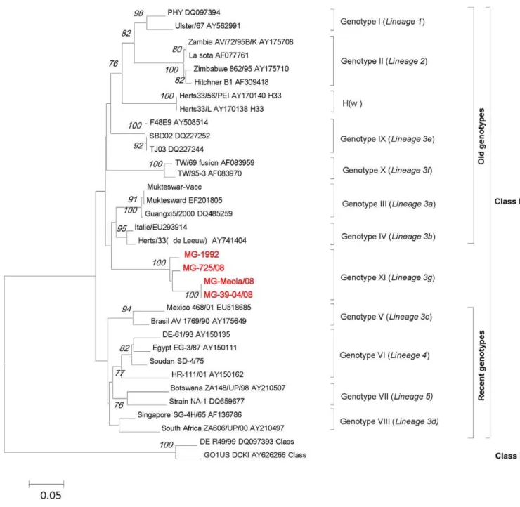
The phylogenetic analysis of F gene (Figure 2) shows that the MG strains are grouped together. They are closer to the ‘‘old’’ genotype IV but may be distant enough to constitute a new genotype, proposed here as genotype XI or lineage 3g. This phylogenetic topology created using 374 nt fusion gene fragments was confirmed by phylogenetic analyses based on the full NH gene (data not shown) and on the full-genome sequences of two isolates of the MG group (MG-1992 and MG-725/08) and 86 other full-genome sequences belonging to different genotypes (Figure 3).
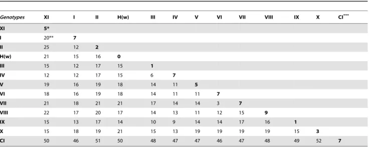
Based on a partial fragment of F gene (374nt), genetic divergences between the MG group and the other ten genotypes (I to X) are presented in Table 3. The F nucleotide sequences divergence within the MG group was 5%. The maximum intra-genotype divergence in old intra-genotypes was 7% (intra-genotypes I and IV) whereas the maximum intra-genotype divergence in recent genotypes was 9% (genotype VIII). The lower genetic divergence found between the MG group and another genotype (genotype IV) was 12%. In contrast, the higher divergence (25%) was observed with the genotype II that includes the La Sota vaccine strain. Since the maximal intra-genotype divergence ever observed does not exceed the minimal divergence of the MG group with other
genotypes, our data support the hypothesis that MG group constitutes a distinct new genotype. This new genotype is further confirmed by the phylogenetic analysis of the full-genome sequences (Figure 3). Surprisingly, the phylogenetic analysis of whole genome sequences also show that the genotype IV does not cluster with the ‘‘old genotypes’’ (I, II, III, IX), as suggested before (Czegledi et al., 2006), but rather with ‘‘recent genotypes’’ (V,VI,VII).
The analysis of the F1/F2protein cleavage site sequence of the
four MG isolates showed that they share an original cleavage site motif formed by five arginines (R) 112R-R-R-R-R116 at the C terminus of F2exhibiting an atypical Q/R substitution at position
114 followed by a phenylalanine (QF117) at position 117 of the F1
amino terminus (Table 2). This motif is considered as a velogenic motif according to OIE [3]. Other virulent-like cleavage motifs with five basic amino acids have been described for genotypes VI, VII and VIII that are formed by a combination of arginine (R) and lysine (K) residues like 112R-R-K-K-R116*F117 or 112 R-R-R-K-R116*F117 [35,36,37,38,39]. The substitution of a glutamine residue by a basic amino acid (R or K), corresponding to nucleotide mutation at positions 4885 or 4886 (CAGRCGG or AAG) is only found in some strains pertaining to the ‘‘recent’’ genotypes VI or VIII.
Like other NDV strains, the complete length of F and HN genes of MG group viruses were 1792 nt and 2002 nt, respectively. The F gene of all MG group isolates showed a single open reading frame (ORF) beginning with a unique double start codons
44
AUGAUG49 and ending at position 1705 followed by the UGA stop codon. Consequently, the open reading frame of the F gene may extend to 1665 nt rather than 1662 nt usually found in other reference strains.
The predicted amino acid sequences of the complete F gene of the four MG isolates was analysed and compared with different strains pertaining to genotypes I to IX (Table 4). This analysis showed that all seven neutralising epitopes critical for both structure and function of the protein positioned at individual residues D72, E74, A75, K78, A79, L343and the stretch of amino acids157 ILRLKESIAATNEAV-HEVTDG171, are conserved [40,41,42]. The twelve cysteines residues placed at positions 25, 76, 199, 338, 347, 362, 370, 394, 399, 401, 424 and 523 [43] and the predicted N-glycosylation sites (Asn-X-Ser/Thr or N-X-S/T) where X is any amino acid except aspartic acid or proline, located at positions85N-R-T87,191N-K-T193,
366
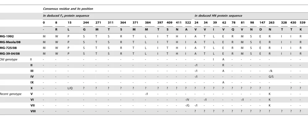
N-T-S368,447N-I-S449,471N-N-S473and541N-N-T543[44,45] are also conserved in the MG group. However, the analysis of predicted amino acid sequence of a F protein fragment (residues 0–553 for MG isolates or 1–553 for the other strains) revealed substitutions in the MG isolates found also in other strains clustered in the old genotypes (I–IV and IX) like T16RI, E104RG, Q195RR as well as substitutions found in recent genotypes (V to VIII) like V81RL, A106RV, V118RI and V350RI. In addition, the MG group has fifteen specific amino acid residues in F protein which are not found in any other genotype (Table 4). Twelve (80%) of these amino acids are located in the head of trimeric NDV-F protein M0R-, W8RR, P15RL, S272RN, M289RL, S311RT, R364RS, T371RM, L384RM, I397RT, T409RS and H411RN. The three remaining substitutions S244RG, K476RN, and I522RA are located in the stalk of the F-trimeric structure (Figure 3). Interestingly, the three MG strains recently isolated in 2008 share eight additional amino acids (Q4RK, F12RP, S28RL, R73RK, S79RA, E110RG, V202RI, S258RN and H337RY)
compared with the strain isolated in 1992.
Table 2.F gene referential NDV strains used in this study.
Strain Class or Genogroup ICPIavalue Cleavage site
Identification Accession number r--- F2R * rF1---R
112 113 114 115 116 117 118
R R Q R R * F I
DE R49/99 DQ097393 Cl I (6) nd G - - G - * L V
GO1US DCKI AY626266 Cl I (6) nd E - - E - * L V
Herts33/56 (PEI) AY170140 H33 (w)b nd - - - - - * -
-Herts33 (L) AY170138 H33 (w)c nd - - - - - * -
-Ulster/67 AY562991 I (1) 0,0 G K - G - * L
-Ethiopie Panvac (2/P2) AY175720 I (1) 0,0 G K - G - * L
-PHY-LMV42/66 DQ097394 I (1) 0,0 G K - G - * L
-Hitchner B1 AF309418 II (2) 0,2–0,5 G - - G - * L
-La Sota AF077761 II (2) 0,2–0,5 G - - G - * L
-Zimbabwe AV862/95 AY175710 II (2) nd G - - G - * L
-Zambie AV 72/95 AY175708 II (2) nd G - - G - * L
-Mukteswar EF201805 III (3a) 1,4 - - - * -
-Guangxi5/2000 DQ485259 III (3a) nd - - - * -
-Herts/33 (de Leeuw) AY741404 IVea (3b) 1,99 - - - * -
-BG 60–81 AF402129 IVea (3b) nd - - - * -
-BG 5–67 AF402104 IVbg (3b) nd
SIMF/64 AJ243390 IVbg (3b) nd - - - * -
-Soudan 72 AV 2203 AY135753 IVit (3b) nd - - - * -
-DE-191/77 AF525378 IVit (3b) nd - - - * -
-IT-48/68 AF297969 IVit (3b) nd - - - * -
-MA-307/77c EU604259 IV (3b) nd - - - - - * - V
MA-13/02c DQ096598 IV (3b) nd - - - - - * - V
Tanzania AV 1300/95 AY175687 V (3c) nd - - - K - * - V
Mexico468/01 EU518685 V (3c) nd - - - K - * - V
Brasil AV1769/90 AY175649 V (3c) nd - - - K - * - V
HR-111/01 AY150162 VI (4) nd K - - K - * -
-Soudan SD-4/75 AY151384 VI (4) nd - - - K - -
-Egypte EG-3/87 AY150111 VI (4) nd - - - K - -
-DE 61/93 AY150135 VI (4) nd - - K K - * -
-Strain NA DQ659677 VII (5) nd - - - K - * -
-MZ 13/94 AF136775 VII (5) nd - - - K - * -
-Botswana ZA148/UP/98 AY210507 VII (5) nd - - - K - * -
-South Africa ZA606/UP/00 AY210497 VIII (3d) nd - - - K - * -
-Singapore SG-4H/65 AF136786 VIII (3d) nd - - - K - * - V
F48E9 AY508514 IX (3e) 1,89 - - - * -
-SBD02 DQ227252 IX (3e) nd - - - * -
-TJ03 DQ227244 IX (3e) nd - - - * -
-TW/69 AF083959 X (3f) nd - - - K - * -
-TW/95-3 AF083970 X (3f) 1,68 - - - K - * -
-MG-1992 HQ266603 XI (3g) 1,9 - - R - - * - V
MG-725/2008 HQ266602 XI (3g) 1,9 - - R - - * - V
MG-39-4/2008 HQ266605 XI (3g) nd - - R - - * - V
MG-Meola/2008 HQ266604 XI (3g) nd - - R - - * - V
a: ICPI intracerebral pathogenicity index; b: Weybridge line [77];
protein is composed of 571 aa and has the feature of virulent NDV strains [14,42,46,47]. In this study, different HN sequence strains pertaining to genotypes or subgenotypes available in Genbank (I to VII and IX) with different lengths were aligned with the MG group sequences (data not shown). The results showed that all the previously described neutralising epitopes 193LSGCRDHSH201, R263, D287, K321,332GR333,346DEQDYQIR353, K356, N481, D494,
513
RITRVSSSS521, G/D569 [48,49,50] are not modified in MG group strains. The three amino acid E401, R416, Y526essential for receptor binding site [51,52] and the neuraminidase activity site represented by functional triarginyl cluster at positions174RI175, R416, R498, the amino acid sequence in region234NRKSCSI/V/ L240[53] as well as the regions314FXXYGGV/L/M320-399GA/ SEGRI/V/L405involved in hemagglutinating activity [54], were likewise conserved in the MG strains. In addition, the MG strains have preserved the thirteen cysteines residues in the linear sequence of the ectodomain 123, 172, 186, 196, 238, 247, 251, 344, 455, 461, 465, 531, 542 [43] and the eleven sialic acid receptor binding sites R174, I175, E258, Y299, Y317, E401, R416, R498, Y526, R516, E547[52]. Except the cysteine residue at position 123, all these amino acid cited before are located in the globular head on the HN spike. Otherwise, the analysis of HN amino acid sequences showed that the four MG strains contain also eleven specific amino acid residues that are different from those of any other NDV strain (Table 4). Seven of these A24RV, T34RV, L39RI, ERV62, RRQ78, M81RV and S98RN are located in the 124 first N-terminal amino acids, including the cytomere (position 1 to 20), the transmembrane region (position 21 to 49) and the stalk (position 50 to 124) of HN protein. The four last characteristic amino acids E147RD, R263RN, I328RT, R539RK are localized on the globular head of the protein (Figure 4). In addition, the MG-725/08 strain has six additional specific substitutions H3RR, T64RS, V117RA, R377RK, A380RT and A510RS when compared with the three other MG strains (Meola/ 08 MG 39-04/08 and MG-1992).
The analysis of recombination events by the RDP 3.0b software [34] was performed among Malagasy strains and other full sequences of APMV-1 of class II (genotypes I, II, III, IV, V, VI, VII and XI) and one strain of class I. No recombination events were detected in the Malagasy strains.
Discussion
In this study, complete genome or complete genes of F and HN proteins of four NDV strains isolated in Madagascar (MG group strains) were sequenced and analysed. These strains are the first
Madagascar isolates characterised at the molecular level. Among them, the strains MG 1992 and MG39-04/08, isolated in 1992 and 2008 respectively, were recovered from vaccinated chickens.
The salient findings of this work are the strong evidence that MG isolates constitute a new genotype closer to the old genotypes that were circulating 50 years ago and responsible for the first pandemic of ND in the world. The MG group strains are branched to the genotype IV cluster from which strains have never been detected since 2002. [12]. The partial sequence of the F gene (374 nt at positions 47 to 420) is regarded as a standard criterion for NDV genotyping of isolates present in various parts of the world [55]. Based on percentage of genetic divergence of this F gene fragment, the maximum intra-genotype nucleotide variations within any old genotype (below 7%) is lower than the minimum nucleotide distance found between these genotypes and the MG group (12% to 25%). This is clearly supporting that MG isolates are closely related to genotype IV but distant enough to constitute a new genotype or lineage, named here genotype XI or lineage 3g in reference to the two last genotypes IX [42] and X [14] (lineages 3e and 3f, respectively) identified in Asia. The cluster topology of the MG group strains based on the F gene was confirmed by the phylogenetic analysis based on full-genome sequences of two isolates of the MG group (1992 and MG-725/08) and 86 other full-genome sequences belonging to different genotypes.
Whole genome sequencing of two MG strains revealed a genome size of 15,192 nucleotides meaning that this group has an insertion of six nucleotides into the 59NCR of the nucleoprotein gene, characteristic of recent genotype strains. In addition, MG isolates have an arginine at position 114 (R114) of the F protein cleavage site found in some recent isolates but never in old strains. MG viruses have also a substitution V118RI, which is considered to be a feature of the genotype V [56,57] that belongs to the recent genotypes cluster. In contrast, MG isolates share a molecular substitution characteristic of old genotypes as E104RG, in F protein [42,58]. Although Yu et al [58] claimed that E104RG substitution caused a dramatic change responsible for the evolution of the old genotypes towards the recent genotypes, we do not found such mutation in the MG strains nor in the strains of genotypes IX and X, phylogenetically closer to the old genotypes. Moreover, the phylogenetic analysis based on full-genome sequences unexpectedly showed that the genotype IV, and the newly proposed genotype XI as well, are not branched in the ‘‘old genotypes’’ cluster (I, II, III, IX), as observed in phylogenetic trees constructed based on the 374 nt fragment of F gene. All together, our results can suggest that genotypes IV and XI are intermediates Figure 1. Alignment showing the inserted sequence at in the position 1647 in 59non-coding region of NP gene.
between old and new genotypes or that the current system of genotypes or lineages clustering based on the 374 nt fragment of F gene is probably inadequate to classify NDV isolates.
All MG isolates show the same fusion protein cleavage motif
112
R-R-R-R-R*F117. To our knowledge, this atypical virulent-like cleavage site has never been reported before in APMV-1 strains. However, it is found in other paramyxoviruses (Rubulavirus) like the simian virus type 5 [45,59]. In addition, the presence of a phenylalanine at position 117 (QF117) in the MG isolates was previously described as being a possible contributor to neurological effects [10]. Thereafter, the length of amino acid sequence of the
haemagglutinin-neuraminidase protein (571 aa) is also characteristic of virulent strains [46,60,61]. The prediction of virulence based on the F cleavage site pattern was confirmed byin vivotests that has resulted in a high ICPI value (1.9), close to the maximum of 2, with the two strains tested (MG-1992 and MG-725/08). In spite of the established virulence of the MG-725/08 strain, it was recovered both from cloacal and tracheal swabs from an unvaccinated and apparently healthy chicken. However, the possibility that this chicken was sampled during the incubation period of the infection or was partially protected by a previous infection with an avirulent or vaccine strain that circulate in the field cannot be ruled out. Figure 2. Phylogenetic tree (unrooted) of nucleotide sequences based on a 374-nt sequence (position 47–421 nt) of the F gene. Phylogenetic relationships of MG group strains with previously published sequences in Genbank. The evolutionary history was inferred using the Neighbor-Joining method [33]. All results are based on the pairwise analysis. Analyses were conducted using the Kimura 2-parameter method in MEGA4 [31,32] with 1,000 bootstraps [76]. The isolates from Madagascar that were subjected to analysis in this work are in bold and red. Genotype or lineage groupings are indicated on the right.
Based on neutralising tests and cross-protective analyses, it is accepted that APMV-1 exists as a single serotype [62]. Therefore, the genetic variations of the virus are not expected to result in vaccination failure. However, different levels of cross-protection have been observed in chickens vaccinated with the vaccine strain La Sota belonging to the genotype II and challenged with different wild-type strains [55]. Sporadic cases of ND in commercial farms vaccinated with this vaccine were previously reported in China [7,58], in southern California and adjacent states [63], in Mali [64], Cameroon, Nigeria and Burkina Faso [65]. Whether these observations are related to a reduction of vaccine efficacy or an improper vaccine use in the field remains unclear. It is however possible that NDV strains responsible for ND sporadic outbreaks in vaccinated chickens can escape the immune responses [16,18,66] and thus contribute to the emergence of new genotypes [42]. This vaccine failure has been announced by Alexander et al., [67] during the third panzootic of ND (1981–1983) in Europe in which the pigeons immunized with live NDV B1 vaccine were not well protected against pigeon paramyxovirus isolates (genotype VI). It is also known that the immune pressure imposed by the vaccination may be selecting virulent variant forms of NDV [68]. Many authors have demonstrated that current vaccines prevent disease but cannot stop viral shedding [47,63]. The NDV strains currently circulating (subgenotype VIId prevalent in Asia, Europe and Africa) and the genotype XI predominant in Madagascar have a significant genetic distance (21% to 25%, respectively), with the widely used vaccine strain La Sota pertaining to the genotype II. These genetic differences and a consequent suboptimal vaccination may be responsible for sporadic cases of ND in vaccinated poultry flocks in Madagascar. In contradiction with
this, multiple sequence alignments of the four MG isolates made with several published sequences including the La Sota strains and HB1, members of genotype II (data not shown) revealed that all the neutralising epitopes identified so far and the predicted attachment receptor sites in the F and HN glycoproteins are conserved. However, all MG group strains possess fifteen and eleven original amino acid substitutions on F and HN proteins, respectively. Some of these substitutions occurred in the globular head of the proteins. Furthermore, 5 out of the 8 mutations in the head of the F protein are concentrated in a short invariable region at position 364–411, suggesting that the observed mutations are actually selected over time by the host immune responses. It is supposed that these substitutions should have resulted in antigenic drift and major changes in the protein conformation [69], particularly the changes in amino acid polarity as T34RV, E62RV in HN protein and S244RG in F protein. The HN/F protein interaction for fusion promotion involves the HN head region (aa 124 to 151) and the F protein heptad repeat 2 (HR2) at position 454 to 492 [70,71]. In these regions, the MG strains show amino acids substitutions at position E147RD, K476RN of HN and F proteins, respectively. Morrison and Gravel [72] have demonstrated that amino acid substitutions in the head region domain of HN protein as L133RI or A140RL were responsible for an enhanced or diminished virus attachment activity, respectively. The possibility that other mutations like E147RD e/or K476RN in the same or related functional region may also contribute to virulence or immune evasion cannot be ruled out. In addition, the MG isolates display other specific mutations, some of them affecting the head of HN. It is tempting to postulate that these modifications (F or HN genes) may play a role in virulence or Figure 3. Phylogenetic tree of the nucleotide sequences based on a 14977nt (NP/P/M/F/HN/L genes).The evolutionary history was inferred using the Neighbor-Joining method [33]. All results are based on the pairwise analysis. Analyses were conducted using the Kimura 2-parameter method in MEGA4 [31,32] with 1,000 bootstraps [76]. The isolates from Madagascar that were subjected to analysis in this work are in blue [22]. Genotype and the lineage groupings are indicated on the right.
doi:10.1371/journal.pone.0013987.g003
Table 3.Matrix estimate of genetic divergence (%) between nucleotide sequences (374 nt) of NDV strains.
Genotypes XI I II H(w) III IV V VI VII VIII IX X CI***
XI 5*
I 20** 7
II 25 12 2
H(w) 21 15 16 0
III 15 12 17 15 1
IV 12 12 17 15 6 7
V 19 16 19 18 14 11 5
VI 18 16 19 18 14 11 11 7
VII 21 18 21 21 17 14 14 3 7
VIII 22 17 20 17 14 13 11 12 15 9
IX 15 13 17 14 10 9 14 14 17 16 1
X 15 18 19 21 15 13 19 19 19 19 15 3
CI 50 46 51 50 48 47 47 46 47 48 49 52 7
*Intragenotype or intrasubgenotype: Maximal value of Nucleotide genetic divergence. **Intergenotype: Minimal value of genetic divergence.
***Class I.
All results are based on the pairwise analysis of 61 NDV sequences including the 4 MG isolates (genotype XI). The nucleic acid analyse were conducted using Kimura 2-parameter method in MEGA4 [31,78]. All positions containing alignment gaps and missing data were eliminated only in pairwise sequence comparisons (Pairwise deletion option). There were a total of 374 nt positions in the final dataset.
Table 4.Residue substitutions specific in deduced F0protein and HN protein sequence of the MG group.
Consensus residue and its position
In deduced F0protein sequence in deduced HN protein sequence
0 8 15 244 271 311 364 371 384 397 409 411 522 24 34 39 62 78 81 98 147 263 328 430 539
- R L G M T S M M T S N A V V I V Q V N D N T T K
MG-1992 M W P S T S R T L I T H I A T L E R M S E R I I R
MG-Meola/08 M W P S T S R T L I T H I A T L E R M S E R I I R
MG-725/08 M W P S T S R T L I T H I A T L E R M S E R I I R
MG-39-04/08 M W P S T S R T L I T H I A T L E R M S E R I I R
Old genotype I - - - I A - - -
-II - - - -/I - - R - - -
-III - - - -/I - - A - - - - -/k -
-IV - - - -/I - - - G/S -
-IX - - - A - - -
-X - - L/Q ? ? ? ? ? ? ? ? ? ? ? ? ? ? ? ? ? ? ? ? ?
Recent genotype V - - - -/I - - - K -
-VI - - - -/V - -/I - - - -/I - - K -
-VII - - - -/G -/I - - - K -
-VIII - - - ? ? ? ? ? ? ? ? ? ? ? ?
? : Sequence not available.
doi:10.1371/journal.pone.0013987.t004
New
Genotype
APMV-1
ONE
|
www.plos
one.org
9
November
2010
|
Volume
5
|
Issue
11
|
emergence of escape mutants and finally the apparent lack of vaccine efficacy observed in the field.
The number of unique motifs found in all MG isolates, including the atypical F protein cleavage site R-R-R-R-R*F-V, also suggests a monophyletic origin of these viruses. Under this hypothesis, the maximum genetic variation observed within MG isolates (5%) is compatible with a divergence from a common ancestor over 50 years, considering that the rate of nucleotide change is approximately 1% per decade under natural field conditions in epizootic periods [12,57]. This scenario is in agreement with the first introduction of an old genotype of NDV in Madagascar in 1946. However, the higher genetic variation (12%) observed between the genotype XI and its closely related genotype IV suggests that the common ancestor of these two genotypes may have emerged several decades before the introduction of its virus progeny in Madagascar. As mentioned before, the accumulation of multiple aa substitutions in F or HN proteins may also result from the immune pressure that can contribute to increase the phylogenetic distance between the MG group and its progenitor genotype. The presence of this selection pressure at specific amino acid sites are recognised as adaptive evolution [47,73]. This abnormal evolution rate of genotype XI with regard to genotype IV may originate from combined characteristics of current poultry production systems including the commercial farm with host genetic homogeneity, intensive and/or improperly executed vaccination programs and backyard poultry breeding with high density of birds (allowing close animal-to-animal contact, and favouring transmission of highly virulent virus over milder forms) [74]. Moreover, high phylogenetic and antigenic distances between vaccines and circulating strains may facilitate the evolution of virulent NDV [75].
In conclusion, all four isolates from Madagascar were clustered in a new genotype XI, presumably deriving from an ancestor close to genotype IV introduced in the 50’s and possibly resulting from a self-contained evolution due to geographical and ecological characteristics of this island. The particular evolution of genotype XI in Madagascar reinforces the idea that this island, known for its outstanding flora and fauna biodiversity, is also a unique ‘‘natural ecosystem’’ for micro-organisms. Other genotypes like genotypes I, II, III and VII were also detected in the island (data not shown). These genotypes may have been introduced more recently in the island, probably as a consequence of poultry import or use of live vaccine strains. The co-circulation of different genotypes at the same time and other introductions of new viruses in relation with the worldwide intensification of animal movements, may contribute in the future to dramatically increase the complexity of the situation.
Supporting Information
Table S1 Primers used in this study.
Found at: doi:10.1371/journal.pone.0013987.s001 (0.07 MB DOC)
Table S2 Accession numbers of the sequences used in this study. Found at: doi:10.1371/journal.pone.0013987.s002 (0.12 MB DOC)
Author Contributions
Conceived and designed the experiments: OFM PG EA RSdA. Performed the experiments: OFM PG FXB DK RSdA. Analyzed the data: OFM PG FXB EA DK RL DM MK AAA VJ RSdA. Contributed reagents/ materials/analysis tools: HRA VC RR JJR MK. Wrote the paper: OFM EA RSdA.
References
1. Alexander D (2000) A review of avian influenza in different bird species. Vet Microbiol 74: 3–13.
2. Sinkovics JG, Horvath JC (2000) Newcastle disease virus (NDV): brief history of its oncolytic strains. Journal of Clinical Virology 16: 1–15.
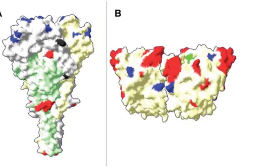
Figure 4. F-NDV trimer (a) and HN-NDV dimer (b) surface representations of the MG strains.The location of neutralising epitopes is shown in red, the amino acid substitutions specific to the MG group are in blue in the globular head and in black in the stalk region. The figures were generated by Swiss PDB-viewer, using the X-ray structure of NDV F protein, position 33aa to 454aa for F protein [45] and 124aa to 569aa for HN protein [51].
3. OIE (2009) Newcastle disease. Chapter 2.3.14. OIE Manual of Standards for Diagnostic Tests and Vaccines, in Manual of Diagnostic Tests and Vaccines for Terrestrial Animals: Mammals, Birds and Bees. Office International des Epizooties, Paris (May 2009). pp 576–589.
4. Mayo MA (2002) Virus taxonomy. Arch Virol 147: 1071–1076.
5. Mayo MA (2002) A summary of taxonomic change recently approved by ICTV. Arch Virol 147: 1655–1656.
6. Peeters BPH, Gruijthuijsen YK, de Leeuw OS, Gielkens ALJ (2000) Genome replication of Newcastle disease virus: involvement of the rule-of-six. Archives of Virology 145: 1829–1845.
7. Cho S-H, Kim S-J, Kwon H-J (2007) Genomic sequence of an antigenic variant Newcastle disease virus isolated in Korea. Virus Genes 35: 293–302. 8. Czegle´di A, Ujva´ri D, Somogyi E, Wehmann E, Werner O, et al. (2006) Third
genome size category of avian paramyxovirus serotype 1 (Newcastle disease virus) and evolutionary implications. Virus Research 120: 36–48.
9. Huang Z, Panda A, Elankumaran S, Govindarajan D, Rockemann DD, et al. (2004) The Hemagglutinin-Neuraminidase Protein of Newcastle Disease Virus Determines Tropism and Virulence. J Virol 78: 4176–4184.
10. Kattenbelt JA, Meers J, Gould AR (2006) Genome sequence of the thermostable Newcastle disease virus (strain I-2) reveals a possible phenotypic locus. Veterinary Microbiology 114: 134–141.
11. Herczeg J, Wehmann E, Bragg RR, Travassos Dias PM, Hadjiev G, et al. (1999) Two novel genetic groups (VIIb and VIII) responsible for recent Newcastle disease outbreaks in Southern Africa, one (VIIb) of which reached Southern Europe. Archives of Virology 144: 2087–2099.
12. Czegle´di A, Eacute, Di A, Herczeg J, Hadjiev G, et al. (2002) The occurrence of five major Newcastle disease virus genotypes (II, IV, V, VI and VIIb) in Bulgaria between 1959 and 1996. Epidemiology and Infection 129: 679–688. 13. Aldous EW, Mynn JK, Banks J, Alexander DJ (2003) A molecular
epidemiological study of avian paramyxovirus type 1 (Newcastle disease virus) isolates by phylogenetic analysis of a partial nucleotide sequence of the fusion protein gene. Avian Pathology 32: 239–257.
14. Tsai H-J, Chang K-H, Tseng C-H, Frost KM, Manvell RJ, et al. (2004) Antigenic and genotypical characterization of Newcastle disease viruses isolated in Taiwan between 1969 and 1996. Veterinary Microbiology 104: 19–30. 15. Bogoyavlenskiy A, Berezin V, Prilipov A, Usachev E, Lyapina O, et al. (2005)
Molecular Characterization of Virulent Newcastle Disease Virus Isolates from Chickens during the 1998 NDV Outbreak in Kazakhstan. Virus Genes 31: 13–20.
16. Wang Z, Liu H, Xu J, Bao J, Zheng D, et al. (2006) Genotyping of Newcastle Disease Viruses Isolated from 2002 to 2004 in China. Annals of the New York Academy of Sciences 1081: 228–239.
17. Kim LM, King DJ, Curry PE, Suarez DL, Swayne DE, et al. (2007) Phylogenetic Diversity among Low-Virulence Newcastle Disease Viruses from Waterfowl and Shorebirds and Comparison of Genotype Distributions to Those of Poultry-Origin Isolates. J Virol 81: 12641–12653.
18. Liu H, Wang Z, Wu Y, Zheng D, Sun C, et al. (2007) Molecular epidemiological analysis of Newcastle disease virus isolated in China in 2005. Journal of Virological Methods 140: 206–211.
19. Lien Y-Y, Lee J-W, Su H-Y, Tsai H-J, Tsai M-C, et al. (2007) Phylogenetic characterization of Newcastle disease viruses isolated in Taiwan during 2003– 2006. Veterinary Microbiology 123: 194–202.
20. Alexander DJ (2003) Newcastle disease virus, other avian paramyxoviruses, and pneumovirus infections. In: Disease of poultry, 11th ed.YM Saif, HJ Barnes, JR Glisson, AM Fadly, LR McDougald, DE Swayne. Iowa State University Press Ames, IA. pp 63–87.
21. Gotoh B, Ohnishi Y, Inocencio NM, Esaki E, Nakayama K, et al. (1992) Mammalian subtilisin-related proteinases in cleavage activation of the paramyxovirus fusion glycoprotein: superiority of furin/PACE to PC2 or PC1/PC3. J Virol 66: 6391–6397.
22. Lomniczi B, Wehmann E, Herczeg J, Ballagi-Pordany A, Kaleta EF, et al. (1998) Newcastle disease outbreaks in recent years in western Europe were caused by an old (VI) and a novel genotype (VII). Arch Virol 143: 49–64.
23. Herczeg J, Wehmann E, Bragg RR, Travassos Dias PM, Hadjiev G, et al. (1999) Two novel genetic groups (VIIb and VIII) responsible for recent Newcastle disease outbreaks in Southern Africa, one (VIIb) of which reached Southern Europe. Arch Virol 144: 2087–2099.
24. Abolnik CHR, Bisschop SP, Parker ME, Romito M, Viljoen GJ (2004) A phylogenetic study of South African Newcastle disease virus strains isolated between 1990 and 2002 suggests epidemiological origins in the Far East. Archives of Virology 149: 603–619.
25. Yang CY, Shieh HK, Lin YL, Chang PC (1999) Newcastle disease virus isolated from recent outbreaks in Taiwan phylogenetically related to viruses (genotype VII) from recent outbreaks in western Europe. Avian Dis 43(1): 125–130. 26. Rajaonarison JJ (1991) Production de vaccin contre la maladie de Newcastle a`
Madagascar. PANVAC Debre Zeit Addis Ababa. pp 135–137.
27. Koko M, Maminiaina OF, Ravaomanana J, Rakotonindrina S (2006) Aviculture villageoise a` Madagascar : enqueˆte e´pide´miologique. In Improving farmyard poultry production in Africa: interventions and their economic assessment. TECDOC-1489. AIEA, Vienne. pp 157–163.
28. Maminiaina OF, Koko, Ravaomanana J, Rakotonindrina SJ (2007) Epide´mio-logie de la maladie de Newcastle en aviculture villageoise a` Madagascar. Rev Sci Tech Off Int Epi 26: 691–700.
29. Zou J, Shan S, Yao N, Gong Z (2005) Complete Genome Sequence and Biological Characterizations of A Novel Goose Paramyxovirus-SF02 Isolated in China. Virus Genes 30: 13–21.
30. Thompson J, Gibson T, Plewniak F, Jeanmougin F, Higgins D (1997) The CLUSTAL_X windows interface: flexible strategies for multiple sequence alignment aided by quality analysis tools. Nucl Acids Res 25: 4876–4882. 31. Tamura K, Dudley J, Nei M, Kumar S (2007) MEGA4: Molecular Evolutionary
Genetics Analysis (MEGA) Software Version 4.0. Mol Biol Evol 24: 1596–1599. 32. Kimura S, Hatakeyama M, Konagaya A (2004) Inference of S-system Models of Genetic Networks from Noisy Time-series Data. Chem-Bio Informatics Journal 4: 1–14.
33. Saitou, Nei (1987) The neighbor-joining method: A new method for reconstructing phylogenetic trees. Molecular Biology and Evolution 4: 406–425. 34. Martin S, Sambade A, Rubio L, Vives MC, Moya P, et al. (2009) Contribution of recombination and selection to molecular evolution of Citrus tristeza virus. J Gen Virol 90: 1527–1538.
35. Huovilainen A, Ek-Kommonen C, Manvell R, Kinnunen L (2001) Phylogenetic analysis of avian paramyxovirus 1 strains isolated in Finland. Archives of Virology 146: 1775–1785.
36. Meulemans G, Berg TPvd, Decaesstecker M, Boschmans M (2002) Evolution of pigeon Newcastle disease virus strains. Avian Pathology 31: 515–519. 37. Terregino C, Cattoli G, Grossele B, Bertoli E, Tisato E, et al. (2003)
Characterization of Newcastle disease virus isolates obtained from Eurasian collared doves(Streptopelia decaocto)in Italy. Avian Pathology 32: 63–68. 38. Ujva´ri D, Wehmann E, Kaleta EF, Werner O, Savic V, et al. (2003)
Phylogenetic analysis reveals extensive evolution of avian paramyxovirus type 1 strains of pigeons (Columba livia) and suggests multiple species transmission. Virus Research 96: 63–73.
39. Lee YJ, Sung HW, Choi JG, Kim JH, Song CS (2004) Molecular epidemiology of Newcastle disease viruses isolated in South Korea using sequencing of the fusion protein cleavage site region and phylogenetic relationships. Avian Pathology 33: 482–491.
40. Toyoda T, Sakaguchi T, Imai K, Inocencio NM, Gotoh B, et al. (1987) Structural comparison of the cleavage-activation site of the fusion glycoprotein between virulent and avirulent strains of newcastle disease virus. Virology 158: 242–247.
41. Yusoff K, Nesbit M, McCartney H, Meulemans G, Alexander DJ, et al. (1989) Location of Neutralizing Epitopes on the Fusion Protein of Newcastle Disease Virus Strain Beaudette C. J Gen Virol 70: 3105–3109.
42. Liu XF, Wan HQ, Ni XX, Wu YT, Liu WB (2003) Pathotypical and genotypical characterization of strains of Newcastle disease virus isolated from outbreaks in chicken and goose flocks in some regions of China during 1985–2001. Archives of Virology 148: 1387–1403.
43. Seal BS (2004) Nucleotide and predicted amino acid sequence analysis of the fusion protein and hemagglutinin-neuraminidase protein genes among New-castle disease virus isolates. Phylogenetic relationships among the Paramyxovir-inae based on attachment glycoprotein sequences. Functional & Integrative Genomics 4: 246–257.
44. Panda A, Elankumaran S, Krishnamurthy S, Huang Z, Samal SK (2004) Loss of N-Linked Glycosylation from the Hemagglutinin- Neuraminidase Protein Alters Virulence of Newcastle Disease Virus. J Virol 78: 4965–4975.
45. Chen L, Gorman JJ, McKimm-Breschkin J, Lawrence LJ, Tulloch PA, et al. (2001) The Structure of the Fusion Glycoprotein of Newcastle Disease Virus Suggests a Novel Paradigm for the Molecular Mechanism of Membrane Fusion. 9: 255–266.
46. Romer-Oberdorfer A, Werner O, Veits J, Mebatsion T, Mettenleiter TC (2003) Contribution of the length of the HN protein and the sequence of the F protein cleavage site to Newcastle disease virus pathogenicity. J Gen Virol 84: 3121–3129.
47. Miller PJ, Kim LM, Ip HS, Afonso CL (2009) Evolutionary dynamics of Newcastle disease virus. Virology 391: 64–72.
48. Iorio RM, Glickman RL, Riel AM, Sheehan JP, Bratt MA (1989) Functional and neutralization profile of seven overlapping antigenic sites on the HN glycoprotein of Newcastle disease virus: monoclonal antibodies to some sites prevent viral attachment. Virus Research 13: 245–261.
49. Iorio RM, Syddall RJ, Sheehan JP, Bratt MA, Glickman RL, et al. (1991) Neutralization map of the hemagglutinin-neuraminidase glycoprotein of New-castle disease virus: domains recognized by monoclonal antibodies that prevent receptor recognition. J Virol 65: 4999–5006.
50. Yusoff K, Nesbit M, Samson ACR, Emmerson PT (1988) Location of epitopes within the haemagglutinin-neuraminidase (HN) protein of newcastle disease virus by sequencing the HN genes of monoclonal antibody-resistant mutants. Virus Research 11: 12–12.
51. Crennell S, Takimoto T, Portner A, Taylor G (2000) Crystal structure of the multifunctional paramyxovirus hemagglutinin-neuraminidase. Nat Struct Mol Biol 7: 1068–1074.
52. Connaris H, Takimoto T, Russell R, Crennell S, Moustafa I, et al. (2002) Probing the Sialic Acid Binding Site of the Hemagglutinin-Neuraminidase of Newcastle Disease Virus: Identification of Key Amino Acids Involved in Cell Binding, Catalysis, and Fusion. J Virol 76: 1816–1824.
54. Lamb RA, Kolakofsky D (1996) Paramyxoviridae: the viruses and their replication. In: Fields BN, Knipe DM, Howley PM, eds. In Fundamental Virology. Philadelphia: Lipincott–Raven. pp 1177–1204.
55. Qin Z, Xu H, Ouyang W, Wang Y, Wang L, et al. (2008) Correlation of the neutralization index in chicken embryo with the homologies of F and HN gene of different Newcastle-disease isolates. Wei Sheng Wu Xue Bao 48: 226–233. 56. Lomniczi B, Wehmann E, Herczeg J, Ballagi-Porda´ny A, Kaleta EF, et al. (1998)
Newcastle disease outbreaks in recent years in Western Europe were caused by an old (VI) and a novel genotype (VII). Archives of Virology 143: 49–64. 57. Wehmann E, Ujva´ri D, Mazija H, Velhner M, Ciglar-Grozdanic I, et al. (2003)
Genetic analysis of Newcastle disease virus strains isolated in Bosnia-Herzegovina, Croatia, Slovenia and Yugoslavia, reveals the presence of only a single genotype, V, between 1979 and 2002. Veterinary Microbiology 94: 269–281.
58. Yu L, Wang Z, Jiang Y, Chang L, Kwang J (2001) Characterization of Newly Emerging Newcastle Disease Virus Isolates from the People’s Republic of China and Taiwan. J Clin Microbiol 39: 3512–3519.
59. Millar NS, Chambers P, Emmerson PT (1986) Nucleotide sequence analysis of the haemagglutinin-neuraminidase gene of Newcastle disease virus. J Gen Virol 67(Pt 9): 1917–1927.
60. OIE (2004) Newcastle disease.» Chapter 2.1.15. OIE Manual of Standards for Diagnostic Tests and Vaccines, in Manual of Diagnostic Tests and Vaccines for Terrestrial Animals: Mammals, Birds and Bees. Office International des Epizooties, Paris (2004). pp 270–282.
61. Ujvari D, Wehmann E, Kaleta E, Werner O, Savic V, et al. (2003) Phylogenetic analysis reveals extensive evolution of avian paramyxovirus type 1 strains of pigeons (Columba livia) and suggests multiple species transmission. Virus Res 96: 63–73.
62. Alexander DJ, Manvell RJ, Lowings JP, Frost KM, Collins MS, et al. (1997) Antigenic diversity and similarities detected in avian paramyxovirus type 1 (Newcastle disease virus) isolates using monoclonal antibodies. Avian Pathol 26: 399–418.
63. Kapczynski DR, King DJ (2005) Protection of chickens against overt clinical disease and determination of viral shedding following vaccination with commercially available Newcastle disease virus vaccines upon challenge with highly virulent virus from the California 2002 exotic Newcastle disease outbreak. Vaccine 23: 3424–3433.
64. Servan de Almeida R, Maminiaina OF, Gil P, Hammoumi S, Molia S, et al. (2009) Africa, a reservoir of new virulent strains of Newcastle disease virus? Vaccine 27: 3127–3129.
65. Snoeck C, Ducatez M, Owoade A, Faleke O, Alkali B, et al. (2009) Newcastle disease virus in West Africa: new virulent strains identified in non-commercial farms. Archives of Virology 154: 47–54.
66. Qin Z, Sun L, Ma B, Cui Z, Zhu Y, et al. (2008) F gene recombination between genotype II and VII Newcastle disease virus. Virus Research 131: 299–303. 67. Alexander DJ, Russell PH, Parsons G, Elzein EMEA, Ballouh A, et al. (1985)
Antigenic and biological characterisation of avian paramyxovirus type I isolates from pigeons - an international collaborative study. Avian Pathology 14: 365–376.
68. Miller PJ, Decanini, EduardoLucio, Afonso, Claudio L (2010) Newcastle disease: Evolution of genotypes and the related diagnostic challenges. Infection, Genetics and Evolution 10: 26–35.
69. Xu M, Chang S, Ding Z, Gao HW, Wan JY, et al. (2008) Genomic analysis of Newcastle disease virus strain NA-1 isolated from geese in China. Archives of Virology 153: 1281–1289.
70. Gravel, Morrison (2003) Interacting Domains of the HN and F Proteins of Newcastle Disease Virus. J Virol 77: 11040–11049.
71. Zaitsev V, von Itzstein M, Groves D, Kiefel M, Takimoto T, et al. (2004) Second Sialic Acid Binding Site in Newcastle Disease Virus Hemagglutinin-Neuramin-idase: Implications for Fusion. J Virol 78: 3733–3741.
72. Gravel KA, Morrison TG (2003) Interacting domains of the HN and F proteins of newcastle disease virus. J Virol 77: 11040–11049.
73. Han G-Z, He C-Q, Ding N-Z, Ma L-Y (2008) Identification of a natural multi-recombinant of Newcastle disease virus. Virology 371: 54–60.
74. Miller PJ, Decanini EL, Afonso CL (2009) Newcastle disease: Evolution of genotypes and the related diagnostic challenges. Infection, Genetics and Evolution. doi:101016/jmeegid200909012.
75. Miller PJ, King DJ, Afonso CL, Suarez DL (2007) Antigenic differences among Newcastle disease virus strains of different genotypes used in vaccine formulation affect viral shedding after a virulent challenge. Vaccine 25: 7238–7246. 76. Felsenstein J (1985) Confidence limits on phylogenies: An approach using the
bootstrap. Evolution 39: 783–791.
77. Czegle´di A, Wehmann E, Lomniczi B (2003) On the origins and relationships of Newcastle disease virus vaccine strains Hertfordshire and Mukteswar, and virulent strain Herts’33. Avian Pathology 32: 271–276.