Molecular analysis of the N gene of canine distemper virus in dogs in Brazil
[Análise Molecular do Gene N do Vírus da Cinomose em Cães do Brasil]
J.G. Castilho, P.E. Brandão, P.Carnieli Jr, R.N. Oliveira, C.I. Macedo, Z.M.P. Peixoto, M.L. Carrieri, I. Kotait.
Instituto Pasteur Avenida Paulista, 393 01311-000 - São Paulo, SP
ABSTRACT
Eleven central-nervous-system samples collected from stray dogs between 2000 and 2004 were found positive by RT-PCR, which amplified a 480bp fragment of the N gene of canine distemper virus (CDV). Phylogenetic analysis based on partial N-gene sequences showed four major clusters. All dog strains segregated into cluster I, with a mean nucleotide identity of 95.8% and 95.6% with the Onderstepoort and Lederle vaccine strains, respectively. Cluster II contained all the raccoon-related strains, cluster III Orient strains and Cluster IV the Onderstepoort and Lederle vaccine strains, with a mean nucleotide identity of 99.7% between them. This is the first report of phylogenetic analysis of CDV strains in Brazil.
Keywords: dog, canine distemper virus, RT-PCR, phylogeny
RESUMO
Onze amostras de sistema nervoso central de cães coletados entre 2000 e 2004 foram positivas pela RT-PCR, a qual amplificou um fragmento de 480pb do gene N do vírus da cinomose canina (VCC). A análise filogenética baseada na seqüência parcial do gene N mostrou quatro principais agrupamentos genéticos. Todas as amostras de cães segregaram no agrupamento I, com identidade média de nucleotídeos de 95,8% e 95,6% com as amostras vacinais Onderstepoort e Lederle, respectivamente. O agrupamento II agregou todas as amostras relacionadas aos guaxinins. O agrupamento III agregou amostras orientais e o agrupamento IV agregou as amostras vacinais Onderstepoort e Lederle, com identidade média de nucleotídeos de 99,7% entre elas. Este é o primeiro relato de análise filogenética de amostras de VCC no Brasil.
Palavras-chave: cão, vírus da cinomose canina, RT-PCR, filogenia
INTRODUCTION
Canine distemper virus (CDV) is a highly contagious pathogen that occurs worldwide and causes a fatal disease called canine distemper in carnivores (Scagliarini et al., 2003). The fatality rate of the disease is second only to that of rabies (Deem et al., 2000). CDV is a member of the
genus Morbillivirus in the family
Recebido em 20 de dezembro de 2005 Aceito em 10 de maio de 2007 E-mail: juliana.castilho@uol.com.br
Paramixoviridae and causes generalized infection with prominent respiratory, gastrointestinal and nervous signs (Scagliarini et al., 2003).
(Blixenkrone-Moller et al., 1993; Gemma et al., 1996). These findings suggest that there are antigenic differences in some geographically distinct wild-type CDV and that a vaccine virus strain may be responsible for the resurgence of the disease (Harder and Osterhaus, 1997). Epidemiological and serological studies suggest that these “new” geographic wild-types have altered antigenicities compared with the same “old” geographic wild-type (Yoshida et al., 1998).
Genetic sequencing and subsequent phylogenetic analysis are methods that not only indicate the evolutionary relationship between samples collected in different geographical regions but also help to understand the antigenic differences between different biological samples. Nucleic-acid sequencing reveals the nucleotide sequence present in the gene being studied and thus makes it possible to predict the structure and function of the protein coded by the gene. When such studies are carried out with samples of pathogens, such as CDV, or even with vaccine strains, the results can help to control a particular pathogenic agent (Bellini et al., 1986; Sharma et al., 1992).
RNA viruses have high replication and mutation rates, and the latter are the primary source of genetic variations (Tsimring et al., 1996). The virus’ ecological environment consists of basically two components: the internal one, represented by the cell itself, and the external one, represented by the uniqueness of the host within the ecological environment. These two components modulate the adaptive responses of the RNA viruses (Domingo and Holland, 1997). The mechanisms that are involved in fixing or removing a genetic mutation are the same ones that act in other living creatures, namely, natural selection, genetic drift (Felsenstein, 1971; Haigh, 1978) and population bottlenecks (Clarke et al., 1993). The different mutations that occur in virus populations are selected at random, and because of geographical barriers, these mutations become restricted to specific regions. All the mechanisms that cause genetic variations produce virus populations that can be identified according to their geographical origin (Clarke et al., 1993).
In Brazil, CDV has been diagnosed by histopathology and fluorescent antibody test (FAT) (Silva et al., 1999; Headly and Graça, 2000). Nonetheless, developments in molecular
techniques revealed the suitability of methods based on the nucleocapsid protein (N) gene (Frisk et al., 1999; Kim et al., 2001; Shin et al., 2004; Józwik and Frymus, 2005) for CDV detection as well as for molecular epidemiology studies (Scagliarini et al., 2003; Keawcharoen et al., 2005).
The N nucleoprotein is the most abundant structural viral protein and has been shown to possess regulatory functions such as transcription and replication, as well as encapsidation of the RNA genome into an RNase-resistant nucleocapsid (Stettler and Zurbriggen, 1995). Some variation of the N gene among CDV isolates has been demonstrated, despite the fact that this protein makes up the conserved region of the CDV genome (Parks et al., 1992; Curran et al., 1993).
The objective of the present study was to assess the molecular genetic diversity of Brazilian strains of CDV by analysis of partial nucleotide sequences of the most conserved region of the N gene using the reverse transcriptase polymerase chain reaction (RT-PCR) method developed for CDV detection.
MATERIALS AND METHODS
The study used central nervous system (CNS) samples from 11 stray dogs from 10 municipalities in the state of São Paulo, southeastern Brazil (Table 1), collected between 2000 and 2004 and sent without any information on clinical signs to the Pasteur Institute as part of the Rabies Epidemiological Surveillance Program. These samples had previously been found positive by Sellers’ staining (Tierkel and Atanasiu, 1996).
The Lederle/VR128 strain, which was kindly provided by Laboratório Biovet, was grown in Vero cells and used as a positive control for the RT-PCR.
CDV-forward (5’-ACTGCTCCTGATACTGC-3’) and CDV-reverse (5’-TTCAACACCAAC(T/C)CCC-3’), respectively.
Total RNA was extracted from CNS samples, Lederle strain (positive control) and ultra-pure water(negative control) with TRIzol1, according to the manufacturer’s instructions. Next, 5µl of extracted RNA were added to the reverse-transcription (c-DNA synthesis) mix containing 1x First Strand Buffer1, 40U RNAseOUT1, 1.3mM of each dNTP, 50pmols of each primer, 8.5mM DTT, 200U Superscript II Reverse Transcriptase1, and RNAse/DNAse-free water to a final volume of 47µl and submitted to 42°C for 60min.
For the PCR amplification, 10µl of cDNA were added to a mix containing 1x PCR Buffer1, 50pmols of each primer, 0.2mM dNTP, 2.4mM
MgCl2, 2.5U/µl Taq DNA polymerase
Recombinant1, and RNAse/DNAse-free water to a final volume of 102 µl and submitted to an initial denaturation at 94ºC for 5 min followed by 35 cycles of 94ºC for 45s, 48ºC for 45s and 72ºC for 90s, and a final cycle for extension of 72ºC for 10min. The PCR products were analyzed by electrophoresis on 1% agarose gel stained with ethidium bromide.
The amplified DNA fragments were purified with GFX PCR DNA and Gel Band Purification Kit2 and submitted to bi-directional sequencing with DYEnamic ET Dye Terminator2 according to the manufacturer’s instructions; the sequences were resolved in a MegaBACE DNAsequencer2.
The final sequence of each sample, 447bp long, was aligned by the CLUSTAL/W method with Bioedit software (Hall, 1999) with homologous sequences retrieved from GenBank (Table 1) and the alignment used to build a Neighbor-Joining distance tree with the Kimura 2-parameter model and 1,000 bootstrap replicates with Mega 2.1 (Kumar et al., 2001).
RESULTS
All of the 11 CNS samples collected from Brazilian dogs, which had previously been found
1InvitrogenTM, USA. 2Amershan BiosciencesTM.
3Strains that originated in China and Taiwan.
positive by Sellers’ staining, were also positive for canine distemper virus by RT-PCR, as was the Lederle vaccine strain. No amplification was found in the negative control and no extra band was detected in any of the reactions (data not shown).
Phylogenetic analysis showed that the sequences of the 11 Brazilian dog strains produced a single cluster (cluster I) (Fig. 1) with an average identity (ID) of 99.2%. The Onderstepoort and Lederle vaccine strains segregated into the same cluster (cluster IV, ID= 99.7%). The ID of clusters I and IV (I x IV) was 95.9%, and the ID of cluster I with the Onderstepoort strain was 95.8% and with the Lederle strain 95.6%. Cluster II (raccoon strains, ID= 99.6%) had a 99.4% identity with cluster I (I x II), and cluster III (Orient strains3, ID= 99.6%) had a 98.8% identity with cluster I (I x III). The German dog strain 25/44-Hans segregated into cluster I with a 99.2% identity with the other samples in cluster I.
Furthermore, as seen in Fig. 1, cluster I is subdivided into clusters IA and IB, supported by bootstrap values of 83 and 58, respectively, with an ID of 99.3% between them. All the previous results are summarized in Table 2.
DISCUSSION
A growing knowledge of molecular biology has led to the development of several N gene-based RT-PCR methods for the detection of CDV (Frisk et al., 1999). However, RT-PCR combined with nested PCR has also been used to detect CDV RNA in dogs and has been shown to be a more sensitive method (Kim et al., 2001; Shin et al., 2004; Józwik and Frymus, 2005).
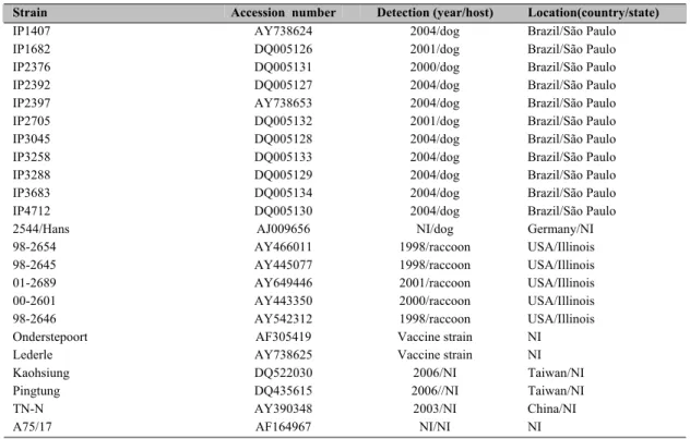
Table 1. Strain, accession number, detection and location of CDV strains used for phylogenetic analysis in this study.
Figure 1. Neighbor-joining phylogenetic tree based on partial sequences of the 5’ half of the CDV N gene. Cluster I: Brazilian dog strains; cluster II: raccoon strains; cluster III: Orient strains and cluster IV: fixed strains. Numbers above each node are 1,000 bootstrap replicate values.
Strain Accession number Detection (year/host) Location(country/state)
IP1407 AY738624 2004/dog Brazil/São Paulo
IP1682 DQ005126 2001/dog Brazil/São Paulo
IP2376 DQ005131 2000/dog Brazil/São Paulo
IP2392 DQ005127 2004/dog Brazil/São Paulo
IP2397 AY738653 2004/dog Brazil/São Paulo
IP2705 DQ005132 2001/dog Brazil/São Paulo
IP3045 DQ005128 2004/dog Brazil/São Paulo
IP3258 DQ005133 2004/dog Brazil/São Paulo
IP3288 DQ005129 2004/dog Brazil/São Paulo
IP3683 DQ005134 2004/dog Brazil/São Paulo
IP4712 DQ005130 2004/dog Brazil/São Paulo
2544/Hans AJ009656 NI/dog Germany/NI
98-2654 AY466011 1998/raccoon USA/Illinois
98-2645 AY445077 1998/raccoon USA/Illinois
01-2689 AY649446 2001/raccoon USA/Illinois
00-2601 AY443350 2000/raccoon USA/Illinois
98-2646 AY542312 1998/raccoon USA/Illinois
Onderstepoort AF305419 Vaccine strain NI
Lederle AY738625 Vaccine strain NI
Kaohsiung DQ522030 2006/NI Taiwan/NI
Pingtung DQ435615 2006//NI Taiwan/NI
TN-N AY390348 2003/NI China/NI
A75/17 AF164967 NI/NI NI
NI = No information available
IP 27 05 IP 23 76 IP 16 82 IP 23 97 IP 14 07M IP 32 88 IP 47 12 IP 23 92 25 44 /H an s IP 30 45 IP 36 83 IP 32 58 98 -265 4 98 -264 6 98 -264 5 00 -260 1 01 -268 9 A 7 5/17 P ing tu ng K a ohs iu ng TN -N Le de rle O nd ers te poo rt 6 5
9 6 8 8 7 4 7 9 5 8 8 3
5 0 7 4 8 2
1 0 0
A
B
I – BRAZILIAN DOG STRAINS
II – RACCOON STRAINS
III – ORIENT STRAINS
The Ngene has approximately 1,683 nucleotides with highly conserved regions in the first 2/3 of the gene among members of the genus Morbillivirus (Parks et al., 1992; Curran et al., 1993; Frisk et al., 1999), and this characteristic makes the gene a good molecular target for CDV detection and sequencing. However, as described by Yoshida et al. (1998), some degree of variation of the N gene among CDV isolates has been demonstrated, also making this gene suitable for differentiating between strains of CDV and for phylogeny-based molecular epidemiology.
In this study, the 480bp that were sequenced belong to the central region of the N gene, which is the most conserved region (Parks et al., 1992) and thus displays the greatest similarity between samples. Analysis of a conserved area provides sufficient resolution to reach reliable conclusions (Russo, 2001).
Phylogenetic analysis showed that the 11 Brazilian CDV strains segregated into a single cluster (cluster I, bootstrap= 82 ID= 99.2%) separate from the cluster that includes the Onderstepoort and Lederle vaccine strains (cluster IV, bootstrap= 100, ID= 99.75) (Fig. 1 and Table 2). The nucleotide sequences of the 11 Brazilian CDV strains showed IDs of 95.8% and 95.6% when compared with Onderstepoort and Lederle vaccine strains, respectively. Analysis of the N gene of wild-type strains in Japan also indicated separate clusters for the wild-type strains and vaccine strains (Yoshida et al., 1998).
Two other clusters were formed: cluster II (raccoon-strains, bootstrap= 74 and ID= 99.6%) and cluster III (Orient-strains, bootstrap= 96 and ID= 99.6%). This analysis shows that not only cluster IV but also clusters II and III diverge from cluster I. Intracluster IDs (I x II, I x III and I x IV) are shown in Table 2.
As shown in Fig. 1, cluster I can be separated into two clusters, IA and IB (bootstrap= 83 and ID intra-clusters IA x IB= 99.3%). Cluster IB (bootstrap= 58, ID= 99.5%) contains sequences related to the Brazilian strains IP3045, IP3258 and IP 3683 and the German strain 2544/Hans. It could be suggested that there are two lineages of CDV circulating in Brazilian dogs in the area surveyed, one of which (IB) is similar to the 2544/Hans lineage isolated in Germany.
Table 2. Mean identities of the nucleotides in clusters 1-4 and mean values of intracluster identities.
Clusters Mean identities (ID)
Cluster I 99.2
Cluster II 99.6
Cluster III 99.6
Cluster IV 99.7
Cluster I A 99.2
Cluster I B 99.5
Clusters I A x I B 99.3
Clusters I x II 99.4
Clusters I x III 98.8
Clusters I x IV 95.9
Clusters I x Hans 99.2
Lederle x I 95.6
Onderstepoort x I 95.8
As the N gene is highly conserved in CDV strains and variations were found in the N-gene fragments of the Brazilian samples, it can be inferred that greater polymorphisms can be found in more variable genes such as the H and F genes, which determine the synthesis of the proteins of the same name involved in receptor-binding and induction of neutralizing antibodies (Blixenkrone-Moller et al., 1992).
Although other CDV-related sequences can be found in GenBank, these were not included in this article because the regions of the genome they cover are not the same as those in the samples investigated in this study. All the samples deposited in GenBank to date that could be aligned with the 11 N-gene sequences of Brazilian dogs generated by RT-PCR using the CDV primers described in this article were used. The vast majority of the N-gene sequences deposited in GenBank are small and cover the 3’-terminal of the gene. This area has greater variability and is therefore not suitable for this study, the aim of which was to analyze the most conserved area of the N gene of CDV.
REFERENCES
BELLINI W.J.; ENGLUND G.; RICHARDSON C.D.; et al. Matrix genes of measles virus and canine distemper virus: cloning, nucleotide sequences, and deduced amino acid sequences. J. Virol., v.58, p.408-416, 1986.
BLIXENKRONE-MOLLER, M.; SVANSSON, V.; APPEL, M. et al. Antigenic relationship between field isolates of morbilliviruses from different carnivores. Arch. Virol., v.123, p.279-294, 1992.
BLIXENKRONE-MOLLER, M.; SVANSSON, V.; HAVE, P. et al. Studies on manifestations of canine distemper virus infection in an urban dog population. Vet. Microbiol., v.37, p.163-173, 1993.
CLARKE, D.; DUARTE, E.; MOYA, A. et al. Genetic bottlenecks and population passages cause profound fitness differences in RNA viruses. J. Virol., v.67, p.222-228, 1993.
CURRAN, J.; HOMANN, H.; BUCHHOLZ, C. et al. The hypervariable C-terminal tail of Sendai Paramyxovirus nucleocapsid protein is required for template function but not for RNA encapsidation. J. Virol., v.67, p.4358-4364, 1993.
DEEM, S.L.; SPELMAN, L.H.; YATES, R.A. et al. Canine distemper in terrestrial carnivores: a review. J. Zoo Wildl. Med., v.31, p.441-451, 2000.
DOMINGO, E.; HOLLAND, J.J. RNA virus mutations and fitness for survival. Annu. Rev. Microbiol., p.151-158, 1997.
FELSENSTEIN, J. The rate of loss of multiple alleles in finite haploid populations. Theor. Pop. Biol., v.2, p.391-403, 1971.
FRISK, A.L.; KONIG, M.; MORITIZ, A. et al. Detection of canine distemper virus nucleoprotein RNA by reverse transcription-PCR using serum, whole blood, and cerebrospinal fluid from dogs with distemper. J. Clin.
Microbiol., v.37, p.3634-3643, 1999.
GEMMA, T.; WATARI, T.; AKIYAMA, K. et al. Epidemiological observations on recent outbreaks of canine distemper in Tokyo area. J. Vet. Med. Sci., v.58, p.547-550, 1996.
HAIGH, J. The accumulation of deleterious genes in a population--Muller's Ratchet. Theor. Pop. Biol., v.14, p.251-267, 1978.
HALL, T.A. BioEdit: a user-friendly biological sequence alignment editor and analysis program for Windows 95/98/NT. Nucl. Acids Symp. Ser., v.41, p.95-98, 1999. HARDER, T.C.; OSTERHAUS, A.D.M.E. Canine distemper virus a morbillivirus in search of new host. Trends
Microbiol., v.5, p.120-124, 1997.
HEADLY, S.A.; GRAÇA, D.L. Canine distemper: epidemiological findings of 250 cases. Braz. J. Vet. Res. Anim. Sci., v.3, p.136-140, 2000.
JÓZWIK, A.; FRYMUS, T. Comparison of the immunofluorescence assay with RT-PCR and nested PCR in
the diagnosis of Canine distemper. Vet. Res. Commun., v.29, p.347-359, 2005.
KEAWCHAROEN, J.; THEAMBOONLERS, A.; JANTARADSAMEE, P. et al. Nucleotide sequence analysis of nucleocapsid protein gene of canine distemper virus isolates in Thailand. Vet. Microbiol., v.105, p.137-142, 2005.
KIM, Y.H.; CHO, K.W.; YOUN, H.Y. et al. Detection of canine distemper virus (CDV) through one step RT-PCR combined with nested PCR. J. Vet. Sci., v.2, p.59-63, 2001.
KUMAR, S.; TAMURA, K.; JAKOBSEN, I.B. et al. MEGA2: Molecular Evolutionary Genetics Analysis software, Arizona State University: Tempe, 2001.
PARKS, G.D.; WARD, C.D.; LAMB, R.A. Molecular cloning of the NP and L genes of simian virus 5: Identification of highly conserved domains in Paramyxovirus NP and L proteins. Virus Res., v.22, p.259-279, 1992.
RUSSO, C.A.M. Como escolher genes para problemas filogenéticos específicos. In: MATIOLI, S.R. (Ed.). Biologia
molecular e evolução. Ribeirão Preto: Holos Editora, 2001, p.
130-136.
SCAGLIARINI, A. ; BATTILANI, M.; CIULLI, S. et al. Molecular analysis of the NP gene of Italian CDV isolates.
Vet. Res. Commun., v.27, p.355-357, 2003.
SHARMA, B.; NORRBY, E.; BLIXENKRONE-MOLLER, M. et al. The nucleotide and deduced amino acid sequence of the M gene of phocid distemper virus (PDV). The most conserved protein of morbilliviruses shows a uniquely close relationship between PDV and canine distemper virus. Virus Res., v.23, p.13-25, 1992.
SHIN, Y.J.; CHO, K.O.; CHO, H.S. et al. Comparison of one-step RT-PCR and a nested PCR for the detection of canine distemper virus in clinical samples. Aust. Vet. J., v.82, p.83-86, 2004.
SILVA, L.H.K.; PEIXOTO, Z.M.P; CUNHA, E.M.C. et al. Emprego das técnicas de coloração de Sellers e imunofluorescência direta no diagnóstico diferencial da raiva e cinomose em amostras de origem canina. Rev. Bras. Med. Vet., v.21, p.232-236, 1999.
STETTLER, M.; ZURBRIGGEN, A. Nucleotide and deduced amino acid sequences of the nucleocapsid protein of the virulent A75/17-CDV strain of canine distemper virus.
Vet. Microbiol., v.44, p.211-217, 1995.
TIERKEL, E.S.; ATANASIU, P. In: MESLIN, F.X., KAPLAN, M.M., KOPROWSKI, H. (Eds.). Laboratory
techniques in rabies. Geneva: World Health Organization,
1996. p.157-174,
TSIMRING L.S.; LEVINE, H.; KESSLER, D.A. RNA virus evolution via a fitness-space model. Phys. Rev. Letters, v.23, p. 4440-4443, 1996.