Epithelial Cell Mitochondrial Dysfunction and
PINK1 Are Induced by Transforming Growth
Factor- Beta1 in Pulmonary Fibrosis
Avignat S. Patel1, Jin Woo Song2*, Sarah G. Chu1, Kenji Mizumura6, Juan C. Osorio1, Ying Shi1, Souheil El-Chemaly1, Chun Geun Lee3, Ivan O. Rosas1,4, Jack A. Elias5, Augustine M. K. Choi6, Danielle Morse1
1Division of Pulmonary and Critical Care Medicine, Brigham and Women’s Hospital and Harvard Medical School, Boston, Massachusetts, United States of America,2Department of Pulmonary and Critical Care Medicine, Asan Medical Center, University of Ulsan, College of Medicine, Seoul, South Korea,3Section of Pulmonary and Critical Care Medicine, Yale University School of Medicine, New Haven, Connecticut, United States of America,4Pulmonary Fibrosis Program, Lovelace Respiratory Research Institute, Albuquerque, New Mexico, United States of America,5Alpert Medical School, Brown University, Providence, Rhode Island, United States of America,6Department of Medicine, Weill Cornell Medical College, New York, New York, United States of America
Abstract
Background
Epithelial cell death is a major contributor to fibrogenesis in the lung. In this study, we sought to determine the function of mitochondria and their clearance (mitophagy) in alveolar epithe-lial cell death and fibrosis.
Methods
We studied markers of mitochondrial injury and the mitophagy marker, PTEN-induced puta-tive kinase 1 (PINK1), in IPF lung tissues by Western blotting, transmission electron micros-copy (TEM), and immunofluorescence.In vitroexperiments were carried out in lung epithelial cells stimulated with transforming growth factor-β1 (TGF-β1). Changes in cell function were measured by Western blotting, flow cytometry and immunofluorescence.In vivoexperiments were performed using the murine bleomycin model of lung fibrosis.
Results
Evaluation of IPF lung tissue demonstrated increased PINK1 expression by Western blot-ting and immunofluorescence and increased numbers of damaged mitochondria by TEM. In lung epithelial cells, TGF-β1 induced mitochondrial depolarization, mitochondrial ROS, and PINK1 expression; all were abrogated by mitochondrial ROS scavenging. Finally,Pink1 -/-mice were more susceptible than control -/-mice to bleomycin induced lung fibrosis.
a11111
OPEN ACCESS
Citation:Patel AS, Song JW, Chu SG, Mizumura K, Osorio JC, Shi Y, et al. (2015) Epithelial Cell Mitochondrial Dysfunction and PINK1 Are Induced by Transforming Growth Factor- Beta1 in Pulmonary Fibrosis. PLoS ONE 10(3): e0121246. doi:10.1371/ journal.pone.0121246
Academic Editor:Oliver Eickelberg, Helmholtz Zentrum München, GERMANY
Received:May 12, 2014
Accepted:February 11, 2015
Published:March 18, 2015
Copyright:© 2015 Patel et al. This is an open access article distributed under the terms of the
Creative Commons Attribution License, which permits unrestricted use, distribution, and reproduction in any medium, provided the original author and source are credited.
Data Availability Statement:All relevant data are within the paper.
Conclusion
TGF-β1 induces lung epithelial cell mitochondrial ROS and depolarization and stabilizes the key mitophagy initiating protein, PINK1. PINK1 ameliorates epithelial cell death and may be necessary to limit fibrogenesis.
Introduction
Mitochondria play a vital metabolic role but can also cause cellular damage via the toxic bypro-ducts of oxidative phosphorylation[1,2]. Damaged mitochondria trigger apoptosis through re-lease of cytochrome c into the cytosol[3] and activation of cell death pathways. Cellular survival therefore depends upon efficient clearance of dysfunctional mitochondria. A selective form of autophagy, dubbed mitophagy, is a major avenue for the removal of these mitochon-dria. During mitophagy, mitochondria are sequestered in double-membrane vesicles and delivered to lysosomes for degradation. The pathways for generating mitophagy overlap considerably with those required for general autophagy; however, in mammals, mitochondria-specific mechanisms have been recently identified and are primarily mediated by PTEN-Induced Putative Kinase 1 (PINK1)[4] and the E3 ubiquitin ligase, Parkin[5].
PINK1 is a serine/threonine kinase that contains a mitochondrial targeting sequence[6]. In healthy mitochondria, PINK1 is constitutively imported to the inner membrane, where it is cleaved by several proteases and ultimately proteolytically degraded[7,8,9]. Loss of the mitochon-drial membrane potential gradient impedes the import of PINK1 to the inner membrane, stabiliz-ing it on the mitochondrial outer membrane where PINK1 serves as a marker of mitochondrial damage[10]. Accumulation of PINK1 on the surface of depolarized mitochondria triggers recruit-ment of Parkin from the cytosol. Through mechanisms that are not yet fully elucidated, Parkin stimulates mitophagy by activating components of the general autophagic machinery[11].
While the role of mitophagy in pulmonary fibrosis has not been investigated, ROS generated from mitochondria are thought to promote fibrogenesis in the lung. Most data are derived from studies of asbestosis, where H2O2production by mitochondria of alveolar macrophages has been shown to contribute to disease pathogenesis[12]. ROS production in structural lung cells such as epithelial cells[13] is also likely to play an important role. Additionally, mitochondria-derived ROS can trigger Nalp3 inflammasome activation, which has recently been implicated in the development of lung fibrosis[14,15].
The relationship between transforming growth factor-β1 (TGF-β1) and oxidative stress is a palimpsest with multiple layers and bidirectional signaling. TGF-β1 is known to stimulate ROS production[16], and oxidative stress can activate latent TGF-β1[17,18], setting up a profibro-genic cycle. Although TGF-β1 can increase ROS production by several distinct mechanisms, the mechanism most relevant to our studies is enhanced mitochondrial ROS production via complex IV inhibition in lung cells[19]. Genetic disruption of mitochondrial complex III-generated ROS production was recently shown to attenuate TGF-β1-induced profibrotic gene expression[16], further implicating mitochondrial ROS as important players in the devel-opment of lung fibrosis.
Given the known relationship between TGF-β1 and mitochondrial ROS production, and given the known contribution of ROS to fibrogenesis, we asked whether mitochondrial clear-ance is altered in idiopathic pulmonary fibrosis (IPF), and whether disruption of the critical mitophagy protein, PINK1, would alter the outcomes ofin vivo andin vitromodels of lung fibrosis.
design, data colelction and analysis, decision to publish, or preparation of the manuscript.
Materials and Methods
Ethics Statement
This study involves the analysis of human clinical samples. Lung tissue procurement was com-pleted under Protocol #2012-P-001134 which was approved by the Institutional Review Board of Brigham and Women’s Hospital. Informed written consent was obtained from all study par-ticipants. All animals were housed in accordance with guidelines from the American Associa-tion for Laboratory Animal Care. Protocols were approved with InstituAssocia-tional Animal Use and Care Committee of Brigham & Women’s Hospital and Harvard Medical School (Protocol #04551). All experiments were carried out in a manner to minimize suffering.
Human Subjects
All IPF subjects met diagnostic criteria of the American Thoracic Society / European Respirato-ry Society /Japanese RespiratoRespirato-ry Society / Latin American Thoracic Association. Human lung tissues were procured from patients undergoing lung transplantation or donor lungs that were not implanted.
Animal Experiments
We thank Dr. Jie Shen (Harvard Medical School/BWH) for providing thePink1-/-mice[20]. Pink1-/-mice and their littermate controls (male, 8–10 weeks) were treated with 3 U/kg
intra-tracheal bleomycin sulfate (RPI Corp., Mount Prospect, IL). The mice were sacrificed and lungs harvested on Day 21. Hydroxyproline assay was performed as previously described[21].
Cell Culture
Beas-2B cells (ATCC, Manassas, VA) were cultured in DMEM (Invitrogen, Carlsbad, CA). After overnight serum starvation, cells were treated with 5 ng/mL TGF-β1 (Sigma, St. Louis, MO) for 24h unless otherwise indicated. For certain experiments, cells were co-treated with mitoTEMPO (200μM; Enzo, Farmingdale, NY). Type II alveolar epithelial cells were isolated
as previously described[21]. Carbonyl cyanide m-chlorophenyl hydrazone (CCCP) (Sigma) was used as a positive control for inducing mitophagy.
RNA interference
PINK1 siRNA (Fisher, Pittsburgh, PA) was transfected using Lipofectamine RNAiMAX (Invi-trogen) according to manufacturer’s protocol.
Western Blotting
Protein was extracted from lung homogenate or cell lysate using RIPA buffer or Mitochondria Isolation Kit (Pierce, Rockford, IL), separated on NuPAGE 4–12% Bis-Tris gels (Invitrogen) and transferred to PVDF membranes by electroblotting. Primary antibodies used were PINK1 (Novus, Littleton, CO),β-actin (Sigma), COX-4 (Santa Cruz Biotechnology, Dallas, TX), phospho-DRP1 (Ser 616; Cell Signaling, Beverly, MA), VDAC (Cell Signaling). Secondary anti-bodies were HRP-conjugated goat anti-rabbit or goat anti-mouse (Santa Cruz).
Flow cytometric analysis
Mitochondrial ROS was measured by MitoSOX (Invitrogen) staining (5μM). For study of
cells were detected using 10μl/ml Annexin V (BioVision, Milpitas, CA). Data were acquired
with BD FACS Canto II flow cytometer and analyzed with FlowJo (Treestar, Ashland, OR).
Transmission Electron Microscopy and Fluorescence Microscopy
Lung tissue sections were fixed in 1.25% formaldehyde, 2.5% glutaraldehyde and 0.03% picric acid in 0.1 M Sodium cacodylate and embedded in epon. Images were obtained using a Tecnai G2 Spirit BioTWIN TEM and AMT2k CCD camera. Immunofluorescence was performed with CellLight BacMAM GFP-Mito, RFP-Lyso vectors (Life Technologies), PINK1(Novus) and LC3B(Sigma) antibodies, AlexaFluor 488(Invitrogen), Cy3(Jackson ImmunoResearch Lab., West Grove, PA) and DAPI(Invitrogen) dyes. Images were taken and processed with Olympus FluoView FV1000 confocal microscope. Image analysis of colocalization was performed with ImageJ.
Statistical analysis
Results were expressed as mean ± SD from at least three independent experiments. Differences in measured variables between experimental and control group were assessed by using the Stu-dent’s t-test. Statistically significant difference was accepted at p<0.05.
Results
IPF lung tissue demonstrates increased PINK1 expression and
damaged mitochondria
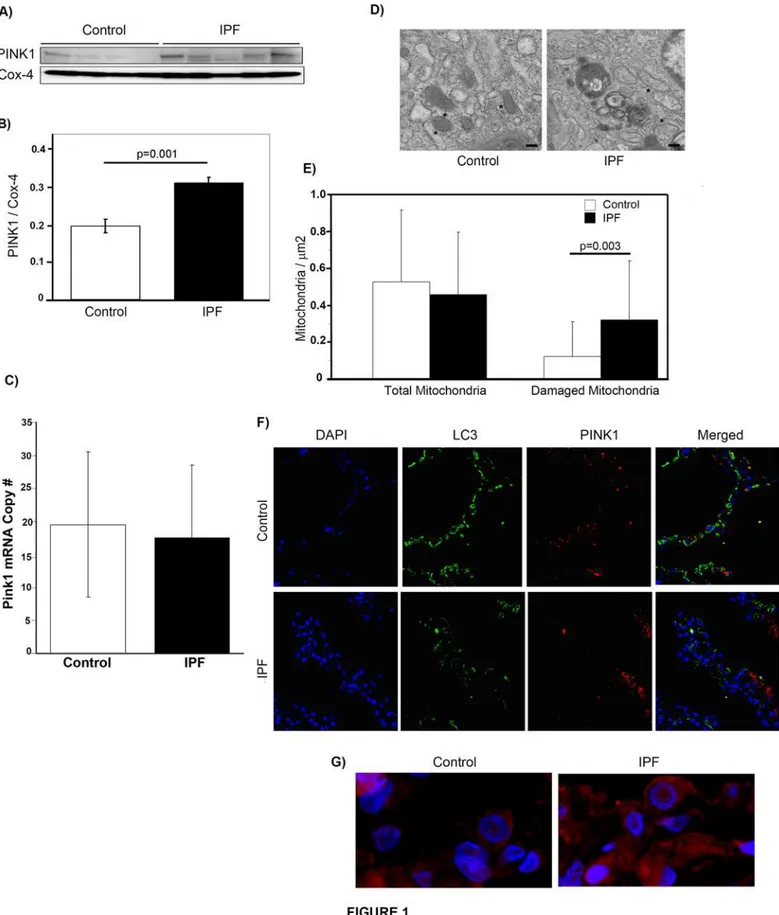
PINK1 accumulation on the outer membrane of mitochondria is the key signal for triggering mitophagy[8]. In order to determine whether PINK1 expression is higher in lungs of IPF pa-tients, we performed Western blotting of the mitochondrial fraction from lung tissue homoge-nate. As shown inFig. 1A & B, PINK1 expression was higher in mitochondria isolated from IPF lung. In order to determine if the change in expression was due to upregulated gene expres-sion, we performed qPCR on whole lung homogenate from control and IPF tissue (Fig. 1C). This demonstrated no change in mRNA levels, suggesting that the increased protein expression is secondary to stabilization of PINK1 in IPF. We further analyzed IPF lungs for evidence of mitochondrial dysfunction by examining mitochondrial morphology by transmission electron microscopy (Fig. 1D). A quantitative analysis (Fig. 1E) was performed by counting and analyz-ing mitochondria in 47 and 27 randomly selected EM fields in the IPF and control samples re-spectively. The number of mitochondria was normalized to area. We found a significantly increased number of mitochondria with swollen appearance or disorganized cristae in IPF lung samples when compared to control lung.
In order to better assess the cellular distribution of PINK1 in IPF and control lungs, we per-formed immunofluorescent staining for PINK1 in conjunction with LC3, an important compo-nent of autophagosomal membranes. In keeping with our previous findings[21], LC3 levels were higher in control lungs than IPF lungs (Fig. 1F). PINK1 expression was detected in both IPF and control lung, however, the most striking finding of our immunostaining was the differ-ence in PINK1 cellular distribution in IPF lungs when compared with control tissue. In IPF, in-tense staining was seen throughout the cytoplasm of individual cells, whereas in controls, the staining was restricted to scattered cytoplasmic puncta. An example of this is shown in the high power (60x) magnification view inFig. 1G. For this study, we did not stain with markers of various lung cell types, the populations of which may vary between healthy and
TGF-
β
1 induces PINK1 expression in epithelial cells
Given the central role of TGF-β1 in the development of fibrosis, we examined whether expo-sure of cells in culture to active TGF-β1 would affect expression of PINK1. Because immuno-fluorescent staining of human lung (shown inFig. 1) revealed high expression of PINK1 in epithelium, we focused on epithelial cells for ourin vitrostudies.
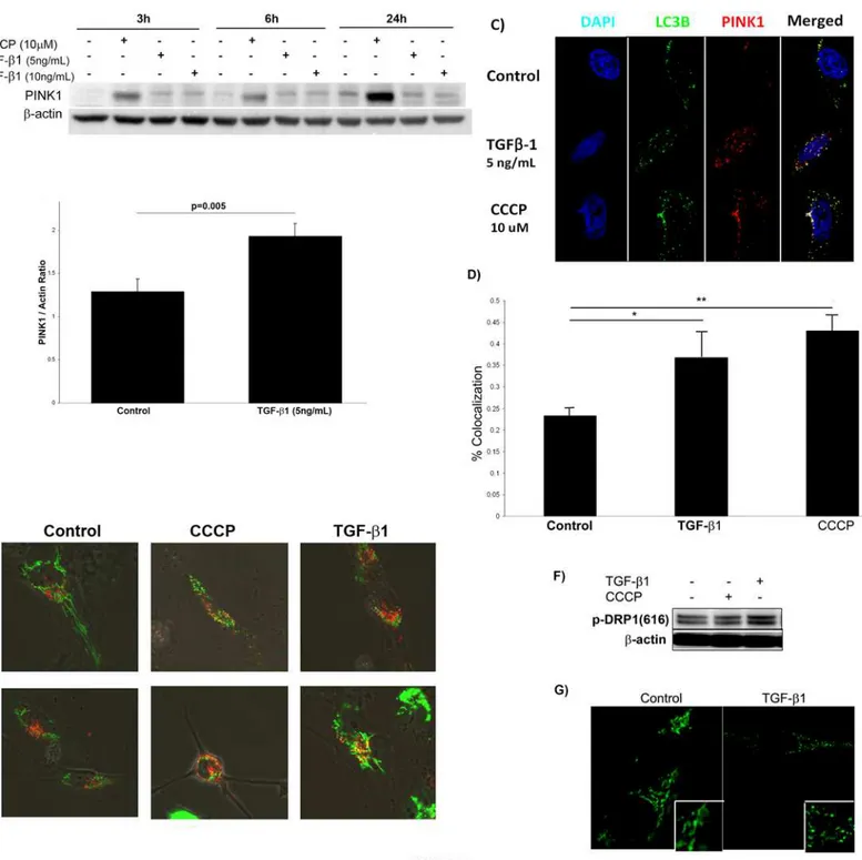
Fig. 2Ashows PINK1 expression in Beas-2B cells treated with 5 or 10 ng/mL of TGF-β1 at timepoints up to 24 hours and demonstrates that TGF-β1 induced PINK1 expression at the early timepoints of 3 hours and 6 hours but less so at 24 hours. The mitochondrial uncoupler CCCP was used as a positive control. CCCP is a nonspecific ionophore that causes severe loss of mitochondrial membrane potential[22].Fig. 2Bshows densitometry for PINK1 expression at 6 hours by Western blot from 3 independent experiments.
If the increase in PINK1 induced by TGF-β1 correlates with enhanced mitophagy, we would expect recruitment of LC3 to the mitochondria decorated with PINK1.Fig. 2Cshows that TGF-β1 does not increase LC3 levels (consistent with previous findings [21]), but co-localization of PINK1 and LC3 can be appreciated. Interestingly, there is less co-co-localization of PINK1 and LC3 in TGF-β1 exposed cells compared with control cells exposed to CCCP (Fig. 2D), implying that TGF-β1 may be a less efficient promoter of mitophagy than CCCP.
In order to more directly evaluate the fate of mitochondria after TGF-β1 treatment, we transfected Beas-2B cells with CellLight BacMAM vectors (Life Technologies) that labeled mi-tochondria with GFP and lysosomes with RFP. After transfection, cells were stimulated with TGF-β1 and analyzed by confocal microscopy at 6hrs and 24hrs (Fig. 2E). In the control cells, mitochondrial and lysosomal puncta are separate. In the CCCP treated control, there is signifi-cant association, suggesting that mitochondria are being recruited to the autolysosome. The TGF-β1 treated cells also demonstrate an association between mitochondria and lysosomes. Although an understanding of the relationship between mitochondrial fission/fusion dy-namics and mitophagy is still evolving, reduced capacity for fusion has been associated with en-hanced mitophagy[23]. On the other hand, mitochondrial fission appears to be necessary for mitophagy initiation[24]. Dynamin-related protein 1 (DRP1) is a major component of the mi-tochondrial fission machinery in humans; when phosphorylated at Ser 616, DRP1 activity in-creases. TGF-β1 stimulation of Beas-2B cells resulted in enhanced phosphorylation of DRP1 (Fig. 2F) with concomitant evidence of mitochondrial fission. This can be appreciated in
Fig. 2G, where cells stimulated with TGF-β1 for 24 hours demonstrated greater fragmentation of the cytosolic areas stained with MitoTracker Green. Increased mitochondrial fission in the setting of increased PINK1 is consistent with cells preparing to initiate mitophagy in response to TGF-β1 stimulation.
TGF-
β
1 induces mitochondrial depolarization
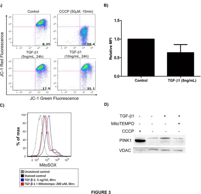
The major trigger for PINK1 stabilization on the surface of mitochondria is thought to be mito-chondrial depolarization[25]. To determine whether TGF-β1 induces mitochondrial depolari-zation, Beas-2B cells were stimulated with TGF-β1 and subsequently stained with JC-1 (Fig. 3A and B). Flow cytometric analysis revealed the expected marked decrease in red fluorescence with CCCP treatment, the positive control, reflecting mitochondrial depolarization. In the cells
lung tissue from control (n = 5) and IPF (n = 5) samples. D) Representative transmission electron micrographs from control and IPF lung tissue showing mitochondria (*). Magnif = 18500x; scale bars = 200nm. E) Quantification of total and abnormal mitochondria. Total mitochondria perμm2in control and IPF was 0.52±0.39 vs. 0.46±0.34. Abnormal mitochondria perμm2in control and IPF was 0.12±0.19 vs. 0.32±0.32,p= 0.003. F) Confocal
immunofluorescence against PINK1(red) and LC3 (green) in control and IPF lung (magnification 10x). G) Magnified view (60x) of confocal immunofluoresence with DAPI (blue) and PINK1 (red) in control and IPF lung.
Fig 2. TGF-β1 increases PINK1 expression and induces mitochondrial fissionin vitro.A) TGF-β1 induced PINK1 expression of Beas-2B cells in a time-and dose-dependent manner. B) Quantification by densitometry of PINK1 expression in Beas-2B cells stimulated with TGF-β1 for 6 hours. C) Confocal microscopy of Beas-2B cells stimulated with TGF-β1 (6h) showed that TGF-β1 induces formation of PINK1 puncta (red) (magnification 120x). D) Quantification of colocalization of LC3 and PINK1 punctae (*p = 0.035,**p = 0.001). E) Confocal microscopy of Beas-2B cells transfected with vectors staining mitochondria (green) and lysosomes (red) and with TGF-β1 stimulation for 6 hours (magnification 60x). F) Western blot of Beas-2B cells stimulated with TGF-β1 (6hrs) showed increased expression of pDRP1 (ser616). G) Confocal microscopy of Beas-2B cells stimulated with TGF-β1 (5 ng/mL, 24 hrs) showed more fragmentation (fission) of mitochondria stained with MitoTracker Green (magnification 120x).
treated with TGF-β1, JC-1 red fluorescence decreased in a dose-dependent manner, indicating disruption of mitochondrial membrane potential by TGF-β1.
Bursts of mitochondrial ROS associated with altered membrane potential have also been shown to induce mitophagy[26]. TGF-β1 stimulation of Beas-2B cells resulted in
Fig 3. TGF-β1 induces mitochondrial depolarization.A) Beas-2B cells stimulated with TGF-β1 and CCCP (positive controls) were stained with JC-1 for 15 min and analyzed by flow cytometric analysis. TGF-β1 treated cells showed decreased red fluorescence (mitochondrial depolarization). B) Quantification of relative MFI for experiment in A (p = 0.06). C) Beas-2B cells stimulated with TGF-β1 were stained with MitoSOX for 10 min and analyzed by flow cytometry. TGF-β1 treated cells showed increased red fluorescence (mitochondrial ROS production) and mitochondria-specific antioxidant (MitoTEMPO) reversed the effect of TGF-β1. D) Western blot of the mitochondrial fraction of Beas-2B cells stimulated with TGF-β1 (5ng/mL) +/- MitoTEMPO (200μM) showing decreased PINK1 expression in presence of MitoTEMPO.
mitochondrial ROS production as shown inFig. 3C. Following exposure to TGF-β1, cells were stained with MitoSOX to detect mitochondrial superoxide and subsequently analyzed by flow cytometry. TGF-β1 treated cells exhibited increased fluorescence intensity (corresponding with mitochondrial ROS production), and a mitochondria-specific antioxidant (MitoTEMPO) re-versed the effect of TGF-β1, reconfirming the mitochondrial origin of the ROS. We also evalu-ated modulation of PINK1 expression in TGF-β1 stimulated Beas-2B cells resulting from mitochondrial ROS quenching by MitoTEMPO. At baseline, TGF-β1 induced PINK1 expres-sion in the mitochondrial fraction, but this upregulation is abrogated in the presence of Mito-Tempo (Fig. 3D). We used VDAC as a mitochondrial housekeeping protein and found no effect on its expression by TGF-β1. This suggests that PINK1 induction by TGF-β1 is depen-dent on the ability to generate mitochondrial ROS.
Loss of PINK1 potentiates TGF-
β
1 induced ROS and cell death
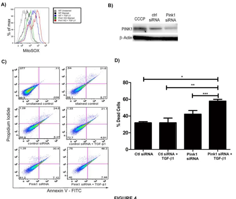
TGF-β1 induced epithelial cell death is thought to be a central mechanism for the development of IPF [27]. While TGF-β1 can induce cell death through a variety of pathways[28], mitochon-drial depolarization is a likely contributor to TGF-β1-induced cell death, given its prominent role in apoptotic programs. It is believed that mitophagy represents an anti-apoptotic force by removing damaged mitochondria along with their accelerated production of ROS and release of pro-apoptotic proteins[29]. To determine whether loss of PINK1 modulates mitochondrial ROS, we stimulated primary type II alveolar epithelial cells fromPink1-/-andPink1WTmice with TGF-β1. Mitochondrial ROS were measured by MitoSOX staining using flow cytometry (Fig. 4A); this demonstrated amplified mitochondrial ROS in the absence of PINK1. To test whether inhibition of mitophagy would also enhance epithelial cell death induced by TGF-β1, we inhibited PINK1 expression in Beas-2B cells using siRNA (Fig. 4B) and then exposed the cells to TGF-β1 for 24 hours. The cells were then analyzed by flow cytometry using Annexin V and propidium iodide (PI) to quantify cell death. As shown inFig. 4C and D, the loss of PINK1 expression resulted in exaggerated cell death in response to TGF-β1.
Absence of PINK1 exacerbates bleomycin-induced lung fibrosis
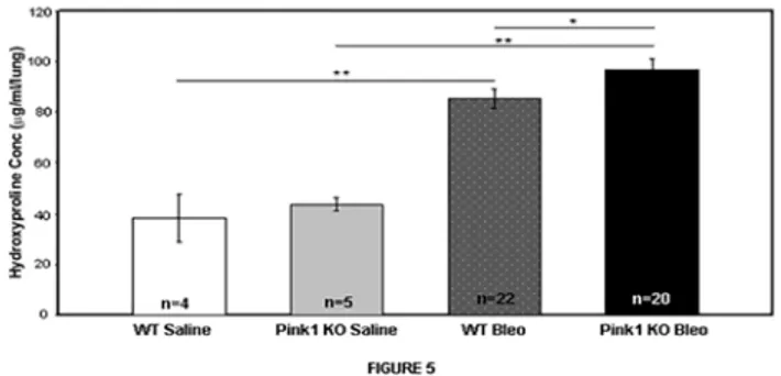
In light of the findings above, we hypothesized that upregulation of PINK1 by TGF-β1 repre-sents an epithelial cell pro-survival response, and that inhibition of mitophagy through genetic elimination ofPink1would worsen experimental lung fibrosis in response to bleomycin. To test this hypothesis, we exposedPink1-/-mice and littermate controls to intratracheal bleomy-cin or saline and harvested lungs for hydroxyproline measurement after 21 days. As shown in
Fig. 5, mice with loss of PINK1 expression developed a more severe fibrotic response to bleo-mycin based on hydroxyproline concentrations (85.4μg/mL vs. 97.0μg/mL, p = 0.05),
al-though the difference was modest at 10%. Since our hypothesis was that the absence of PINK1 would cause more epithelial cell death, we performed TUNEL staining on lung tissue sections from day 21 (data not shown). We did not find a significant difference in the number of TUNEL positive cells in thePink1-/-mice compared to WT.
Discussion
Prevention of PINK1 upregulation by TGF-β1 enhances epithelial cell death, and genetic dele-tion ofPink1modestly exacerbates the murine pulmonary fibrotic response to bleomycin.
Our findings add to those presented in the recent publication by Bueno, et al. [30] by eluci-dating the role of TGF-β1 in mitochondrial dynamics. In their work, the authors found similar evidence of mitochondrial dysfunction in IPF lung tissue and type II alveolar epithelial cells (AEC) from IPF samples. Furthermore,in vitroexperiments demonstrated decreased cell via-bility in the absence of PINK1.In vivo,Pink1-/-mice were more susceptible to fibrosis and were found to have increased AECII apoptosis. Interestingly, aging and ER stress were key potentia-tors of mitochondrial dysfunction.
Fig 4. Loss of PINK1 augments TGF-β1 induced cell death.A) Measurement of mitochondrial ROS in murine type II alveolar epithelial cells fromPink1 -/-andPink1WTmice by flow cytometry and MitoSOX staining. B) Western blot against PINK1 demonstrated knockdown of expression with PINK1 siRNA. C) Beas-2B cells were treated with siRNA (50 nM, 24hrs). Then, they were stimulated with TGF-β1 (5 ng/mL, 24hrs) and analyzed by Annexin V/PI flow cytometry. D) Quantification of cell death from experiment in C (*p = 0.0005,**p = 0.0119,***p = 0.032). Loss of PINK1 exaggerated cell death in cells treated with TGF-β1 relative to transfection with control siRNA.
The most straightforward interpretation of our findings is that TGF-β1 induces epithelial cell death at least in part through disruption of mitochondrial membrane integrity, but this ef-fect is counterbalanced by the upregulation of PINK1 and subsequent induction of mitophagy. If mitophagy is successful in clearing dysfunctional mitochondria, then cell death is attenuated. If, on the other hand, mitophagy is inhibited via deletion of PINK1, cell death and fibrosis can proceed unchecked.
This interpretation raises a number of new questions, the most important of which is whether the machinery of mitophagy is impaired or functioning normally in the lungs of pa-tients who develop IPF. Although we propose that mitophagy is reduced in IPF, the evidence provided is circumstantial at this point. One piece of such evidence is that autophagosome for-mation and LC3 activation are reduced by TGF-β1 and in IPF lung[21,31,32]. Selective and non-selective autophagy rely on common downstream machinery, and one would therefore ex-pect mitophagy to be impacted in the same manner as non-selective autophagy in IPF. Our immunofluorescence images inFig. 2suggest that while TGF-β1 is a potent inducer of mito-chondrial depolarization, it does not induce mitophagyin vitroas efficiently as our positive control (CCCP). The level of mitophagy in the fibrotic lung might therefore be insufficient to clear defective mitochondria and rescue the mitochondrial pool. This hypothesis is supported by our EM studies of IPF lung demonstrating significant numbers of damaged mitochondria. In neurodegenerative diseases, such as Parkinson’s disease and Alzheimer’s disease, studies have shown a similar accumulation of dysfunctional mitochondria leading to neuronal injury and death[33,34]. A more recent investigation in myocardial infarction has also demonstrated that attenuated mitophagy with accumulation of abnormal mitochondria promotes myocyte apoptosis and ventricular remodeling after cardiac ischemia[35]. The study by Bueno, et al. found type II AECs are similarly more susceptible to apoptosis when mitochondrial function is impaired either due to aging or induction of ER stress[30]. Thus, the paradigm of impaired mitophagy leading to increased cell death and eventual organ dysfunction may be important in the pathogenesis of multiple different diseases. The lack of definitive evaluation of mitophagy in human lung tissue is the major limitation of our work. Future studies analyzing IPF tissue for colocalization of mitochondrial and autophagy markers as well as PINK1 and mitochondri-al markers could partimitochondri-ally address this limitation and expand the understanding of PINK1-dependent and-inPINK1-dependent mechanisms of mitophagy.
This work has several additional limitations partly stemming from the immature state of the mitophagy field. First, this study focused on the effects of TGF-β1 on PINK1 expression and PINK1 modulation of epithelial cell apoptosis but it does not shed light on other aspects of
Fig 5. Loss of PINK1 aggravates bleomycin induced lung fibrosis.Pink1-/-mice and their littermate controls (male, 8–10 weeks old) were treated with 3U/kg of intratracheal bleomycin sulfate or saline on day 1 and sacrificed on Day 21.Pink1-/-mice in the bleomycin group showed higher levels of hydroxyproline compared to controls (85.4μg/mL vs. 97.0μg/mL;*p = .05,**p<0.001).
mitophagy regulation in fibrosis. It is possible that alterations in PINK1 in fibrosis are related to mitophagy independent processes, such as proteasomal stress which is also present in IPF [36,37]. Furthermore, although PINK1 is one of the few known mitophagy-specific regulators, other more relevant markers or effectors may be discovered in future. Another known effector protein is the ubiquitin ligase, Parkin. Although Parkin has not yet been studied in lung fibro-sis, given its close interaction with PINK1[11], it should be the subject of future investigations. Second, we do not know to what extent the genetic deletion ofPink1actually inhibits mito-phagy in the lung, and currently, we do not have effective tools to monitor mitophagic activity. In fact, there is evidence that mitophagy can be rescued even in the absence of PINK1[38] and that loss of PINK1 can even promote mitophagy by coordinating autophagy and mitochondrial fission machinery[39]. This may account for the modest findings in our bleomycin experi-ments and points to the need for otherin vivomodels, as well as assessment of specific changes in matrix protein expression and histology, to obtain a better understanding of the role of mito-chondrial dysfunction and mitophagy in lung fibrosis. Furthermore, neurological studies indi-cate that CNS dysfunction inPink1-/-mice is age dependent[40] and Bueno, et al. also demonstrate the age-dependence of mitochondrial function in lung epithelial cells[30]. Thus, the young age ofPink1-/-animals in our experiments may also be moderating the effect on lung fibrosis. Finally, it is certainly possible that deletion ofPink1has off-target effects in the lung beyond the turnover of mitochondria. Because of these limitations, the work we present here should be considered a first step in elucidating what is sure to be a complex relationship between mitophagy and lung fibrosis.
In conclusion, this study demonstrates TGF-β1 induces mitochondrial depolarization trig-gering PINK1 stabilization. The ability of cells to respond to this mitochondrial dysfunction is certainly a key factor in the development of pulmonary fibrosis. This study lays a foundation upon which to build a more complete understanding of mitochondrial dynamics and mito-phagy in idiopathic pulmonary fibrosis.
Author Contributions
Conceived and designed the experiments: ASP JWS SE CGL JAE AMC IOR DM. Performed the experiments: ASP JWS KM SGC JCO YS. Analyzed the data: ASP JWS KM DM. Contributed reagents/materials/analysis tools: KM CGL JAE. Wrote the paper: ASP JWS SE AMC IOR DM.
References
1. Saraste M. Oxidative phosphorylation at the fin de siecle. Science. 1999; 283: 1488–1493. PMID: 10066163
2. Wallace DC. A mitochondrial paradigm of metabolic and degenerative diseases, aging, and cancer: a dawn for evolutionary medicine. Annu Rev Genet. 2005; 39: 359–407. PMID:16285865
3. Parsons MJ, Green DR. Mitochondria in cell death. Essays Biochem. 2010; 47: 99–114. doi:10.1042/ bse0470099PMID:20533903
4. Valente EM, Abou-Sleiman PM, Caputo V, Muqit MM, Harvey K, Gispert S, et al. Hereditary early-onset Parkinson's disease caused by mutations in PINK1. Science. 2004; 304: 1158–1160. PMID:15087508
5. Kitada T, Asakawa S, Hattori N, Matsumine H, Yamamura Y, Minoshima S, et al. Mutations in the parkin gene cause autosomal recessive juvenile parkinsonism. Nature. 1998; 392: 605–608. PMID:9560156
6. Youle RJ, Narendra DP. Mechanisms of mitophagy. Nat Rev Mol Cell Biol. 2011; 12: 9–14. doi:10. 1038/nrm3028PMID:21179058
7. Meissner C, Lorenz H, Weihofen A, Selkoe DJ, Lemberg MK. The mitochondrial intramembrane prote-ase PARL cleaves human Pink1 to regulate Pink1 trafficking. J Neurochem. 2011; 117: 856–867. doi: 10.1111/j.1471-4159.2011.07253.xPMID:21426348
9. Greene AW, Grenier K, Aguileta MA, Muise S, Farazifard R, Haque ME, et al. Mitochondrial processing peptidase regulates PINK1 processing, import and Parkin recruitment. EMBO Rep. 2012; 13: 378–385. doi:10.1038/embor.2012.14PMID:22354088
10. Lazarou M, Jin SM, Kane LA, Youle RJ. Role of PINK1 binding to the TOM complex and alternate intra-cellular membranes in recruitment and activation of the E3 ligase Parkin. Dev Cell. 2012; 22: 320–333. doi:10.1016/j.devcel.2011.12.014PMID:22280891
11. Narendra D, Tanaka A, Suen DF, Youle RJ. Parkin is recruited selectively to impaired mitochondria and promotes their autophagy. J Cell Biol. 2008; 183: 795–803. doi:10.1083/jcb.200809125PMID: 19029340
12. Osborn-Heaford HL, Ryan AJ, Murthy S, Racila AM, He C, Sieren JC, et al. Mitochondrial Rac1 GTPase import and electron transfer from cytochrome c are required for pulmonary fibrosis. J Biol Chem. 2012; 287: 3301–3312. doi:10.1074/jbc.M111.308387PMID:22157762
13. Carnesecchi S, Deffert C, Donati Y, Basset O, Hinz B, Preynat-Seauve O, et al. A key role for NOX4 in epithelial cell death during development of lung fibrosis. Antioxid Redox Signal. 2011; 15: 607–619. doi:10.1089/ars.2010.3829PMID:21391892
14. Dostert C, Petrilli V, Van Bruggen R, Steele C, Mossman BT, Tschopp J. Innate immune activation through Nalp3 inflammasome sensing of asbestos and silica. Science. 2008; 320: 674–677. doi:10. 1126/science.1156995PMID:18403674
15. Xu JF, Washko GR, Nakahira K, Hatabu H, Patel AS, Fernandez IE, et al. Statins and pulmonary fibrosis: the potential role of NLRP3 inflammasome activation. Am J Respir Crit Care Med. 2012; 185: 547–556. doi:10.1164/rccm.201108-1574OCPMID:22246178
16. Jain M, Rivera S, Monclus EA, Synenki L, Zirk A, Eisenbart J, et al. Mitochondrial reactive oxygen spe-cies regulate transforming growth factor-beta signaling. J Biol Chem. 2013; 288: 770–777. doi:10. 1074/jbc.M112.431973PMID:23204521
17. Pociask DA, Sime PJ, Brody AR. Asbestos-derived reactive oxygen species activate TGF-beta1. Lab Invest. 2004; 84: 1013–1023. PMID:15122306
18. Liu RM, Gaston Pravia KA. Oxidative stress and glutathione in TGF-beta-mediated fibrogenesis. Free Radic Biol Med. 2010; 48: 1–15. doi:10.1016/j.freeradbiomed.2009.09.026PMID:19800967
19. Yoon YS, Lee JH, Hwang SC, Choi KS, Yoon G. TGF beta1 induces prolonged mitochondrial ROS generation through decreased complex IV activity with senescent arrest in Mv1Lu cells. Oncogene. 2005; 24: 1895–1903. PMID:15688038
20. Kitada T, Pisani A, Porter DR, Yamaguchi H, Tscherter A, Martella G, et al. Impaired dopamine release and synaptic plasticity in the striatum of PINK1-deficient mice. Proc Natl Acad Sci U S A. 2007; 104: 11441–11446. PMID:17563363
21. Patel AS, Lin L, Geyer A, Haspel JA, An CH, Cao J, et al. Autophagy in idiopathic pulmonary fibrosis. PLoS One. 2012; 7: e41394. doi:10.1371/journal.pone.0041394PMID:22815997
22. Cai Q, Zakaria HM, Simone A, Sheng ZH. Spatial parkin translocation and degradation of damaged mi-tochondria via mitophagy in live cortical neurons. Curr Biol. 2012; 22: 545–552. doi:10.1016/j.cub. 2012.02.005PMID:22342752
23. Chen H, Chan DC. Mitochondrial dynamics—fusion, fission, movement, and mitophagy—in neurode-generative diseases. Hum Mol Genet. 2009; 18: R169–176. doi:10.1093/hmg/ddp326PMID: 19808793
24. Twig G, Shirihai OS. The interplay between mitochondrial dynamics and mitophagy. Antioxid Redox Signal. 2011; 14: 1939–1951. doi:10.1089/ars.2010.3779PMID:21128700
25. Matsuda N, Sato S, Shiba K, Okatsu K, Saisho K, Gauiter CA, et al. PINK1 stabilized by mitochondrial depolarization recruits Parkin to damaged mitochondria and activates latent Parkin for mitophagy. J Cell Biol. 2010; 189: 211–221. doi:10.1083/jcb.200910140PMID:20404107
26. Wang Y, Nartiss Y, Steipe B, McQuibban GA, Kim PK. ROS-induced mitochondrial depolarization initiates PARK2/PARKIN-dependent mitochondrial degradation by autophagy. Autophagy. 2012; 8: 1462–1476. doi:10.4161/auto.21211PMID:22889933
27. Thannickal VJ, Horowitz JC. Evolving concepts of apoptosis in idiopathic pulmonary fibrosis. Proc Am Thorac Soc. 2012; 3: 350–356.
28. Siegel PM, Massague J. Cytostatic and apoptotic actions of TGF-beta in homeostasis and cancer. Nat Rev Cancer. 2003; 3: 807–821. PMID:14557817
29. Kim I, Rodriguez-Enriquez S, Lemasters JJ. Selective degradation of mitochondria by mitophagy. Arch Biochem Biophys. 2007; 462: 245–253. PMID:17475204
31. Mi S, Li Z, Yang HZ, Liu H, Wang JP, Ma YG, et al. Blocking IL-17A promotes the resolution of pulmo-nary inflammation and fibrosis via TGF-beta1-dependent and-independent mechanisms. J Immunol. 2011; 187: 3003–3014. doi:10.4049/jimmunol.1004081PMID:21841134
32. Araya J, Kojima J, Takasaka N, Ito S, Fujii S, Hara H, et al. Insufficient autophagy in idiopathic pulmo-nary fibrosis. Am J Physiol Lung Cell Mol Physiol. 2013; 304: L56–69. doi:10.1152/ajplung.00213. 2012PMID:23087019
33. Wood-Kaczmar A, Gandhi S, Yao Z, Abramov AY, Miljan EA, Keen G, et al. PINK1 is necessary for long term survival and mitochondrial function in human dopaminergic neurons. PLoS One. 2008; 3: e2455. doi:10.1371/journal.pone.0002455PMID:18560593
34. Baloyannis SJ. Mitochondrial alterations in Alzheimer's disease. J Alzheimers Dis. 2006; 9: 119–126. PMID:16873959
35. Hoshino A, Matoba S, Iwai-Kanai E, Nakamura H, Kimata M, Nakaoka M, et al. p53-TIGAR axis attenu-ates mitophagy to exacerbate cardiac damage after ischemia. J Mol Cell Cardiol. 2012; 52: 175–184. doi:10.1016/j.yjmcc.2011.10.008PMID:22044588
36. Muqit MM, Abou-Sleiman PM, Saurin AT, Harvey K, Gandhi S, Deas E, et al. Altered cleavage and lo-calization of PINK1 to aggresomes in the presence of proteasomal stress. J Neurochem. 2006; 98: 156–169. PMID:16805805
37. Baker TA, Bach HHt, Gamelli RL, Love RB, Majetschak M. Proteasomes in lungs from organ donors and patients with end-stage pulmonary diseases. Physiol Res. 2014; 63: 311–319. PMID:24564596
38. Park J, Lee SB, Lee S, Kim Y, Song S, Kim S, et al. Mitochondrial dysfunction in Drosophila PINK1 mu-tants is complemented by parkin. Nature. 2006; 441: 1157–1161. PMID:16672980
39. Dagda RK, Cherra SJ 3rd, Kulich SM, Tandon A, Park D, Chu CT. Loss of PINK1 function promotes mitophagy through effects on oxidative stress and mitochondrial fission. J Biol Chem. 2009; 284: 13843–13855. doi:10.1074/jbc.M808515200PMID:19279012