Assessing Bacterial Diversity in the
Rhizosphere of
Thymus zygis
Growing in the
Sierra Nevada National Park (Spain) through
Culture-Dependent and Independent
Approaches
Javier Pascual1,2¤a, Silvia Blanco1, Marina García-López2, Adela García-Salamanca1, Sergey A. Bursakov1¤b, Olga Genilloud2, Gerald F. Bills2¤c, Juan L. Ramos1¤d, Pieter van Dillewijn1*
1Estación Experimental del Zaidín, Spanish National Research Council (CSIC), Granada, Spain,
2MEDINA Foundation, Centre of Excellence for Innovative Medicines Research, Granada, Spain
¤a Current address: Department of Microbial Ecology and Diversity Research, Leibniz Institute DSMZ-German Collection of Microorganisms and Cell Cultures, Braunschweig, DSMZ-Germany
¤b Current address: Federal State Budget Scientific Institution Center of Experimental Embryology and Reproductive Biotechnologies, Moscow, Russia
¤c Current address: Texas Therapeutics Institute, The Brown Foundation Institute for Molecular Medicine, University of Texas Health Science Center at Houston, Houston, Texas, United States of America ¤d Current address: Biotechnology Technological Area, Abengoa Research, Sevilla, Spain
Abstract
Little is known of the bacterial communities associated with the rhizosphere of wild plant spe-cies found in natural settings. The rhizosphere bacterial community associated with wild thyme,Thymus zygisL., plants was analyzed using cultivation, the creation of a near-full length
16S rRNA gene clone library and 454 amplicon pyrosequencing. The bacterial community was dominated byProteobacteria(mostlyAlphaproteobacteriaandBetaproteobacteria), Actino-bacteria,Acidobacteria, andGemmatimonadetes. Although each approach gave a different
perspective of the bacterial community, all classes/subclasses detected in the clone library and the cultured bacteria could be found in the pyrosequencing datasets. However, an exception caused by inconclusive taxonomic identification as a consequence of the short read length of pyrotags together with the detection of singleton sequences which corresponded to bacterial strains cultivated from the same sample highlight limitations and considerations which should be taken into account when analysing and interpreting amplicon datasets. Amplicon pyrose-quencing of replicate rhizosphere soil samples taken a year later permit the definition of the core microbiome associated withThymus zygisplants. Abundant bacterial families and
pre-dicted functional profiles of the core microbiome suggest that the main drivers of the bacterial community in theThymus zygisrhizosphere are related to the nutrients originating from the
plant root and to their participation in biogeochemical cycles thereby creating an intricate rela-tionship with this aromatic plant to allow for a feedback ecological benefit.
OPEN ACCESS
Citation:Pascual J, Blanco S, García-López M, García-Salamanca A, Bursakov SA, Genilloud O, et al. (2016) Assessing Bacterial Diversity in the Rhizosphere ofThymus zygisGrowing in the Sierra Nevada National Park (Spain) through Culture-Dependent and Independent Approaches. PLoS ONE 11(1): e0146558. doi:10.1371/journal.pone.0146558
Editor:Gabriele Berg, Graz University of Technology (TU Graz), AUSTRIA
Received:September 30, 2015
Accepted:December 19, 2015
Published:January 7, 2016
Copyright:© 2016 Pascual et al. This is an open access article distributed under the terms of the
Creative Commons Attribution License, which permits unrestricted use, distribution, and reproduction in any medium, provided the original author and source are credited.
Data Availability Statement:All 16S rRNA gene sequences were submitted to GenBank/EMBL/DDBJ under the accession numbers JX840944-JX841091 for cultured bacteria and JX114334-JX114519 for the 16S rRNA gene clone library. The pyrosequencing data was submitted to the SRA database under accession numbers SRR957690 and SRR2924985.
Introduction
The rhizosphere, or the soil under the influence of plant roots [1], is considered one of the most diverse microbial habitats with respect to species richness and community size [2]. The rhizosphere bacterial community can affect plant health by playing important roles in nutrient acquisition, protection against adverse environmental conditions and plant pathogens, and in plant growth promotion through, for instance, the production of plant hormones [2–4]. Within the soil, rhizosphere bacteria also participate in soil formation and the biogeochemical cycling of carbon, nitrogen, phosphorus, and other elements [5]. Moreover, these microorgan-isms may also be involved in other important processes including the removal or degradation of toxic and/or recalcitrant organic contaminants [6,7].
The importance of the rhizosphere has led to widespread interest in understanding the diversity and function of the microbial communities which make up this microbiome. As a result, most studies regarding rhizosphere microbial diversity have centred on crop plants or model plants such asArabidopsisspp. However, although some progress is being made, the rhi-zosphere of non-cultivated plant species remains largely unknown [5]. Members of the genera
Thymusconstitute aromatic plants typical of Mediterranean shrublands which may participate in vegetation succession [8] and as seeders for re-vegetation after forest fires [9]. In addition, the essential oils and volatile compounds derived from thyme species are valuable for the phar-maceutical, food and perfume industries due to their medicinal, anti-oxidant, fragrance and culinary properties. Within this context, the wild thyme speciesThymus zygisL. are particu-larly interesting as a source for essential oils with high contents of thymol, a monoterpene phe-nol with antifungal and antibacterial activities [10]. Nonetheless, aside from studies of the arbuscular mycorrhizal communities associated with this plant [11] little is known of the diver-sity of the microbial communities present in its rhizosphere.
The aim of the present study was to analyze the bacterial diversity naturally present in the rhizosphere associated with wildThymus zygisL. Since the assembly of microbial communities in the rhizosphere can be affected by human activities such as the input of fertilizers and pesti-cides [5], a pristine site in the Sierra Nevada National Park located in the southeast of the Ibe-rian Peninsula was used as the study site to reduce possible anthropogenic effects. To
thoroughly study these rhizosphere bacterial communities, data obtained through cultivation methods, a near-full length 16S rRNA gene clone library and 454 amplicon pyrosequencing were integrated and compared. This together with further amplicon sequencing has revealed the core rhizosphere soil bacterial community associated withThymus zygis. Altogether, these results are discussed within the context of the technical constraints of the methodologies used and the possible functional roles of the bacterial communities found with regard to their adap-tion to this rhizosphere niche.
Material and Methods
Rhizosphere soil sampling, processing and physicochemical
characteristics
Three apparently healthyThymus zygisL. plants were collected at an elevation of approxi-mately 2000 m previous to their seasonal flowering period in the Sierra Nevada National Park, Granada, Spain (36° 57´ 55.4´´N 3° 20´ 19.4´´W) in April 2010 and again at the same location in May 2011. Individually growing, mature (woody, 11–15 cm tall) plants were selected within a distance of 1 to 13 m from each other. Permits for sampling were obtained from the authori-ties of the Sierra Nevada National Park whom likewise facilitated access to protected areas. From each individual plant, roots with soil were taken and stored in sterile plastic bags for
funds. Additional financial support was obtained from the Network of National Parks (OAPN) within the Spanish Ministry of Agriculture, Food and the Environment (MAAMA) (project 590/2012) and the Junta de Andalucía (project P12-BIO-772). The funders had no role in study design, data collection and analysis, decision to publish, or preparation of the manuscript.
transport to the laboratory where samples were further processed (maximum time between sampling and processing was 12 h).
To obtain the rhizosphere soil sample, roots from each plant were separated from the shoot and soil not adhering to the roots removed. For the sampling period of April 2010, roots from three plants were pooled while for the sampling period of May 2011 roots from each of three different plants were treated separately. Treatment consisted of washing the root material twice with 100 mL sterile PBS at room temperature by vigorous shaking for 5–10 minutes in a closed container. After removing roots, the resulting slurry from both washes were mixed in a centri-fuge container and centricentri-fuged for 15 minutes at 8000 g. Almost all of the supernatant was removed and the remaining rhizosphere soil was used as the source for rhizosphere bacterial isolates and metagenomic DNA. The physicochemical characteristics of the soil in which the plants were growing in 2010 were determined by the Andalusian soil analysis laboratory (Laboratorio Agroalimentario de Atarfe, Granada, Spain) using standard international meth-ods. The soil of the sampling site had a sandy loam texture, nearly neutral pH, an electrical con-ductivity (salinity) of 0.04 mmhos cm-1, 1.24% humic matter, 1.6% carbonates and a total nitrogen concentration of 0.082% (S1 Table).
Isolation, cultivation and molecular identification of cultivable bacteria
Rhizosphere soil from the samples taken in 2010 (1 g wet weight) was dispersed in 100 mL of sterile diluent (VL70 medium without added growth substrate or vitamins) in 250-mL Erlen-meyer flasks by stirring for 30 min and serial dilutions spread (five replicates at each dilution) onto the surface of the isolation medium with sterile glass spreading rods. The medium used for the isolations was gellan gum-solidified VL70 containing as growth substrates a mixture of peptone-casein, 0.025% (w/v) [12,13]. The culture medium was supplemented with 0.1% (w/v) cycloheximide to inhibit fungi. Isolation plates were incubated for six weeks at 18°C and 60% relative humidity in the dark. After incubation, 148 bacterial colonies were randomly picked and transferred to new Petri dishes filled with R2A medium (Becton-Dickinson, Sparks, MD, USA), a relatively low nutrient medium, albeit more complex than VL70 medium. Pure cul-tures were frozen in glycerol 20% (v/v) at -80°C for long-term storage.
The total number of viable bacterial cells per gram of wet rhizosphere soil was determined by microscopic counts of preparations stained using the LIVE/DEADBacLight bacterial viability kit (Molecular Probes Inc., Eugene, Oreg, USA) according to manufacturer's instructions. The col-ony forming units (CFUs) per g of wet rhizosphere soil was also determined after six weeks of incubation. Cultivability was calculated as the percentage of CFUs recovered from the total num-ber of viable bacterial cells. The taxonomic identities of isolates were assigned by 16S rRNA gene sequence analysis. Bacterial genomic DNAs were extracted by microwave lysis [14] by suspend-ing a few colonies from each strain in 750μL of MilliQ water in a microcentrifuge tube and
irra-diation at maximum power in a microwave oven in three sessions of alternating 45 s pulses with 30 s recovery intervals. For recalcitrant bacteria, DNAs were alternatively isolated by using a GeneJET Genomic DNA Purification Kit (Thermo Fisher Scientific Inc., Waltham, MA, USA). For nearly full-length amplification of the 16S rRNA gene, the primer pair FD1 (5´-AGA GTTTGATCCTGGCTCAG-3´) and RP2 (5´- ACGGCTACCTTGTTACGACTT-3´) was used [15]. PCR mixtures were composed of 5.0μL PCR buffer (10×), 4.0μL MgCl2(final
concentra-tion 2 mM), 1.0μL dNTPs (10 mM each), 1.0μL each forward and reverse primers (10 mM),
0.3μL Taq polymerase (5 UμL-1Qbiogene) and 5.0μL DNA solution in a total volume of 50μL.
Metagenomic DNA extraction, construction of a 16S rRNA gene clone
library and 454 pyrosequencing
Metagenomic DNA was extracted from all rhizosphere soil samples using the Fast DNA Spin Kit for Soil (MP Biomedicals, LLC., Solon, OH, USA) following the manufacturer´s instruc-tions. DNA was further purified by separation on 1.25% agarose gels amended with 2% polyvi-nylpyrrolidone and isolation from the excised bands using QIAQUICK Gel Extraction Kit (Qiagen, Hilden, Germany). DNA integrity was checked by agarose gel electrophoresis and quantified spectrophotometrically.
For the 16S rRNA gene clone library, approximately 1450 bp long amplification products were obtained using universal primers GM3F (5’-AGAGTTTGATCMTGGC-3’) and GM4R (5’-TACCTTGTTACGACTT-3’) [17]. Metagenomic DNA obtained from the pooled rhizo-sphere soil taken in 2010 was amplified in 50-μL reaction volumes with 2.5 UTaqDNA
poly-merase (Roche Molecular Biochemicals, Indianapolis, IN, USA), 20 ng of metagenomic DNA, 250μM of each dNTP, 1.5 mM MgCl2, 200 nM of each primer and the appropriate buffer
sup-plied by the manufacturer. The amplification program was as follows: 1 cycle of 5 min at 95°C; 30 cycles of 45 s at 95°C, 45 s at 45°C, and 2 min at 72°C; and, finally, 1 cycle of 10 min at 72°C. PCR were run in duplicate, and the resulting amplicons were further pooled prior to purifica-tion by running the PCR amplicons on 1% (w/v) agarose gels. Amplicons were excised and purifying using the QIAQUICK Gel Extraction Kit (Qiagen, Hilden, Germany). The resulting DNA was ligated in triplicate into the pGEM-T plasmid vector (Promega Corp., Madison, WI, USA) and subsequently transformed into competent cells ofE.colistrain DH5α. Using an
automated colony picking robot (Qpix2, GENETIX), from the approximately 8000, white transformed colonies growing on LB plates supplemented with 50μg mL-1(w/v) ampicillin
and 10μg mL-1X-gal, 384 (approximately 5%) were randomly picked and sequenced using the
M13 forward (5’-GACGTTGTAAAACGACGGCCAG-3’) and M13 reverse (5’ -GAGGAAA-CAGCTATGACCATG-3’) primers by the sequencing service provided by Secugen (Madrid, Spain). Sequences belonging to the 16S rRNA gene clone library were analyzed systematically. Nucleotide sequences were manually checked, forward and reverse complementary sequences were assembled and trimmed of vector sequences using DNA Baser Sequence Assembler soft-ware (Heracle BioSoft S.R.L., Pitesti, Romania). Chimera sequences were detected and further removed by the web-based version of the USEARCH 6.0 chimera detection tool [18].
For pyrosequencing, the V123 region of the 16S rRNA gene was amplified from the pooled rhizosphere metagenomic DNA sample of 2010 or the separate DNA samples from 2011 using the eubacterial primers atE.coliposition 8 (5´-AGAGTTTGATCMTGGCTCAG-3´) and posi-tion 532 (5´-TACCGCGGCKGCTGGC-3´) or 8 (5´- TCAGAGTTTGATCCTGGCTCAG-3´) and position 532 (5´- CACCGCGGCKGCTGGCAC-3´) [19], respectively, appended with the 454 A or B fusion sequence together with a 4 bp key tag and 10 bp barcode. PCR were per-formed with 20 ng of metagenomic DNA, 200μM of each of the four dNTPs, 1.5 mM of
MgCl2, 200 nM of each primer, 2.5 U of Taq DNA polymerase (Roche), and the appropriate
was performed prior to bidirectional pyrosequencing with the Roche GS FLX Titanium instru-ment. Partial 16S rRNA gene sequences were trimmed according to the initial data processing step in the RDP pyrosequencing pipeline [20] with default parameters (max number of N's = 0, minimum average quality score = 20 and minimum sequence length = 350 bp). Chimera sequences were detected and removed in the same fashion as the 16S rRNA gene sequences retrieved from the clone library.
Bacterial diversity, richness and taxonomic distribution of taxa
For most of the 16S rRNA gene sequence analysis, the online Ribosomal Database Project (RDP) release 11, update 3 was used [20,21]. For taxonomic-based analysis the RDP Classifier tool was used at confidence level of 80% for all six datasets. In the case of pyrosequencing data-sets, the 16S rRNA gene copy number of each taxon was corrected in order to obtain more accurate abundance estimates [22]. The similarities of cultured bacteria and clone library sequences with closest type strains were determined using the SeqMatch tool of RDP [20]. After independently aligning each sequence dataset with the RDP Infernal aligner, a fast sec-ondary-structure aware aligning algorithm [23,24], OTUs were defined at the species level (3% sequence divergence) [25] using the Complete Linkage Clustering tool of RDP [20]. For Beta diversity analyses between pyrosequencing datasets (2010 vs. 2011), operational taxonomic units (OTUs) were identified using the open reference OTU picking pipeline implemented in QIIME V1.8.0 [26]. For the OTU picking, the algoritm usearch61 with a 97% clustering iden-tity and the Greengenes database release 13.8 were used. For multivariable analysis weighted Unifrac distances were calculated and visualized by Principal Coordinate Analysis (PCoA). For the prediction of functional and metabolic profiles of the bacterial community based on the 16S rRNA gene sequences from each dataset the newly developed open-source R package Tax4Fun was used [27] after data processing with QIIME [26] and the SILVA database 119 [28] as required for this tool. The taxonomic profiles were normalized by the 16S rRNA gene copy number and the functional reference profiles were computed based on 400 bp reads. Box-plots were generated using ggplot2 package in R (http://had.co.nz/ggplot2/).
All 16S rRNA gene sequences were submitted to GenBank/EMBL/DDBJ under the acces-sion numbers JX840944-JX841091 for cultured bacteria and JX114334-JX114519 for the 16S rRNA gene clone library. The pyrosequencing data were submitted to the SRA database under accession numbers SRR957690 and SRR2924985.
Results
Cultured bacterial community
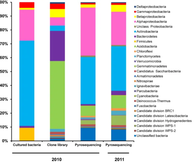
strains which corresponded to the generaMethylobacterium(Alphaproteobacteria) and Nocar-dioides-Streptomyces(Actinobacteria) (S2 Table). Within the cultured bacteria collection, most (61.5%) belong to the phylumActinobacteria, almost all of which fall into the subclass Actino-bacteridaeand only 1 isolate into the subclassRubrobacteridae. Isolates belonging to the phy-lumProteobacteriafell within three different classes:Alphaproteobacteria(22.3%),
Gammaproteobacteria(4.1%) andBetaproteobacteria(1.4%). The next most common phyla wereFirmicutes(9.5%), of which all were identified asBacilli, and the phylumBacteroidetes
(1.4%) which were equitably classified asSphingobacteriiaorFlavobacteriia(Fig 1).
16S rRNA gene clone library
A total of 384 clones (approximately 5% of all of the transformed colonies obtained) were sequenced from the 16S rRNA gene clone library generated from the rhizosphere soil DNA of the 2010 pooled sample. After quality filtering, only 184 high quality near-full length and non-chimeric reads could be assembled and used for downstream analysis. Sequence lengths ranged from 1077 to 1092 bp. The nucleotide sequence of the cloned fragments of the 16S rRNA gene
Fig 1. Relative abundance of the different bacterial phyla and proteobacterial classes identified through culture-dependent and culture
independent (clone library and 454 pyrosequencing) methodologies targeting the 16S rRNA gene.Bacterial sequences were classified with the RDP classifier tool (Release 11, Update 3), selecting 80% as the confidence threshold and adjusting the copy number of 16S rRNA operons in the case of the pyrosequencing.
could be classified into 10 bacterial phyla, 15 classes, 15 orders, 23 families and 37 genera (S1 Fig). However, 3, 3, 12, 3, and 11 clones could not be classified at the phylum, class, order, fam-ily or genus taxonomic ranks, respectively. Of the classified clone sequences, 48.9% could be affiliated to the phylaAcidobacteria, 21.7% toBacteroidetes, 16.8% toProteobacteria, and 3.8% toActinobacteria. Other minority phyla were the“candidatusSaccharibacteria”(1.6%), Planc-tomycetes(2.2%),Nitrospirae(1.1%),Ignavibacteriae(1.1%),Armatimonadetes(0.5%) and candidate division WPS-2 (0.5%). TheAcidobacteriaclones fell into four classes: Gp1, Gp4, Gp6 and Gp7, with Gp6 and Gp4 being the most numerous. TheBacteroidetesclasses observed belonged toCytophagia,FlavobacteriiaandSphingobacteriia, with the latter as the most abun-dant. Among theProteobacteria,AlphaproteobacteriaandBetaproteobacteriawere the most predominant classes followed byGammaproteobacteriaandDeltaproteobacteria, respectively. Within the phylumActinobacteria, clones were affiliated to the classesAcidimicrobiaeand
Rubrobacteridae(Fig 1). Out of 184 clones, 13, 89 and 55 shared similarity values between 97– 95%, 97–85% and<85% with their respective closest type strain (S3 Table). These results show the high degree of taxonomic novelty present in this bacterial community, mainly at higher tax-onomic ranks.
16S rRNA gene amplicon pyrosequencing
The pyrosequencing-based analysis of the V123 region of the 16S rRNA gene from metage-nomic DNA from the 2010 rhizosphere pooled sample resulted in the recovery of 17,948 high quality non-chimeric sequences from the 27,909 reads initially included in the pipeline. The average read length was 497 ± 18.05 bp. After the taxonomic normalization by the 16S rRNA gene copy number, a total number of 8751 sequences were retained. Pyrosequencing revealed the presence of 16 phyla or candidate divisions, 39 bacterial classes, 44 bacterial orders, 96 fam-ilies or 250 different genera in the rhizosphere soil sample (S1 Fig). Of all the sequences, 9.4% of the pyrotags could not be classified at the phylum or candidate division. Only one read could be assigned to chloroplasts (Eukaryota) and was not analyzed further. The most common phyla wereProteobacteria(39.5% of all pyrotags),Actinobacteria(33.9%),Acidobacteria
(7.2%),Gemmatimonadetes(3.1%),“candidatusSaccharibacteria”(1.3%),Bacteroidetes(1.1%),
Planctomycetes(1.0%),Verrucomicrobia(1.0%) andChloroflexi(1.0%). Representatives of the phyla/candidate divisions WPS-1,Armatimonadetes,NitrospiraeandFirmicutes, were detected below 1% each, and WPS-2,ParcubacteriaandHydrogenedentesbelow 0.1%. Among the pro-teobacterias, theAlphaproteobacteriawas the largest class (34.6% of all pyrotags), followed by
Betaproteobacteria,GammaproteobacteriaandDeltaproteobacteriawhich accounted for 2.2%, 1.0% and 0.6%, respectively of the sequences. A group of unclassifiedProteobacteriawas also detected (1.0%). WithinActinobacteria, the subclassActinobacteridaewas the most numerous (19.6%), followed byRubrobacteridae(10.4%) andAcidimicrobidae(1.4%). Less than 0.1% of the reads belonged to classThermoleophilia, and some (1.2%) of theActinobacteriasequences remained unclassified. Representatives of ten classes/subdivisions within the phylum Acidobac-teriawere found, with Gp6 (2.2%), Gp16 (1.7%) and Gp4 (1.4%) as the classes with the most reads. All theGemmatimonadetessequences fall into the only class with taxonomic validation described so far for this phylum, the classGemmatimonadetes(http://www.bacterio.net/). Sequences belonging to the phylumBacteroidetescould be grouped into the classes Sphingobac-teriia(0.7%),Cytophagia(0.2%) andFlavobacteriia(0.01%), whereas 0.2% remained as unclas-sifiedBacteroidetes. Sequences belonging toPlanctomycetesharboured mainly representatives of the classPlanctomycetaciaand also a fewPhycisphaerae. Finally, sequences belonging to
Comparison among the three approaches used for the 2010 rhizosphere
sample
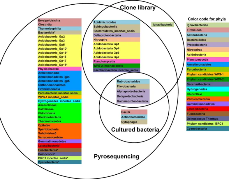
A comparison to determine to what degree the sequences retrieved among the three approaches from the same pooled rhizosphere soil of 2010 were shared at the class/subclass level is represented as a Venn diagram (Fig 2). Of the 17 phyla/ candidate divisions recorded globally in theThymus zygisrhizosphere soil in 2010, only proteobacterias ( Alphaproteobac-teria,BetaproteobacteriaandGammaproteobacteria), actinobacterias belonging to the subclass
RubrobacteridaeandBacteroidetesclassFlavobacteriiawere detected by all three approaches. No taxa were exclusively recovered with the culture-dependent approach. However, members ofBacilli,ActinobacteridaeandCytophagiawhich were successfully cultured were also detected by pyrosequencing but not in the clone library dataset. In fact, no sequences classified as belonging to theFirmicutesphylum were detected in the clone library dataset. More
Fig 2. Venn diagram at the class level.Classification at the rank of class/subclass showing shared and unique taxa identified with each approach. Bacterial classes or in the case ofActinobacteriasubclasses which belong to the same phylum are highlighted with the same color. Asterisks indicate those phyla detected in the 2011 pyrosequencing datasets but not in the 2010 pyrosequencing dataset.
surprisingly, representatives of the lineageIgnavibacteriaeappeared to be unique to the clone library. However, when the sequences of the clones classified asIgnavibacteriae(clones SNNP_2012_60 and SNNP_2012_78) are trimmed to contain only regions V123 (500 bp in length) as if they were pyrotags, the taxonomic assignment changed to become unclassifiable at the phylum level. Therefore, any possible representatives ofIgnavibacteriaepresent in the pyr-osequencing dataset would likewise be identified as unclassified bacteria. Of the remaining 12 phyla/ candidate divisions,Parcubacteria,Hydrogenedetes,Chloroflexi,Verrucomicrobia, Gem-matimonadetesand candidate division WPS-1 lineages were only detected in the pyrosequen-cing reads.
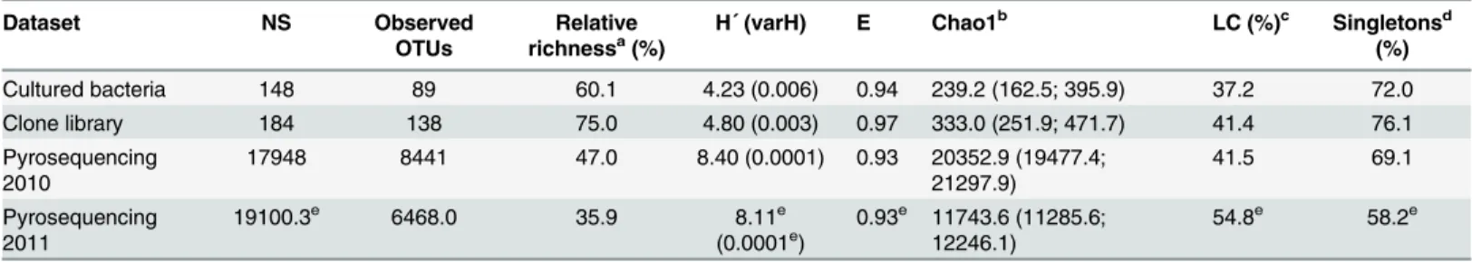
Since the contents of current databases will introduce bias in the number of species and gen-era classified into each phyla/class, the alpha-diversity parameters of each approach were calcu-lated as Operational Taxonomic Units (OTUs) at 97% sequence similarity. The relative richness, calculated as the number of OTUs observed with regard to the total number of sequences within of each dataset, was 60.1%, 75.0% and 47.0% for cultured bacteria, clone library and 2010 pyrosequencing reads, respectively (Table 1). Between 72.0%, 76.1% and 69.1% of these OTUs harboured single representatives, respectively, thus revealing the numeri-cal importance of singletons in the bacterial community in theThymus zygisrhizosphere. When a multiple alignment was performed, 15 clusters at a similarity of 97% grouped single-tons from the pyrosequencing dataset with one or more bacterial isolates. These clusters com-prised easily cultured taxa such asStreptomyces,Nocardioides,Agreia,Williamsia,Patulibacter,
Mycobacterium,Arthrobacter,Paenibacillus,PsychrobacillusandPseudomonas. Diversity based on the Shannon index was higher in the 2010 pyrosequencing dataset than in the cul-tured bacteria and the 16S rRNA gene clone library mainly due to the higher sampling effort offered by the second generation sequencing technology. Evenness values were also almost similar (from 0.93 to 0.97) among the three approaches (Table 1) suggesting that the commu-nity associated with the rhizosphere ofThymus zygisconsisted of a few dominant taxa and many minority groups. This result was in agreement with the large number of singletons detected in the datasets. Rarefaction curves obtained from the sequences of the pyrosequencing dataset showed that a greater sampling effort would still be required to cover the diversity in this rhizosphere soil sample at the level of species (97% cut-off) and genus (95% cut-off) Table 1. Diversity, equitability and richness indices, relative number of singletons and library coverage of OTUs defined at 3% sequence divergence.
Dataset NS Observed
OTUs
Relative
richnessa(%) H´ (varH) E Chao1 b
LC (%)c Singletonsd (%)
Cultured bacteria 148 89 60.1 4.23 (0.006) 0.94 239.2 (162.5; 395.9) 37.2 72.0
Clone library 184 138 75.0 4.80 (0.003) 0.97 333.0 (251.9; 471.7) 41.4 76.1
Pyrosequencing
2010 17948 8441 47.0 8.40 (0.0001) 0.93 20352.9 (19477.4;21297.9) 41.5 69.1
Pyrosequencing
2011 19100.3
e 6468.0 35.9 8.11e
(0.0001e) 0.93
e 11743.6 (11285.6;
12246.1) 54.8
e 58.2e
Abbreviations: E, Shannon Wiener equitability index; H´, Shannon-Wiener index; LC, library coverage; NS, number of sequences for each dataset; OTUs, operational taxonomic units; varH', variance of H´.
aRelative richness, defined as the number of OTUs observed regarding to NS bValues in brackets are lower limit and upper limit Chao1 estimates at 95% con
fidence interval.
cLC, defined as OTUs observed/ Chao1 estimate of OTUs richness dRelative number of singletons regarding to the number of OTUs eStandard deviation lower than 5% of the average value (n = 3)
(S2A–S2D Fig). However, taking into account the recently re-evaluated thresholds by Yarza and colleagues [29] to delimit higher taxonomic ranges, the sampling effort achieved full cover-age at the levels of family (90% cut-off) and class (85% cut-off). In order to evaluate the library coverage (hereafter LC) of the clone library and cultured bacteria datasets, the ratio of the actual number of OTUs observed with the Chao1 estimate of species richness (%) was calcu-lated. According to the LC statistic, when the sampling effort is weighted, both approaches allow access at the species level with comparable diversity as observed with pyrosequencing technology (Table 1). In order to determine to what extent the functional profiles associated with the results obtained by each approach may differ, the open source R package Tax4Fun [27] was used. The results reveal that despite differences at the taxonomic level, the functional profiles for each approach are similar to each other (S4 Table).
Comparison between pyrosequencing replicates
To obtain a better understanding of the bacterial communities present in the rhizosphere of
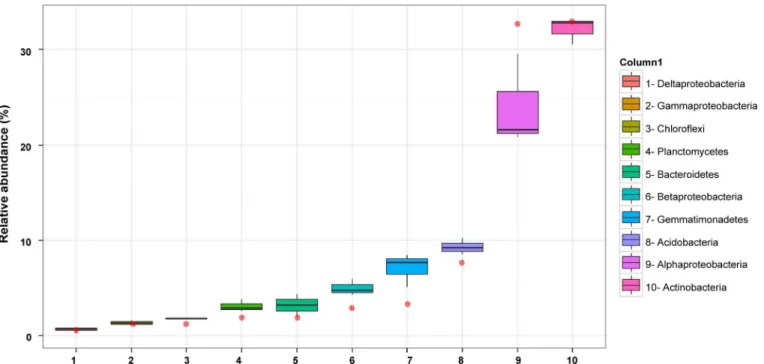
Thymus zygis, additional 454 amplicon sequences were obtained using the same 16S rRNA gene region as for the 2010 sample but instead of using metagenomic DNA from a pooled rhi-zosphere sample, the metagenomic DNA from the rhirhi-zosphere of three different plants sam-pled in 2011 were analysed separately. This resulted in a mean number of 19,100 high quality non-chimeric sequences which corresponded to a mean number of 9,175 sequences after nor-malization for copy number. In general, the taxonomic structures of the bacterial communities observed in the rhizosphere of the three plants collected in 2011 were similar to each other (Fig 3). The mean relative abundance (Fig 1) revealed thatActinobacteria(32.1% of all pyrotags), is the most represented phyla followed byProteobacteria(31.6%),Acidobacteria(9.3%), Gemma-timonadetes(7.0%),Bacteroidetes(3.1%),Planctomycetes(3.1%),Chloroflexi(1.8%), and
Fig 3. Relative abundance of the 10 most abundant phyla/ proteobacterial classes in the pyrosequencing datasets.The sample from 2010 is represented as a red point whereas three replicates from 2011 are represented as box-plots. The boxes represent the interquartile range (IQR) between the first and third quartiles (25th and 75th percentiles, respectively) and the vertical line inside the box defines the median. Whiskers represent the lowest and highest values within 1.5 times the IQR from the first and third quartiles, respectively.
“candidatus Saccharibacteria”(1.4%). Representatives of the candidate division WPS-1, Arma-timonadetes, candidate division WPS-2,Verrucomicrobia, andNitrospirae, andFirmicutes, were detected below 1% each, andParcubacteria, candidate division BRC1, candidate division
Hydrogenedentes,Deinoccocus-Thermus,Cyanobacteria(non-chloroplast) candidate division
Latescibacteria, andFusobacteria, below 0.1%. Several of the latter are often represented by extremely few sequences (from 1 to 6 sequences) and not always shared between all three repli-cate samples. Of the two most abundant phyla, withinActinobacteria, the subclass Actinobac-teridaewas the most numerous (21.3% of all pyrotags), followed byRubrobacteridae(5.8%) andAcidimicrobidae(3.1%). On the other hand, among the proteobacterias, the Alphaproteo-bacteriawas the largest class (23.9%), followed byBetaproteobacteria,Gammaproteobacteria
andDeltaproteobacteriawhich accounted for 4.9%, 1.3% and 0.6%, respectively, of the sequences. A group of unclassifiedProteobacteriawas also detected (0.8%).
In general, the relative abundances shifted with respect to the 2010 dataset with the largest differences observed inProteobacteria,Gemmatimonadetes,Acidobacteria,Planctomycesand
Bacteriodetes(Fig 1,Fig 3). Moreover, ultra-low-represented phyla appeared in some of the replicate samples from 2011 but not in the 2010 sample (Fig 2: asterisks) thereby increasing the total number of different phyla/ candidate divisions detected, but this had a smaller relative effect on the total number of different classes, orders, families and genera (S1 Fig). According to the diversity indices (Table 1), the replicate pyrosequencing samples of 2011 show lower diversity and relative richness than the pyrosequencing dataset from 2010 but also a propor-tionally lower percentage of singleton reads, as could be expected taking into account the higher sampling effort. Nevertheless, and similar to the 2010 sample, between 11 to 13 of the singletons from each replicate dataset clustered at a similarity of 97% with one or more bacte-rial isolates. According to the principal coordinates analysis (PCoA) plot based on weighted UniFrac distances, the three replicates of 2011 and that of 2010 (pooled sample) were placed more or less equidistant from each other, as explained by 63.2%, 26.1% and 10.7% of the observed differences at variable 1, 2 and 3 respectively (S3 Fig). This indicates that the bacterial community showed, at least seasonally, the same taxonomic pattern during two consecutive years and hence it might be a stabilized community. The similarity between all the pyrose-quenced amplicon libraries was also revealed by the number of families shared. This permitting the definition of the core microbiome of the rhizosphere of theThymus zygisplant (Fig 4) which is constituted of a total of 78 different families (accounting for a relative
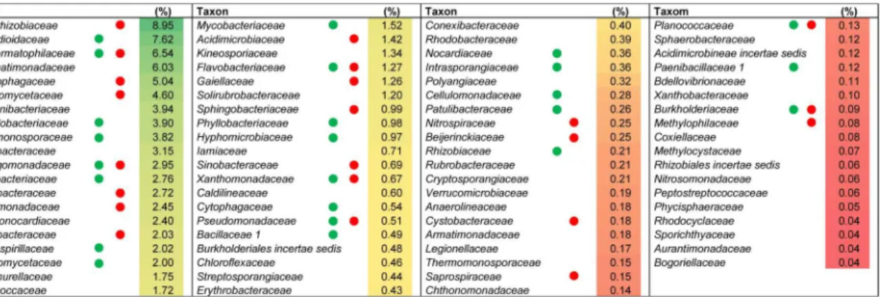
abundance0.14% of the total community). The most highly represented families (more than
Fig 4. Core microbiome of the wild thymus rhizosphere at the family level.They represent bacterial families shared by all the pyrosequencing datasets (2010, 2011_1, 2011_2 and 2011_3). The heat map shows the average value (n = 4) of their relative abundances. Green or red circles indicate coincidence with families detected in cultured bacteria or the clone library, respectively.
3%) found in this core microbiome wereBradyrhizobiaceae,Nocardioidaceae, and Geoderma-tophilaceaefollowed by other families belonging toActinobacteriaorAlphaproteobacteriaas well asGemmatimonadetes,BacteroidetesandPlanctomycetes. Most of the families which make up the core microbiome were also found in the clone library and/or cultured-dependent approach (Fig 4), confirming qualitatively the prevalence of these families independently of the methodology used to study the rhizospheric bacterial community. The functional profile of the core microbiome of the rhizososphere ofThymus zygisinferred with Tax4Fun (S4 Table) sug-gested that the overall functional structure of the community was dominated by KEGG path-ways related to metabolism especially that of carbohydrates (starch, sucrose, amino sugars and nucleotide sugars) and nitrogen-containing compounds such as amino acids and nucleotides (arginine, proline, glycine, serine, threonine, purine and pyrimidine among others). With regard to energy metabolism, genes related with the nitrogen metabolism, oxidative phosphor-ylation, methane metabolism and carbon fixation pathways in prokaryotes dominated. The metabolism of terpenoids, polyketides, lipids, xenobiotics, and glycans were also predicted but at lower abundances. Another dominant KEGG category of the inferred functional profile of the core rhizosphere bacterial community is associated with environmental information pro-cessing, principally in pathways related to membrane transport by ABC transporters and signal transduction by two component systems.
Discussion
In this work, the diversity within the rhizosphere bacterial community associated with a wild thyme species from the Sierra Nevada National Park was explored using culture-dependent and independent approaches. The clone library and multiplex amplicon pyrosequencing per-mit access to the prokaryotic diversity at high resolutions, including low-abundance species of the so-called“rare biosphere”[30,31] and/or bacteria resistant toin vitroculturing strategies belonging to uncultured“dark matter”clades [32,33]. On the other hand, the cultured bacteria complement the molecular approaches and will permit further in-depth metabolic, physiologi-cal and genomic characterization and thereby the possibility to obtain a better understanding of their roles in the rhizosphere ofThymus zygisplants. This work is currently in progress.
In this study, the percentage of isolates that could be cultivatedin vitro(cultivability) com-pared to the total viable bacteria present in the sample was 0.60% which is within the range of values (0.01% to 1%) obtained in other solid media based studies [34,35]. Although relatively low diversity was recovered compared to the molecular techniques used in this study, the rela-tive abundance of members of the four phyla detected appears to be proportionally more simi-lar to those observed in the pyrosequencing datasets than to the clone library. This suggests that within the constraints faced for effective cultivation, the media and conditions used in this study to culture microorganismsin vitrosuccessfully recovered a representative subset of the true diversity present, including numerically non-dominant taxa. Moreover, in spite of the dif-ficulty to isolate“uncultured bacteria”a strain belonging to the subclassRubrobacteridaewas successfully isolated [36]. Although this taxon was also detected by both molecular techniques and is ubiquitous in the soil environment, only a few known cultivable representatives of this subclass have been identified [13,36]. In addition to this isolate, 4.7% of the isolates identified in this study shared a 16S rRNA gene similarity of less than 97% with their closest type strains, and thus may constitute new bacterial taxa [37]. In fact, if the sequence identity threshold of 98.65% suggested by Kimet al. [38] is used to differentiate species, 37.8% of all the strains iso-lated from the rhizosphere soil sample in this study may represent new bacterial species.
but with low depth and amplicon pyrosequencing of short 16S rRNA gene fragments at very high depth. Surprisingly, members of several dominant phyla which had been successfully cul-tured were not recovered in the clone library and the relative abundances of dominant taxa were not in agreement with those observed with the pyrosequencing technique. According to the RDP probe match tool [20],a priori, coverage of the primers used to amplify 16S rRNA genes from metagenomic DNA does not account for these differences. However, during the clone library construction other factors such as the soil metagenomic DNA quality, PCR, clon-ing biases durclon-ing ligation and transformation steps, etc. may affect the outcome and thereby distort the perception of the bacterial community structure. As a result other authors have also observed that 16S rRNA gene clone libraries may not represent a complete or accurate picture of the true bacterial community [39,40]. Another unexpected result was the detection of repre-sentatives of the lineageIgnavibacteriaein the clone library but not in the pyrosequencing data-set regardless of the much higher sampling effort of the latter. As pointed out above, when sequences identified asIgnavibacteriaewere trimmed to a fragments resembling both in size and gene regions the 16S rRNA pyrotags, it could no longer be identified. This result highlights the limitation of taxonomic assignment methods and current databases for the correct identifi-cation of short fragments of the 16S rRNA gene.
Although the large differences in the sampling effort between the approaches used do not permit accurate comparative studies, the clone library and isolates may complement qualita-tively the pyrosequencing dataset by corroborating the presence of certain taxa and to permit a more accurate taxonomic assignment of those taxa. In this manner, the cultured isolates can also be used to benchmark data obtained by the molecular approaches by permitting the verifi-cation of singleton sequences. We have observed that 11 to 15 of the singletons detected in each pyrosequencing dataset clustered with isolated bacteria. Therefore, a proportion of the singleton sequences of the pyrosequencing dataset correspond to real but underrepresented bacteria and consequently suggest that conservative strategies that eliminate singletons as sequencing artefacts should take into account this possible loss of information especially when the“rare biosphere”is being sought. This fact also confirms that the cultured approach permits access to the“rare biosphere”[41].
The overall diversity observed in the rhizosphere ofThymus zygisreveals an abundance of
Proteobacteria(predominantlyAlphaproteobacteriaandBetaproteobacteriaclasses), Actino-bacteria,Acidobacteria,Gemmatimonadetes,Bacteroidetes,Planctomycetes,Chloroflexi, and “candidatusSaccharibacteria”. Although the order of predominance may vary, all of the men-tioned phyla together withFirmicutesare usually abundant in soil in general [39,42] and the rhizosphere in particular such as those associated with a number of different plants such as oak [43], aspen [44], potato [45],Arabidopsisspp. [4,46,47], cactus [48], cucumber [49], artic grasses [50], maize [51,52], Japanese barberry [53], cannabis [54], medicinal plants [55,56], rice [57], soybean [58], wheat [59,60] and creosote plants [61], amongst others. Aside from effects due to edaphic characteristics such as soil pH [62], the variations in phyla predominance observed in the rhizosphere bacterial communities associated with the different plants could depend on the nutrients released by the plant in the rhizosphere micro-niche where decompo-sition of plant-derived carbon sources favour faster growingProteobacteriaandBacteriodetes
Deeper analysis of the rhizosphere bacterial communities ofThymus zygisusing amplicon pyrosequencing permitted the definition of a core microbiome for this niche. The most abun-dant families observed within this core microbiome belong to the alphaproteobacterial orders
Rhizobiales,Rhodospirilalles,SphingomonadalesandCaulobacteriales. Many of these appear to dominantly assimilate wheat root derived carbon together with the actinobacterial orders
Micrococcales,Acidimicrobiales, andPropionibacteriales[60]. Besides the generally heterotro-phic nature of the abundant families found in the core microbiome, the presence of Methylo-bacteriaceaeandHyphomicrobiaceaewhich both have methylotrophic members suggests an importance of single carbon compounds such as methane or methanol as C sources in the rhi-zosphere. On the other hand, the actinobacterial familiesMicromonosporaceaeand Pseudono-cacardiaceaehave been associated with senescing wheat roots [59] and in general
Actinobacteriahave been reported to have increased numbers on older roots [65]. Within the alphaproteobacterial and actinobacterial families, abundant genera are found which include important xenobiotic degraders such asSphingomonas,Phenylobacterium, andPatulibacter. Therefore, it appears that at least part of the core microbiome associated with theThymus zygis
rhizosphere may have been selected by the plant in reponse to rhizodeposition composition while others may be related to the degradation of complex molecules associated with older sce-nescing roots or plant derived secondary products. On the other hand, the abundance of the familyBradyrhizobiaceae, which includeBradyrhizobiumand other genera which participate in the nitrogen cycle [66] could indicate an important role for this cycle in this niche. This is also supported by the presence of possible nitrifying genera associated withNitrospiraceaeand that many of the taxa identified in our datasets coincide with those associated with nitrogen cycle genes in the rhizosphere of holm oak [67] growing within the same geographical region. Altogether this suggests that the main drivers of the bacterial community in theThymus zygis
rhizosphere might be related with the plant-bacteria interchange of nutrients and their partici-pation in the biogeochemical cycles.
To obtain a better understanding of the possible gene functions associated with the taxa in the core microbiome, the Tax4Fun prediction tool was used. The major functional category that could be inferred in the rhizosphere ofThymus zygiswas metabolism especially that of car-bohydrates and amino acids which are typical components of root exudates. However, also sec-ondary metabolite degradation functions could be detected including those of geraniol, limonene and pinene which have been associated withThymus zygis[68]. The abundance of functional groups related to the biodegradation and metabolism of xenobiotics such as benzo-ates, aminobenzoate and bisphenol, may also be related to the presence of complex secondary metabolites or polymers with aromatic structures which may be released by the plants into the rhizosphere or form part of lignin in woody roots. A high abundance of functions related to energy metabolism, including nitrogen metabolism, and the abundance of transporters and two component systems imply an exchange of nutrients and signals. Therefore, the functional profile inferred by the prediction tool are similar to those which may be attributed to the more abundant families of the core microbiome especially with regard to the importance of metabo-lism of simple and complex carbon and nitrogen sources which may include methane, xenobi-otics, and secondary metabolites such as terpenoids or complex polymers. This functional profile suggests that the bacterial community shares an intricate relationship with the roots of this aromatic plant, presumably allowing a feedback ecological benefit.
underway in order to elucidate how the identified bacterial families function and respond to environmental changes and benefit wild thyme plant growth.
Supporting Information
S1 Fig. Number of taxa recovered at the different taxonomic levels, from phylum to genus, with each of the three approaches used in this study.Asterisks indicate a standard deviation inferior to 8% for the 2011 pyrosequencing datasets (n = 3).
(TIF)
S2 Fig. Rarefaction curves of 16S rRNA gene sequences from the 454 pyrosequencing data-sets calculated with A) 3%, B) 5%, C) 10% and D) 15% distance cut-offs.
(TIF)
S3 Fig. Principal Coordinate Analysis (PCoA) based on weighted Unifrac distances of bac-terial community inhabiting the rhizosphere soil of wild thyme based on the pyrosequen-cing dataset from 2010 (blue) and the three replicates from 2011 (1–3, red).
(TIF)
S1 Table. Soil physicochemical properties.
(DOCX)
S2 Table. Taxonomic diversity of cultured bacteria based on their 16S rRNA gene sequences.
(DOCX)
S3 Table. Taxonomic diversity of clone library based on their 16S rRNA gene sequences.
(DOCX)
S4 Table. Percentages (>0.5%) of 1, 2 and 3 tier KEGG Orthology (KO) categories
pre-dicted from each 16S rRNA dataset with the Tax4Fun tool.1, Cultured bacteria; 2, Clone library; 3, Pyrosequencing 2010; 4, Pyrosequencing 2010; 5, Core microbiome.
(DOCX)
Author Contributions
Conceived and designed the experiments: JP SB MG-L AG-S SAB OG GFB JLR PvD. Per-formed the experiments: JP SB MG-L AG-S SAB. Analyzed the data: JP SB PvD. Wrote the paper: JP SB MG-L AG-S SAB OG GFB JLR PvD.
References
1. Hartmann A, Rothballer M, Schmid M. Lorenz Hiltner, a pioneer in rhizosphere microbial ecology and soil bacteriology research. Plant Soil. 2007; 312: 7–14. doi:10.1007/s11104-007-9514-z
2. Mendes R, Garbeva P, Raaijmakers JM. The rhizosphere microbiome: Significance of plant beneficial, plant pathogenic, and human pathogenic microorganisms. FEMS Microbiol Rev. 2013; 37: 634–663. doi:10.1111/1574-6976.12028PMID:23790204
3. Berendsen RL, Pieterse CMJ, Bakker PAHM. The rhizosphere microbiome and plant health. Trends Plant Sci. 2012; 17: 478–486. doi:10.1016/j.tplants.2012.04.001PMID:22564542
4. Bulgarelli D, Schlaeppi K, Spaepen S, Ver Loren van Themaat E, Schulze-Lefert P. Structure and func-tions of the bacterial microbiota of plants. Annu Rev Plant Biol. 2013; 64: 807–838. doi:10.1146/ annurev-arplant-050312-120106PMID:23373698
6. Ramos JL, González-Pérez MM, Caballero A, van Dillewijn P. Bioremediation of polynitrated aromatic compounds: plants and microbes put up a fight. Curr Opin Biotechnol. 2005; 16: 275–281. doi:10. 1016/j.copbio.2005.03.010PMID:15961028
7. McGuinness M, Dowling D. Plant-associated bacterial degradation of toxic organic compounds in soil. Int J Environ Res Public Health. 2009; 6: 2226–2247. doi:10.3390/ijerph6082226PMID:19742157
8. Cheng M, Xiang Y, Xue Z, An S, Darboux F. Soil aggregation and intra-aggregate carbon fractions in relation to vegetation succession on the Loess Plateau, China. CATENA. 2015; 124: 77–84. doi:10. 1016/j.catena.2014.09.006
9. Chamorro D, Luna B, Moreno JM. Germination response to various temperature regimes of four Medi-terranean seeder shrubs across a range of altitudes. Plant Ecol. 2013; 214: 1431–1441. doi:10.1007/ s11258-013-0264-0
10. Jordán MJ, Martínez RM, Martínez C, Moñino I, Sotomayor JA. Polyphenolic extract and essential oil quality ofThymus zygisssp.gracilisshrubs cultivated under different watering levels. Ind Crops Prod. 2009; 29: 145–153. doi:10.1016/j.indcrop.2008.04.021
11. Sánchez-Castro I, Ferrol N, Cornejo P, Barea J-M. Temporal dynamics of arbuscular mycorrhizal fungi colonizing roots of representative shrub species in a semi-arid Mediterranean ecosystem. Mycorrhiza. 2012; 22: 449–460. doi:10.1007/s00572-011-0421-zPMID:22124663
12. Joseph SJ, Hugenholtz P, Sangwan P, Osborne CA, Janssen PH. Laboratory cultivation of widespread and previously uncultured soil bacteria. Appl Environ Microbiol. 2003; 69: 7210–7215. doi:10.1128/ AEM.69.12.7210–7215.2003PMID:14660368
13. Davis KE, Joseph SJ, Janssen PH. Effects of growth medium, inoculum size, and incubation time on culturability and isolation of soil bacteria. Appl Env Microbiol. 2005; 71: 826–834. doi:10.1128/AEM.71. 2.826–834.2005
14. Sánchez-Hidalgo M, Pascual J, de la Cruz M, Martin J, Kath GS, Sigmund JM, et al. Prescreening bac-terial colonies for bioactive molecules with Janus plates, a SBS standard double-faced microbial cultur-ing system. Antonie Van Leeuwenhoek. 2012; 102: 361–374. doi:10.1007/s10482-012-9746-7PMID: 22562433
15. Weisburg WG, Barns SM, Pelletier DA, Lane DJ. 16S ribosomal DNA amplification for phylogenetic study. J Bacteriol. 1991; 173: 697–703. PMID:1987160
16. Pascual J, Macián MC, Arahal DR, Garay E, Pujalte MJ. Multilocus sequence analysis of the central clade of the genusVibrioby using the 16S rRNA,recA,pyrH,rpoD,gyrB,rctBandtoxRgenes. Int J Syst Evol Microbiol. 2010; 60: 154–165. doi:10.1099/ijs.0.010702–0PMID:19648344
17. Muyzer G, Teske A, Wirsen CO, Jannasch HW. Phylogenetic relationships ofThiomicrospiraspecies and their identification in deep-sea hydrothermal vent samples by denaturing gradient gel electrophore-sis of 16S rDNA fragments. Arch Microbiol. 1995; 164: 165–172. doi:10.1007/BF02529967PMID: 7545384
18. Edgar RC, Haas BJ, Clemente JC, Quince C, Knight R. UCHIME improves sensitivity and speed of chi-mera detection. Bioinformatics. 2011; 27: 2194–2200. doi:10.1093/bioinformatics/btr381PMID: 21700674
19. Brosius J, Palmer ML, Kennedy PJ, Noller HF. Complete nucleotide sequence of a 16S ribosomal RNA gene fromEscherichia coli. Proc Natl Acad Sci. 1978; 75: 4801–4805. PMID:368799
20. Cole JR, Wang Q, Fish J a., Chai B, McGarrell DM, Sun Y, et al. Ribosomal Database Project: Data and tools for high throughput rRNA analysis. Nucleic Acids Res. 2014; 42: 633–642. doi:10.1093/nar/ gkt1244
21. Wang Q, Garrity GM, Tiedje JM, Cole JR. Naïve Bayesian classifier for rapid assignment of rRNA sequences into the new bacterial taxonomy. Appl Environ Microbiol. 2007; 73: 5261–5267. doi:10. 1128/AEM.00062-07PMID:17586664
22. Stoddard SF, Smith BJ, Hein R, Roller BRK, Schmidt TM. rrnDB: improved tools for interpreting rRNA gene abundance in bacteria and archaea and a new foundation for future development. Nucleic Acids Res. 2015; 43: D593–598. doi:10.1093/nar/gku1201PMID:25414355
23. Nawrocki EP, Eddy SR. Query-dependent banding (QDB) for faster RNA similarity searches. PLoS Comput Biol. 2007; 3: e56. doi:10.1371/journal.pcbi.0030056PMID:17397253
24. Nawrocki EP, Eddy SR. Infernal 1.1: 100-fold faster RNA homology searches. Bioinformatics. 2013; 29: 2933–2935. doi:10.1093/bioinformatics/btt509PMID:24008419
26. Caporaso JG, Kuczynski J, Stombaugh J, Bittinger K, Bushman FD, Costello EK, et al. QIIME allows analysis of high-throughput community sequencing data. Nat Methods. 2010; 7: 335–336. doi:10. 1038/nmeth.f.303PMID:20383131
27. Aβhauer KP, Wemheuer B, Daniel R, Meinicke P. Tax4Fun: predicting functional profiles from metage-nomic 16S rRNA data. Bioinformatics. 2015; doi:10.1093/bioinformatics/btv287
28. Quast C, Pruesse E, Yilmaz P, Gerken J, Schweer T, Yarza P, et al. The SILVA ribosomal RNA gene database project: improved data processing and web-based tools. Nucleic Acids Res. 2013; 41: D590– 596. doi:10.1093/nar/gks1219PMID:23193283
29. Yarza P, Yilmaz P, Pruesse E, Glöckner FO, Ludwig W, Schleifer K-H, et al. Uniting the classification of cultured and uncultured bacteria and archaea using 16S rRNA gene sequences. Nat Rev Microbiol. 2014; 12: 635–645. doi:10.1038/nrmicro3330PMID:25118885
30. Sogin ML, Morrison HG, Huber JA, Mark Welch D, Huse SM, Neal PR, et al. Microbial diversity in the deep sea and the underexplored“rare biosphere”. Proc Natl Acad Sci U S A. 2006; 103: 12115–12120. doi:10.1073/pnas.0605127103PMID:16880384
31. Pedrós-Alió C. The rare bacterial biosphere. Ann Rev Mar Sci. 2012; 4: 449–466. doi:10.1146/ annurev-marine-120710-100948PMID:22457983
32. Marcy Y, Ouverney C, Bik EM, Lösekann T, Ivanova N, Martin HG, et al. Dissecting biological“dark matter”with single-cell genetic analysis of rare and uncultivated TM7 microbes from the human mouth. Proc Natl Acad Sci U S A. 2007; 104: 11889–11894. doi:10.1073/pnas.0704662104PMID:17620602
33. Rinke C, Schwientek P, Sczyrba A, Ivanova NN, Anderson IJ, Cheng J-F, et al. Insights into the phylog-eny and coding potential of microbial dark matter. Nature. 2013; 499: 431–437. doi:10.1038/
nature12352PMID:23851394
34. Amann RI, Ludwig W, Schleifer KH. Phylogenetic identification andin situdetection of individual micro-bial cells without cultivation. Microbiol Rev. 1995; 59: 143–169. PMID:7535888
35. Staley JT, Konopka A. Measurement of in situ activities of nonphotosynthetic microorganisms in aquatic and terrestrial habitats. Annu Rev Microbiol. 1985; 39: 321–346. doi:10.1146/annurev.mi.39. 100185.001541PMID:3904603
36. Davis KER, Sangwan P, Janssen PH.Acidobacteria,RubrobacteridaeandChloroflexiare abundant among very slow-growing and mini-colony-forming soil bacteria. Environ Microbiol. 2011; 13: 798–805. doi:10.1111/j.1462-2920.2010.02384.xPMID:21108723
37. Stackebrandt E, Goebel BM. Taxonomic Note: A place for DNA-DNA reassociation and 16S rRNA sequence analysis in the present species definition in Bacteriology. Int J Syst Bacteriol. 1994; 44: 846– 849. doi:10.1099/00207713-44-4-846
38. Kim M, Oh HS, Park SC, Chun J. Towards a taxonomic coherence between average nucleotide identity and 16S rRNA gene sequence similarity for species demarcation of prokaryotes. Int J Syst Evol Micro-biol. 2014; 64: 346–351. doi:10.1099/ijs.0.059774–0PMID:24505072
39. Janssen PH. Identifying the dominant soil bacterial taxa in libraries of 16S rRNA and 16S rRNA genes. Appl Env Microbiol. 2006; 72: 1719–1728. doi:10.1128/AEM.72.3.1719–1728.2006
40. Spain AM, Krumholz LR, Elshahed MS. Abundance, composition, diversity and novelty of soil Proteo-bacteria. ISME J. 2009; 3: 992–1000. doi:10.1038/ismej.2009.43PMID:19404326
41. Shade A, Hogan CS, Klimowicz AK, Linske M, Mcmanus PS, Handelsman J. Culturing captures mem-bers of the soil rare biosphere. Environ Microbiol. 2012; 14: 2247–2252. doi:10.1111/j.1462-2920. 2012.02817.xPMID:22788977
42. Fierer N, Leff JW, Adams BJ, Nielsen UN, Bates ST, Lauber CL, et al. Cross-biome metagenomic anal-yses of soil microbial communities and their functional attributes. Proc Natl Acad Sci U S A. 2012; 109: 21390–21395. doi:10.1073/pnas.1215210110PMID:23236140
43. Uroz S, Buée M, Murat C, Frey-Klett P, Martin F. Pyrosequencing reveals a contrasted bacterial diver-sity between oak rhizosphere and surrounding soil. Environ Microbiol Rep. 2010; 2: 281–288. doi:10. 1111/j.1758-2229.2009.00117.xPMID:23766079
44. Lesaulnier C, Papamichail D, McCorkle S, Ollivier B, Skiena S, Taghavi S, et al. Elevated atmospheric CO2affects soil microbial diversity associated with trembling aspen. Environ Microbiol. 2008; 10: 926–
941. doi:10.1111/j.1462-2920.2007.01512.xPMID:18218029
45. Inceoǧlu Ö, Al-Soud WA, Salles JF, Semenov A V., van Elsas JD. Comparative analysis of bacterial communities in a potato field as determined by pyrosequencing. PLoS One. 2011; 6: e23321. doi:10. 1371/journal.pone.0023321PMID:21886785
47. Schlaeppi K, Dombrowski N, Oter RG, Ver Loren van Themaat E, Schulze-Lefert P. Quantitative diver-gence of the bacterial root microbiota inArabidopsis thalianarelatives. Proc Natl Acad Sci USA. 2014; 111: 585–592. doi:10.1073/pnas.1321597111PMID:24379374
48. Torres-Cortés G, Millán V, Fernández-González a. J, Aguirre-Garrido JF, Ramírez-Saad HC, Fernán-dez-López M, et al. Bacterial community in the rhizosphere of the cactus speciesMammillaria carnea
during dry and rainy seasons assessed by deep sequencing. Plant Soil. 2012; 357: 275–288. doi:10. 1007/s11104-012-1152-4
49. Tian Y, Gao L. Bacterial diversity in the rhizosphere of cucumbers grown in soils covering a wide range of cucumber cropping histories and environmental conditions. Microb Ecol. 2014; 68: 794–806. doi:10. 1007/s00248-014-0461-yPMID:25027276
50. Teixeira LCRS, Peixoto RS, Cury JC, Sul WJ, Pellizari VH, Tiedje J, et al. Bacterial diversity in rhizo-sphere soil from Antarctic vascular plants of Admiralty Bay, maritime Antarctica. ISME. 2010; 4: 989– 1001. doi:10.1038/ismej.2010.35
51. Garcia-Salamanca A, Molina-Henares MA, van Dillewijn P, Solano J, Pizarro-Tobias P, Roca A, et al. Bacterial diversity in the rhizosphere of maize and the surrounding carbonate-rich bulk soil. Microb Bio-technol. 2013; 6: 36–44. doi:10.1111/j.1751-7915.2012.00358.xPMID:22883414
52. Peiffer J a, Spor A, Koren O, Jin Z, Tringe SG, Dangl JL, et al. Diversity and heritability of the maize rhi-zosphere microbiome under field conditions. Proc Natl Acad Sci U S A. 2013; 110: 6548–6553. doi:10. 1073/pnas.1302837110PMID:23576752
53. Coats VC, Pelletreau KN, Rumpho ME. Amplicon pyrosequencing reveals the soil microbial diversity associated with invasive Japanese barberry (Berberis thunbergiiDC.). Mol Ecol. 2014; 23: 1318–1332. doi:10.1111/mec.12544PMID:24118303
54. Winston ME, Hampton-Marcell J, Zarraonaindia I, Owens SM, Moreau CS, Gilbert JA, et al. Under-standing cultivar-specificity and soil determinants of the cannabis microbiome. PLoS One. 2014; 9: e99641. doi:10.1371/journal.pone.0099641PMID:24932479
55. Köberl M, Schmidt R, Ramadan EM, Bauer R, Berg G. The microbiome of medicinal plants: Diversity and importance for plant growth, quality, and health. Front Microbiol. 2013; 4: 1–9.
56. Jin H, Yang X-Y, Yan Z-Q, Liu Q, Li X-Z, Chen J-X, et al. Characterization of rhizosphere and endo-phytic bacterial communities from leaves, stems and roots of medicinalStellera chamaejasmeL. Syst Appl Microbiol. 2014; 37: 376–385. doi:10.1016/j.syapm.2014.05.001PMID:24958606
57. Edwards J, Johnson C, Santos-Medellín C, Lurie E, Podishetty NK, Bhatnagar S, et al. Structure, varia-tion, and assembly of the root-associated microbiomes of rice. Proc Natl Acad Sci U S A. 2015; 112: E911–920. doi:10.1073/pnas.1414592112PMID:25605935
58. Mendes LW, Kuramae EE, Navarrete AA, van Veen JA, Tsai SM. Taxonomical and functional microbial community selection in soybean rhizosphere. ISME J. 2014; 8: 1577–1587. doi:10.1038/ismej.2014.17 PMID:24553468
59. Donn S, Kirkegaard JA, Perera G, Richardson AE, Watt R. Evolution of bacterial communities in the wheat crop rhizosphere. Environ Microbiol 2015; 17: 610–621. doi:10.1111/1462-2920.12452PMID: 24628845
60. Ai C, Liang G, Sun J, Wang X, He W, Zhou W, et al. Reduced dependence if rhizospheric microbiome on plant-derived carbon in 32-year long-term inorganic and organic fertilized soils. Soil Biol Biochem. 2015; 80: 70–78. doi:10.1016/j.soilbio.2014.09.028
61. Nguyen LM, Buttner MP, Cruz P, Smith SD, Robleto EA. Effects of elevated atmospheric CO2on
rhizo-sphere soil microbial communities in a mojave desert ecosystem. J Arid Environ. 2011; 75: 917–925. doi:10.1016/j.jaridenv.2011.04.028PMID:21779135
62. Lauber CL, Hamady M, Knight R, Fierer N. Pyrosequencing-based assessment of soil pH as a predictor of soil bacterial community structure at the continental scale. Appl Environ Microbiol. 2009; 75: 5111– 5120. doi:10.1128/AEM.00335-09PMID:19502440
63. Fierer N, Bradford M a., Jackson RB. Toward an ecological classification of soil bacteria. Ecology. 2007; 88: 1354–1364. doi:10.1890/05-1839PMID:17601128
64. Basilio A, González I, Vicente MF, Gorrochategui J, Cabello a., González a., et al. Patterns of antimicro-bial activities from soil actinomycetes isolated under different conditions of pH and salinity. J Appl Microbiol. 2003; 95: 814–823. doi:10.1046/j.1365-2672.2003.02049.xPMID:12969296
65. Watt M, Hugenholtz P, White R, Vinall K. Numbers and locations of native bacteria on field grown wheat roots quantified by fluorescence in situ hybridization (FISH). Environ Microbiol, 2006; 8: 871– 884. doi:10.1111/j.1462-2920.2005.00973.xPMID:16623744
putative newBradyrhizobiumspecies and a novel symbiovar (sierranevadense). Syst Appl Microbiol. 2014; 37: 177–185. doi:10.1016/j.syapm.2013.09.008PMID:24268094
67. Cobo-Díaz JF, Fernández-González AJ, Villadas PJ, Robles AB, Toro N, Fernández-López M. Metage-nomic assessment of the potential microbial nitrogen pathways in the rhizosphere of a mediterranean forest after a wildfire. Microb Ecol. 2015; 69: 895–904. doi:10.1007/s00248-015-0586-7PMID: 25732259