Cytology, biochemistry and molecular changes during coffee
fruit development
Renato D. De Castro1 and Pierre Marraccini2,3*
1UCSAL (Universidade Católica do Salvador), Laboratório de Estudos em Meio Ambiente – LEMA, Av. Prof. Pinto de Aguiar 2589, Pituaçu, 41740-090, Salvador, BA, Brazil; 2CIRAD-Centre de Coopération Internationale en Recherche Agronomique pour le Développement-CP, UMR PIA 1096, TA 80/03, Avenue d’Agropolis, 34398 Montpellier Cedex 5, France ; 3IAPAR-Instituto Agronômico do Paraná, LBI-AMG, CP 481, 86001-970, Londrina, PR, Brazil. * Corresponding author: [email protected]
In commercial coffee species (Coffea arabica and Coffeacanephora), fruit development is a lengthy process, characterized by tissue changes and evolutions. For example, soon after fecundation and up to mid development, the fruit is mainly constituted of the pericarp and perisperm tissue. Thereafter, the perisperm gradually disappears and is progressively replaced by the endosperm (true seed). Initially present in a “liquid” state, the endosperm hardens as it ripens during the maturation phase, as a result of accumulation of storage proteins, sucrose and complex polysaccharides representing the main reserves of the seed. The last step of maturation is characterized by the dehydration of the endosperm and the color change of the pericarp. Important quantitative and qualitative changes accompany fruit growth, highlighting the importance of its study to better understand the final characteristics of coffee beans. Following a description of the coffee fruit tissues, this review presents some data concerning biochemical, enzymatic and gene expression variations observed during the coffee fruit development. The latter will also be analyzed in the light of recent data (electronic expression profiles) arising from the Brazilian Coffee Genome Project. Key words: Coffea spp, bean development, cell cycle, endosperm, EST, gene expression, pericarp, perisperm.
Cytology, biochemistry and molecular changes during coffee fruit development: Em espécies comerciais de café (Coffea arabica e Coffea canephora), o desenvolvimento do fruto de café é um processo longo, caracterizado por mudanças e evoluções nos tecidos. Por exemplo, logo após a fecundação e até a metade do desenvolvimento, o fruto é principalmente constituído pelo pericarpo e perisperma. Em seguida, o perisperma gradualmente desaparece e é progressivamente substituído pelo endosperma (semente verdadeira). Inicialmente o endosperma apresenta-se no estado “líquido”, o endosperma endurece durante a fase de maturação, como resultado do acúmulo gradual de proteínas de reserva, sacarose e polissacarídeos complexos representando as principais reservas da semente. O último passo da maturação é caracterizado pela desidratação do endosperma e pela mudança de cor do pericarpo. Importantes alterações quantitativas e qualitativas acompanham o crescimento do fruto, ilustrando a importância do seu estudo para melhor compreender as características finais das sementes de café. Seguindo a descrição dos tecidos do fruto de café, esta revisão apresenta alguns dados relativos às variações bioquímicas, enzimáticas e de expressão gênica durante o desenvolvimento do fruto. A expressão de genes será também analisada em função de dados recentes (perfil de expressão eletrônica) oriundos do Projeto Genoma Brasileiro Café.
Palavras-chave: Coffea spp, ciclo celular, desenvolvimento da semente, endosperma, EST, expressão gênica, pericarpo, perisperma.
INTRODUCTION
The Coffea genus contains around 100 species (Charrier and Berthaud, 1985). Within these species, C. arabica (Arabica) and C. canephora (Robusta) represent respectively about 70 % and 30 % of the coffee market
al., 1999, 2000). It is a self-pollinating species. This in part explains its reduced genetic diversity and also the difficulty in incorporating traits of interest (Charrier and Berthaud, 1985). On the other hand, Robusta coffee is diploid (2n=22 chromosomes) and highly self-incompatible, due to the presence of a gametophytic system of incompatibility controlled by one gene with multiple S-alleles (Berthaud, 1980). This explains the higher genetic diversity within this species and also permits its genetic improvement by conventional breeding techniques (Montagnon, 2000).
At the beverage quality level, coffee from beans of C. arabica is preferred by consumers mainly in view of its lower bitterness and better flavor (see: Genetics of coffee quality, Leroy et al. in this issue). It is now well known that some compounds that accumulate in mature coffee beans play an important role in the quality of the beverage. For example, sucrose, which ranges from 6 % to 8.5 % in Arabica and from 0.9 to 4.9 % in Robusta (Clifford, 1985a; Campa et al., 2004), is considered as one the major contributors to coffee cup quality because its degradation during roasting leads to a wide range of compounds (i.e. aliphatic acids) involved in coffee flavor, either as the volatile aroma compounds, or as non-volatile taste compounds (De Maria et al., 1996; Tressl et al., 1998; Ginz et al., 2000). In contrast, caffeine and chlorogenic acids (CGA) are found at higher levels in Robusta than in Arabica, and are responsible for beverage bitterness (Leloup et al., 1995). Due to the importance of caffeine in human health (Bättig, 1985; Schilter et al., 2001), caffeine metabolism has been studied for some time in coffee (see: Metabolism of alkaloids, Ashihara et al. in this issue). Many genes implicated in its biosynthesis were recently cloned (Moisyadi et al., 1998, 1999; Ogawa et al., 2001; Mizuno et al., 2003a, 2003b; Uefuji et al., 2003) and the
first trials to produce transgenic coffee plants with reduced caffeine content in beans are being carried out (Ogita et al., 2003, 2004).
Except for the large number of studies concerning caf-feine metabolism in coffee, little data are available concern-ing genes, enzymes and metabolic pathways of many other biochemical compounds also important for coffee cup qual-ity (ex. sucrose metabolism) or for human health (ex. diter-penes). Considering that the knowledge of coffee cytology, biochemistry and molecular biology events is essential for an integrated view of coffee seed physiology, the main objective of this article is to review cellular and tissue differentiation, metabolism and gene expression. Each of these subjects is described below in separated sections.
Coffee fruit development: tissue evolution and changes In the genus Coffea, the fruit development covering the time between anthesis and full ripening is variable from few (10 to 12) weeks, as in C. racemosa and pseudozanguebariae, to more than a year for C. liberica. For the species of high economic value, C. arabica requires 6 to 8 months to mature while C. canephora requires 9 to 11 months (for a review, see Cannell 1985). Fruit growth is asynchronous during development, with different proportions of various fruits sizes on the same plant. Although possibly related to the several flowering events that may occur in C. arabica during each production season, a tendency for synchrony was observed during the later stages of maturation when a significantly higher proportion of fruits entered the largest sized ripe ‘cherry’ stage (De Castro et al., 2005). Despite the asynchrony in fruit growth and differences in the length of the reproductive cycle between coffee species, key steps of fruit and seed development appear to be identical, at least between the commercial species.
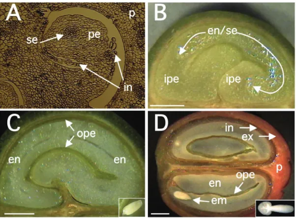
In C. arabica var. Bourbon, for which a detailed study of tissue evolution was reported, a rapid growth of fruits was observed between 0 and 7 WAF (weeks after flowering), reaching its maximum final size at 17 WAF (Wormer, 1966). The initial growth coincided with the sudden development of the perisperm up to 11-12 WAF, at the time when the endosperm had also begun to be identified. Thereafter, the endosperm continued to grow gradually, until completely occupying the space (locule) left by the inner portion of the perisperm at around 19 WAF, as observed for C. arabica var. Acaiá Cerrado (figure 2). At the maturity stage of the coffee fruits, usually occurring around 30 to 35 WAF, only the outer layer of the perisperm tissue remains surrounding Figure 1. Schematic representation of the tissues present at
the endosperm (figures 1and 2), sometimes referred to as the “silver skin” (Mendes, 1941, 1942; Wormer, 1964, 1966; De Castro et al., 2001, 2005; Miriam et al., in this issue). The endosperm actually corresponds to the bean that, after the post-harvest treatments, will form the “green coffee” that is sold in the international markets.
Previous studies have reported detailed information on the histology and cytology of developing coffee beans by light and transmission electron microscopy coupled with specific staining (Dentan, 1985a, 1985b). From the biochemical point of view, exchanges of sugars and organic acids between the perisperm and endosperm tissues were also shown to occur (Rogers et al., 1999b; Marraccini et al., 2001a). In addition, the thin perisperm layer (future silver skin), which is not consumed by the growing endosperm, remains as a greenish tissue, suggesting it could play important functions during seed filling, for example in the translocation of assimilates to the developing endosperm (De Castro et al., 2005). To better appreciate changes occurring during coffee fruit development, a rapid description of coffee fruit tissues and cell cycle events is given below.
Pericarp: The pericarp is composed of several tissues (Aval-lone 1999): the exocarp (peel), the mesocarp and the endo-carp (figure 1). The exoendo-carp persists as a green color colored tissue during most of the coffee fruit development, becoming transiently yellow and then red at the final stage of develop-ment. The change in color is due to the disappearance of chlorophyll pigments and anthocyan anthocyan accumulation during the last stages of fruit maturation (Marín-López et al., 2003). In some natural mutants of C. arabica, also referred to as yellowish (“Amarelo”) cultivars, the exocarp does not be-come red, but apparently remains yellow at the mature stage (Carvalho et al., 1957). Whatever the species considered, the change in color is of great importance since it is the main criterion for fruit maturation, even if the absence of coupling between the maturation of pericarp and endosperm (bean) has also been observed (Guyot et al., 1988).
The mesocarp, also commonly referred to as the “true pulp”, is rich in sugar (both reducing sugars and sucrose) and water. With 0.5-2 mm thickness, it can be divided into the external and internal parts (figure 2D). The former is formed of parenchyma cells with compact and dense cell walls in green fruits that become thinner during maturation, probably due to pectin modifications (Ouguerram, 1999). However, the cytological structure of the internal mesocarp, also referred to as the mucilaginous tissue in mature fruits, is still controversial (Beille, 1947; Menezes and Maneiro, 1955).
In the most internal location, the endocarp (also called parchment layer or “pergaminho”) is a hard and lignified tissue of around 150 µm (Mendes, 1942), and was proposed to protect the coffee seed against digesting enzymes from the gut of frugivorous animals (Urbaneja et al., 1996).
The first data on pericarp growth and development in Arabica were reported by Keller et al. (1971, 1972). Dry weight measured regularly during pericarp development exhibited a biphasic time course, what was also observed when measuring fresh weight (figure 4). Such a process was also seen in Robusta, but with a delay due to the increased maturation time usually observed for this species.
In immature green coffee berries, the exocarp is a pho-tosynthetically active tissue and could contribute to supply the needs of carbohydrates especially during the bean-fill-ing stage (Vaast et al., 2006). Measurements of chlorophyll, carotenoids and anthocyan contents during pericarp matura-tion were followed in C. arabica L. var.Colombia (Marín-López et al., 2003). A great decrease in chlorophyll content was observed between 182 (16 µg.g-1 dry weight, DW) and 203 (7µg.g-1 DW) days after flowering (DAF), followed by a rapid and sudden anthocyan accumulation between 210 and 224 DAF, from 1.5 to 47 µg.g-1 DW.
Kumar and Tieszen (1976) estimated that green coffee berries could provide 100 % of their maintenance requirements and up to 30 % of their growth requirements through their own photosynthetic activity. In a more recent study, it was estimated that berries could produce about 30 % of their own daily respiration costs and contributed to approximately 12 % of their total carbon requirements (Vaast et al., 2005). Therefore, even if large long distance carbohydrate transfers occur in coffee trees (Cannell, 1971), the contribution of the pericarp to the coffee bean development is far from negligible.
Perisperm: The coffee perisperm, which has also been in the past referred to as the “integument” or “spermoderm”, develops from the nucellus of the ovule soon after the fecundation (Mendes, 1942). Its persistence up to the mature stage was still a matter of debate since several articles reported the mature coffee bean as being composed of perisperm (Dentan, 1985a; West et al., 1995) as initially reported by Houk (1936, 1938).
1966). Intense cell divisions and expansion occurring during early stages of perisperm development (0-90 DAF, figure 6A) are characterized by the sudden increase of its fresh weight (figure 4). In further developmental stages (90-150 DAF), the perisperm can be identified as the inner perisperm (ipe), with large cells localized close to the endosperm, and the outer perisperm (ope), formed by smaller cells (figure 5). A growing cuticle cell layer (cc) is also observed between the endosperm and the inner perisperm, and is composed of walls of the inner perisperm cells that gradually disappear as the endosperm grows. From 150 DAF to 200 DAF, the remaining outer perisperm looks like a thin green testa entirely surrounding the endosperm (figures 2C-D). At the mature stage, the perisperm appears as a thin pellicle of about 70 µm thick silver skin, and has been characterized as being formed by sclerenchyma cells organized longitudinally, probably arising from absorbed perisperm cells (Dedecca, 1957), a phenomenon often observed in dried coffee berries or beans as a result of dehydration (De Castro et al., 2001, 2005).
Because of the sporophytic origin of the perisperm, it has been proposed that the maternal genome could be in part responsible of some physical final characteristics of the coffee beans, like the final size of the coffee bean for example (Vishveshwara and Chinnappa, 1965; Rogers et al., 1999b). Recent experiments analyzing enzymatic activities and gene expression in this tissue, clearly suggest the participation of the perisperm in such a way, at least during the time when it is easily separated from the endosperm (Geromel et al., submitted). However, to our knowledge, no maternal effects on this character have been observed in either Arabica (B. Bertrand, personal communication) or Robusta breeding projects (T. Leroy and C. Montagnon, personal communication).
Endosperm: As in other plants, the endosperm of coffee is a triploid tissue with a non-sporophytic origin (Mendes, 1941, Medina, 1964, 1965, De Castro et al., 2001). Cytological observations carried out a few days after anthesis already allows its identification, as the embryo sac (se) (figure 2A). However, it can be easily identified and separated from the perisperm because of its milky color (figure 2B) only after 90 DAF. Up to this stage, endosperm cells are characterized by thin cell walls that then begin to thicken between 130-190 DAF (figure 5B-C), due to the deposition of complex polysaccharides like arabinogalactans and galactomannans (Dentan, 1985b). This period also corresponds to the “storage
phase”, when intense gene expression is also observed (see section “Gene expression during coffee fruit development”).
At the mature stage (around 230 DAF), the endosperm contains polyhedric cells that could be isodiametrically divided into hard external endosperm, with cells of a polygonal shape, and a soft internal endosperm with rectangular cells (figure 5D). Walls of both cell types are crossed by plasmodesmata that should permit exchange of solute compounds between cells (De Melo Ayres, 1954; Dentan, 1985a, 1985b). It is also a hard tissue, due to the high polysaccharide deposition in the cell walls (Wolfrom et al., 1961; Wolfrom and Patin, 1964). The main polysaccharide is a β-(1→4)-D-mannan which is poorly soluble due to its low degree of galactose branchings (Redgwell et al., 2003). As a storage tissue, the mature endosperm accumulates compounds that will serve as nutrient reserves that are mobilized by the embryo during the germinative process of the coffee bean (seed), such as 11S legumin-type storage proteins, which represent around half of soluble proteins (see section "Gene expression during coffee fruit development-genes coding for storage proteins").
Cell cycle: The analysis of cell cycle events serves as a good means for characterizing the phases of histo-differentiation and organogenesis during seed development (De Castro, 1998; Jing et al., 1999). The spatial and temporal profiles of tubulin accumulation correspond to different configurations of microtubular cytoskeleton as cells progress through division or expansion events (figure 6). In developing C. arabica seeds, a sequential pattern of decreasing tubulin accumulation has been observed, first in perisperm cells, then in endosperm and finally in embryos, until undetectable levels are attained in each tissue at the final stages of maturation (Estanislau, 2002; De Castro et al., 2005).
of the perisperm, yielding space for the growth of endosperm (figure 6C), with the seed attaining its final size at around 150 DAF. From 150 DAF onwards, organogenesis was resumed towards the completion of maturation (≈ 255 DAF in the case of C. arabica var. Acaiá Cerrado exemplified here), and was characterized by the complete degradation of the microtubu-lar network and the decrease in tubulin content in all tissues at the final maturation stages (figure 6C-E).
Overall, histo-differentiation is characterized by high contents of tubulin, and intense mitotic and cortical microtu-bular cytoskeletons, that are respectively related to cell divi-sion and expandivi-sion. On the other hand, organogenesis is the result of only cell expansion events, which occur concomi-tantly with the degradation of cortical microtubules and tu-bulin contents once maturation is complete (De Castro, 1998; Jing et al., 1999; De Castro et al., 2000, 2005). The patterns of microtubules and tubulin degradation in endosperm and embryos during coffee seed maturation are similar to those detected during the development of other seeds species such as neem (Azadirachta indicat Juss.), tomato (Lycopersicon esculentum Mill) and cucumber (Cucumis sativum) (Sacandé et al., 1997; De Castro, 1998; De Castro and Hilhorst, 2000; Jing et al., 2000; Sacandé, 2000).
Conclusions: The understanding of coffee bean cytology, tissue evolution and relationships between pericarp and endosperm, are essential to clarify the key steps of coffee fruit development and metabolism. For example, studies performed by incubating leaves of branches bearing coffee fruits at approximately 120 DAF with 14CO
2, clearly showed that the radioactivity was rapidly converted, via photosynthesis, to photoassimilates (presumably mainly sucrose), and that these photoassimilates were actively unloaded into fruit tissues, with most of 14C accumulating in the perisperm (Geromel et al., 2005). When fruits at 120 DAF were feed with 14C-sucrose by applying sucrose solution in a pericarp section, the radioactivity was observed not only in this tissue but also in the endosperm, showing the existence of active transport of sugars between these two tissues (Geromel et al., submitted), as reported previously for caffeine (Baumann and Wanner, 1972). These data also indicate that the endocarp should function as a physical barrier, thereby limiting the diffusion of biochemical compounds between the pericarp and other tissues. In that case, biochemical compounds (i.e. sugars, caffeine) should be transported from source tissues (i.e. leaves and pericarp) initially to the perisperm, where they should
accumulate transiently, before being further downloaded to the endosperm.
Cytological observations have effectively reported the existence of narrow contacts between the perisperm and endosperm tissues, which should facilitate exchanges between these tissues, either by passive diffusion of by active transport that could require specific-transporters. The existence of intense transfer between the perisperm and endosperm should also explain why starch was strongly detected in the outer perisperm but undetectable in inner perisperm cells localized close to the endosperm (Geromel et al., submitted). Large exchanges and diffusion of metabolites are also supposed to occur in the parenchymatous cells of the endosperm and could be facilitated by the presence of plasmodesmata (De Melo Ayres, 1954; Dentan, 1985a). This should help the migration of diterpenes, phenolic compounds and serotonine (5-hydroxytryptamine) from the internal endosperm to its epiderm, as has been reported for the last stages of bean maturation (Dentan, 1985b).
Biochemical and enzymatic changes during coffee fruit development
Therefore, the understanding of metabolic pathways occurring in coffee bean, as well as at the whole plant level, is of great interest particularly in the search of “candidate” genes linked to quality traits in coffee. Some examples of protein evolution and biochemical pathways during coffee fruit development are given below.
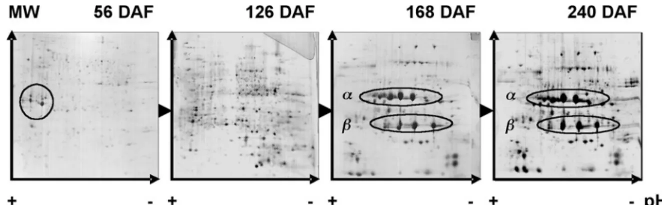
Evolution of proteins during coffee fruit development: Al-though limited, some studies have been made regarding protein evolution during the development of perisperm and endosperm tissues (Marraccini et al., 2001a; Estanislau, 2002). By 2-D gel electrophoresis, it was shown that perisp-erm tissue contains several abundant proteins that differ from those accumulated in the endosperm (figure 3). For example, N-terminal and internal sequencing of acidic proteins of homologous MW (≈30 kDa) accumulating in the perisperm revealed that they correspond to class-III chitinases (Rog-ers and Marraccini, unpublished observations) homologous (62 % of identity, 78 % of homology) to the partial protein (123 amino acid residues) coded by the AR-52 partial cDNA (Rojas-Herrera and Loyola-Vargas, 2002). These proteins are known as PR- (Pathogenesis-Related) proteins that are usu-ally expressed after plant tissue infection (Graham and Stick-len, 1994), but could also play the function of storage pro-teins, as suggested for the P31 class-III chitinase from Musa acuminata (Clendennen et al., 1998; Peumans et al., 2002). In C. arabica, the expression of AR-52 appear to be up-regu-lated in tissues with significant cellular division activity, such as during the induction of somatic embryogenesis from foliar explants (Rojas-Herrera and Loyola-Vargas, 2002), suggest-ing its participation in plant growth and developmental proc-esses, as also proposed for carrot embryos (De Jong et al.,
Figure 3. Evolution of proteins during coffee bean development. Soluble proteins of C. arabica var. Caturra T2308 were extracted from beans (perisperm plus endosperm) at different developmental stages, separated by 2-D gel electrophoresis (pI 3-10 linear) and analyzed after silver-stained (Marraccini et al., 2001a). Chitinases of class III (30 kDa, pI [4-5]) are identified (circle) in the perisperm at 56 DAF. Subunits (α: 32 kDa and β: 20 kDa) of 11S proteins detected in the endosperm during the storage phase (168 DAF) are maintained up to the mature stage (240 DAF) Data from Marraccini et al., (2001a).
Figure 4. Fresh weight evolution of separated tissues (pericarp, endosperm and perisperm) during fruit development of C. arabica var. Iapar 59. A) Curve of fruit fresh weight (expressed in g,®). B) Evolution of pericarp (n), perisperm (°) and endosperm (r) expressed in percentage of total fruit fresh weight (FW) (data from Geromel et al., submitted).
1992) and cucumber (Lawton et al., 1994). More recently, the cleaving activity of chitinases on arabinogalactan proteins (AGPs), leading to the release of Nod factor-like molecules, was also reported to promote plant cell division (Schultze and Kondorosi, 1996; Gough, 2003).
Interestingly, the large accumulation of chitinases in young perisperm also coincides with the detection of high amounts of β-tubulin which, as a component of microtubules, is considered as marker protein indicative of cell division and expansion events (De Castro, 1998; Estanislau, 2002). Indeed, the immunocytochemical detection of mitotic and cortical microtubules confirmed high cellular division and elongation activity in the developing perisperm of C. arabica L. var. Acaiá Cerrado (figure 6).
Fruit weight (g)
% of fruit fresh weight
When β-tubulins were detected by Western-blot experi-ments in the endosperm, their amount decreased gradually from 120 to 210 DAF, becoming undetectable by the latest stages of endosperm development (between 225-255 DAF). In fact, the presence of a mitotic microtubular cytoskeleton up to 120 DAF indicates that the histo-differentiation of the endosperm was accomplished before 120 DAF (Estanislau, 2002). After 150 DAF, only a cortical cytoskeleton was ob-served indicating the predominance of cellular expansion as characteristic of the final steps of plant seed maturation (De Castro, 1998). As also observed for other plant seeds, the degradation of the cytoskeleton and tubulin subunit proteins follow the dehydration process observed in the last stages of endosperm development (Estanislau, 2002).
Caffeine metabolism: Caffeine metabolism will be treated in detail in a special chapter in this issue (see Ashihara in this issue). However, it appears important to us that a rapid overview be made about key points concerning caffeine metabolism.
Figure 5. Histological studies of coffee (C. arabica) tissues at different developmental stages. Endosperm (en), cuticle cell layer (cc), embryo (em) and perisperm (pe) divided in outer (ope) and inner (ipe) layers. Materials were sampled at 90 DAF (A), 130 DAF (B), 150 DAF (C) and mature stage at 230 DAF (D). A, B and D: materials were fixed, embedded in butylmethylmetacrylate, sectioned, fixed on slides according to Baskin et al. (1992) and visualized using Differential Interference Contrast (DIC) microscopy; C: the material was treated and stained with toluidine blue (0.05 %) according to Gerrits and Smid (1983). The torpedo embryo in A was underlined to facilitate identification. Scale in left corner of each document represents 100 μm.
Figure 6. Immunofluorescence (FITC) micrographs showing the microtubular cytoskeleton in coffee (C. arabica var. Acaiá Cerrado MG-1474) seeds. A: Fruits at 90 DAF, showing the intact and growing perisperm, composed of the inner perisperm (ipe) with larger cells and the outer perisperm (ope) with smaller cells. B: Fruits at 90 DAF, showing the initial development of the endosperm (en) which encloses a still undifferentiated globular embryo (em). C: Fruits between 150 and 180 DAF, showing
β-tubulin fluorescence granules as a result of the initial degradation of the cortical microtubular cytoskeleton in the embryo. D: Fruits at 165 DAF showing the endosperm still containing some few granules of β-tubulin, the compact cuticle (cc) layer of cell wall remnants resulting from the consumption of the inner perisperm. E: Radicle of an embryo from mature cherry fruits at 225 DAF, showing the complete absence of microtubular cytoskeleton and β -tubulin granules. Arrows indicate dividing cells with mitotic microtubular cytoskeleton configurations. Bars indicate 10
μm. Data from Estanislau (2002).
In coffee plants, caffeine is actively biosynthesized dur-ing leaflet emergence and then decreases when leaves reach their optimal photosynthetic capacity (Frischknecht et al., 1986; Ashihara et al., 1996; Zheng and Ashihara, 2004). In addition to leaves, the incorporation of 14CO
corresponding gene. Caffeine synthesis should also occur in young (“liquid”) endosperm of C. arabica since it serves as an ideal source for the isolation of N-methyltransferases (Mazzafera et al., 1994; Gillies et al., 1995).
Low levels of caffeine were detected in xylem sap of coffee (C. arabica cv. Catuaí “Vermelho”) plants, suggesting that it is not highly transported within the plant and that a major part of caffeine synthesis occurs directly in the tissues where it is detected (Mazzafera and Gonçalves, 1999). In seeds, absolute amounts of caffeine parallel the dry weight curve, with more or less constant caffeine content during maturation, nearing 1 % on a dry matter basis (DMB) at the time of harvest. It is interesting to note that the major increase in caffeine content in seeds occurred between the developmental stage 6 (8 to 12 WAF) and 7 (16 WAF) in the Arabica model system analyzed (Keller et al., 1972), corre-sponding to the time of rapid endosperm expansion. From the developmental stage 7, and up to the harvest stage (stage 11) the caffeine content in the pericarp was constant, suggesting that its biosynthesis stopped or that it continues but with an active transfer to the bean (Baumann and Wanner, 1972).
Although no data are available for enzymatic activities of caffeine synthesis for individual coffee tissues, the above results suggest the existence of tissue-specific expression of genes coding for N-methyltransferases (see section “Gene expression during coffee fruit development – genes of caffeine metabolism”). In addition, concerted synthesis of caffeine and chlorogenic acids (mainly 5-CQA) was also reported during coffee leaf development (Baumann et al., 1993; Aerts and Baumann, 1994), and could play an important physiological role by limiting the autotoxicity of caffeine, by reducing its diffusion through the membranes (Mösli-Waldhauser and Baumann, 1996; see Baumann in this issue).
Chlorogenic acids: Chlorogenic acids (CGA) constitute a family of esters formed between transcinnamic acids (caffeic and ferulic) and quinic acid (Clifford, 1985b). These phenolic compounds represent an average of 4.1 % and 11.3 % DMB, respectively for Robusta and Arabica (Ky et al., 2001a), and could explain the flavor differences between these species (Clifford, 1985b; Guyot et al., 1996). Recently, the antioxidant capacity of these chlorogenic acids in animal systems was demonstrated (Daglia et al., 2004), which increased the interest to study CGA pathways in plants (Hoffmann et al., 2003; Niggeweg et al., 2004), including coffee (Campa et al., 2005; Melo and Mazzafera, 2005).
By analyzing clones of C. canephora from different geo-graphical regions, Ky et al. (2001a) showed that the origin was a source of intra-specific variations, mainly for percent-age of 5-FQA and 4,5-diCQA. In addition, the increase of the CQA/di-CQA ratio was also reported to be as a good marker for coffee bean maturity (Dentan, 1985b). The reasons for these differences are still unknown but could be controlled in part by the hydroxycinnamoyl-CoA: shikimate / quinate hydroxycinnamoyl-transferase (HCT enzyme), which es-terifies quinic acids with cinnamic acids (see section "Gene expression during coffee fruit development-genes of CGA pathway").
The analysis of chlorogenic acids during the development of both leaves and fruits of C. pseudozanguebariae, showed that CGA contents were equal in leaves and fruits when both tissues were at young developmental stages, i.e. leaf length less than 40 mm and fruits with perisperm predominating (Bertrand et al., 2003). However, when the endosperm begins to develop, a substantial increase of CGA content was observed, mainly characterized by the accumulation of CQAs (3-CQA: 3-caffeoylquinic acid; CQA: 4-caffeoylquinic acid and 5-CQA: 5-4-caffeoylquinic acid) in coffee beans. High quinic acid content was also detected in the initial developmental stages of grain (perisperm and endosperm), between 6 and 16 % of dry weight for Arabica and Robusta coffees (Rogers et al., 1999b; Marraccini et al., 2001a). Towards the end of seed development, these levels were shown to decrease drastically in both species, again arguing for a stimulation of CGA metabolism in the endosperm, mainly oriented towards CQA accumulation (Bertrand et al., 2003).
Trigonelline synthesis: Trigonelline is synthesized through the methylation of nicotinic acid (NAD) by S-adenosyl-L-methionine (SAM)-dependent nicotinate N-methyltransferase (EC2.1.1.7). Its function is still a matter of debate but it might be a reserve compound for NAD that could be used during germination (Shimizu and Mazzafera, 2000). During roasting, trigonelline alkaloid gives rise to many aroma (flavor) compounds, like alkyl-pyridines and pyrroles (De Maria et al., 1994), which explains the interest of breeding programs to increase of trigonelline content in Robusta green beans (Ky et al., 2001b).
Ashihara, 2004, see Ashihara in this issue). Using [car-bonyl-14C]nicotinamide as a precursor, Zheng et al. (2004) showed that trigonelline was synthesized in the pericarp of unripe fruits but declined as fruits ripen. Although no tissue separation was made in this study, most of the trigonelline synthesis activity was detected in the “endosperm” stage of coffee seeds. In fully ripe seeds, high trigonelline accumula-tion was observed even if no synthesis activity was detected. This suggested that net biosynthesis of trigonelline takes place in the pericarp and that trigonelline may be further transported from this tissue to the endosperm. Interestingly, genetic variability was observed in coffee for trigonelline content, with Arabica ranging from 0.88 to 1.77 % of DMB and Robusta with 0.75 to 1.24 % of DMB (Ky et al., 2001a). Different trigonelline contents were also reported for C. pseudozanguebariae (PSE) and C. liberica var. Dewevrei (DEW) respectively with 1.02 % and 0.57 % of DMB (Ky et al., 2001b). Using a PSE x DEW cross between these two species, the same authors identified one nuclear quantitative trait loci (QTL) for trigonelline content and located it on the G linkage map (Ky et al., 2001b). The identification of EST (Expressed Sequenced Tag) sequences coding for nicotinam-idase (EC 3.5.1.19) and nicotinate N-methyltransferase (EC 2.1.1.7) should help to reveal whether the variations of trigo-nelline contents observed in these species can be explained by differential expression of these genes.
Diterpene metabolism: Despite of their important role in flavor stability and release, coffee lipids have been very little studied (see Speer and Kölling-Speer in this issue). They range from 15 % of dry weight in coffee beans of Arabica to 10 % in Robusta commercial species, but are subject to a larger variation depending on origin and varieties, for example with 8-9 % of dry weight in C. liberica to 30 % in C. salvatrix (Picard et al., 1984; Speer et al., 1993; Mazzafera et al., 1998). As cited above, it was proposed that the better cup quality of coffee grown at higher altitudes was linked to higher “fat matter” content of beans (Decazy et al., 2003; Vaast et al., 2006), although it is not known how the lipid composition was affected.
Within lipids, diterpenes (cafestol: C20H28O3 and kahwe-ol: C20H26O3) represent 10 % of coffee oil and are specific of the coffee genus (Speer and Kölling-Speer, 2001). Although no biological functions have been assigned, both cafestol and kahweol are of particular interest, especially in human health where they are considered to stimulate cholesterol synthesis (Urgert et al., 1997; Post et al., 2000; De Roos et al., 2001),
and also reported to give protection against some types of cancer (Lam et al., 1982, Cavin et al., 1998). A great diversity was observed for diterpene content both at the interspecific (De Roos et al., 1997; Mazzafera et al., 1998) and intraspe-cific levels (Speer and Kölling-Speer, 2001), suggesting the existence of genetic polymorphism of the enzymes control-ling the cafestol / kahweol biosynthesis pathways.
Preliminary observations suggested that key steps of the diterpene biosynthesis are restricted to specific tissues in coffee (Kölling-Speer and Speer, 1997). For example, cafestol is the main diterpene detected in leaves of both Arabica and Robusta, whereas kahweol is undetectable in this latter species. Kahweol is also undetected in the pericarp of Arabica fruits, but accumulates to high level in endosperm. Green beans of C. canephora do not contain kahweol but accumulate another diterpene, 16 O-methyl cafestol that is not encountered in Arabica. Based on these results, one might predict the existence of a “kahweol-synthase” enzyme specifically expressed in C. arabica seeds, and of a “cafestol-methylase”, responsible of the 16-OMC biosynthesis, specifically in C. canephora seeds. The identification of genes coding for these enzymes and for those of the isoprenoid biosynthesis pathway (Liu et al., 2005) should be facilitated by the screening of UniGene membranes of the Brazilian Coffee Genome Project (see Vieira et al., in this issue).
Coffee storage proteins and free amino acids: At the cup quality level, amino acids are also important because they react with sugar moieties through “Maillard” reactions (Maillard, 1912) to produce aromatic compounds. Among these, glutamic acid and asparagine are the most abundant, representing near 40 % of free amino acids in coffee beans (Poisson, 1977; Arnold et al., 1994; Arnold and Ludwig, 1996). Both are also the major forms of nitrogen transport in xylem sap of coffee (Mazzafera and Gonçalves, 1999). Some differences exist in the composition of free amino acids in green coffee beans of different species, for example with glutamic acid content being higher in Arabica than in Robusta (Arnold et al., 1994).
electro-phoresis) analysis, Baú et al. (2001) identified the presence of 11S-type storage proteins in various coffee species, except in C. racemosa where storage proteins of lower molecular weight were observed. Independent of the coffee species studied (Arabica or Robusta), 11S storage proteins were not detected in perisperm tissue (figure 3), but specifically at the time of rapid endosperm expansion (Rogers et al. 1999a). Although to a lesser extent, accumulation of 11S storage pro-teins was also reported in both zygotic and somatic embryos (Yuffá et al., 1994).
Comparison of amino acid sequences of 11S protein deduced from cloned full-length cDNAs reveals the existence of some differences that could not explain the variations of pI and of molecular weight usually observed by SDS-PAGE (Acuña et al., 1999; Baú et al., 2001) or 2D-gel electrophoresis (Rogers et al., 1999a). At the amino acid level, 11S storage proteins are relatively rich in glutamine, glycine, leucine and acid glutamic amino acid residues, respectively with 11 %, 10 %, 9 % and 7.5 % of total molecular weight. Their content is relatively low in the sulfur amino acids cysteine (1 %) and methionine (0.6 %), suggesting a limited role of these amino acids as precursors of aromas and flavors in coffee (Grosch, 2001).
When the amino acid composition of 11S proteins was compared with the composition of free amino acids in coffee beans, no obvious correlations were observed (Poisson, 1977; Arnold et al., 1994; Arnold and Ludwig, 1996). This argues for a little influence of 11S storage proteins in the control of the pool of free amino acids. However, differences of 11S-protein profiles (SDS-PAGE and 2D-gel electrophoresis) were observed between Arabica and Robusta, as for Ugandan and Congolese varieties (Rogers et al., 1999a), and also in other species, such as C. liberica and dewevrei (Baú et al., 2001), suggesting the existence of several types of 11S proteins in the coffee genus.
Finally, post-harvest treatments were also reported to influence amino acid contents in coffee beans, for example, the higher levels of L-alanine found in beans of C. arabica processed by the dry method (Ludwig and Arnold, 1996; Casal et al., 2001). The origin of these variations is still unknown but could be due to the action of specific-proteases as described below. Because storage proteins seem to be encoded by a limited number of genes (Acuña et al., 1999; Lin et al., 2005), the analysis of 11S-cDNA sequences from different coffee species and cultivars should be interesting to provide an in depth view of their amino acid composition.
Proteases: In cocoa, specific aroma precursors are derived preferentially from the vicilin-class (7S) globulin storage pro-tein, via its successive degradation by an aspartic protease and a carboxypeptidase (Voigt et al., 1994). Recently, beans from C. arabica cv. Obatã grown in the state of São Paulo (Brazil), in a location with relatively higher annual mean temperature (23.1°C, Adamantina), were shown to contain higher protease activities, total protein and free amino acids than beans from the same cultivar grown in other locations of lower annual mean temperatures (21.8°C, Mococa; 20.7°C, Campinas) (Sil-va, 2004; Silva et al., 2005). Sensorial evaluations showed that beans from Adamantina had lower beverage quality than beans from the other two locations, suggesting a negative correlation between protease activities and cup quality.
It has been considered that there is no clear proof that coffee storage proteins can act as flavor or aroma precursors through their degradation by specific proteases (Montavon et al., 2003). Nevertheless, 2D-gel electrophoresis and N-terminal sequencing studies clearly demonstrate that abundant peptides of 12 to 16 kDa found in protein extracts of mature coffee beans correspond to degradation products of the α subunit of 11S storage proteins (Rogers et al., 1999a). Ludwig et al. (2000) also identified low molecular weight peptides (ranging from 4 to 12.5 kDa) that accumulated differentially in beans of C. arabica and C. canephora. The origin of these polypeptides is unknown, but part of them could also correspond to degradation products of 11S storage proteins by particular peptidases. Such enzymes could also be responsible for higher amino acid contents usually detected in washed coffee beans (Casal et al., 2001; Selmar et al., 2001), which are known to have a better cup quality than beans processed by the dry method. Proteases activities were reported in mature beans of Arabica and Robusta coffee species (Ludwig et al., 2000).
Altogether these observations have increased the interest in the search for protease-encoding cDNA sequences (Ben Amor et al., 2003; Baptista et al., 2005). Hence, more than 1,800 “protease-encoding” ESTs were identified from the Brazilian Coffee Genome Project and arranged in 18, 13, 13 and 10 clusters for cysteine-, aspartic-, metal- and serine-proteases respectively (Baptista et al., 2005). The availability of protease-encoding sequences should now permit the analysis of the influence of environmental conditions and post-harvest treatments on their gene expression.
qual-ity because it is rapidly degraded during roasting, to give anhydro-sugars and other compounds like glyoxal (Clifford, 1985a; De Maria et al., 1994). These molecules are then able to react with amino acids to produce aliphatic acids, hy-droxymethylfurfural and other furans, pyrazine and carbonyl compounds, which are essential contributors to coffee flavor, either as volatile aroma compounds, or as non-volatile taste compounds (Grosch, 2001; Homma, 2001).
Differences of sucrose content in commercial species of coffee are well known, with means of 9.4 % and 6 % DMB of sucrose, respectively for beans of C. arabica and C. robusta (Ky et al., 2001a; Campa et al. 2004). The first attempt to analyze sucrose metabolism in isolated tissues of coffee was made by Rogers et al. (1999b), who showed that reducing sugar (i.e. glucose and fructose) content decreased in the perisperm concomitantly with its disappearance, whereas sucrose content increased gradually within the developing endosperm. In addition, levels of reducing sugars in the perisperm were higher in C. arabica than in C. canephora which could explain in part the differences in sucrose content observed in mature endosperm of these species.
Recently, Geromel et al. (2005) refined this previous work by analyzing sugar contents and enzymatic activities in separated tissues (pericarp, perisperm and endosperm) of developing fruits of C. arabica var. Iapar 59. High hexose to sucrose ratios were observed during early stages of perisperm (90 DAF) and endosperm (120 DAF) development (figure 7), which is a characteristic in tissues with intense cell division activity (Weber et al., 1998). In the perisperm, the inversion of this ratio between 120 to 176 DAF accompanied the gradual disappearance of this tissue, and coincided with the beginning of endosperm expansion. In the latter, the transition from high to low hexose to sucrose ratio observed between 147 and 176 DAF marks the shift between the expansion and storage phase, also characterized by the accumulation of storage proteins and sucrose in the endosperm (Rogers et al., 1999a; Geromel et al., 2005). In the pericarp, reducing sugars and sucrose accumulated slowly to similar levels up to 176 DAF. During the two latter months of fruit maturation (176 to 234 DAF), a large and rapid accumulation of reducing sugars and sucrose was observed, respectively with concentrations near 280 and 180 mg.g-1 DMB at the time of harvest, thus confirming other observations showing a gradual increase of soluble solids during the maturation of the pericarp (Marín-López et al., 2003). Initial data concerning the identification of genes coding for sugar-metabolizing enzymes implicated in this process will be presented below (see section "Gene expression during coffee fruit development-genes of sucrose metabolism").
Polyphenol oxidase (EC 1.10.3.2): The oxidation of polyphenols (mainly CGA) occurs either by an autoxidation process (Cilliers and Singleton, 1991), or enzymatically by the action of polyphenol oxidases (Amorim and Silva, 1968; Mazzafera and Robinson, 2000). This leads to semiquinones and quinones that could play a defensive role by reducing the nutritive value of plant tissues wounded by herbivores (Mazzafera and Robinson, 2000). On the other hand, quinones may also react with amino acids and proteins (Mason, 1955) and thereby play a direct role in coffee cup quality. In this regard, several studies have shown that coffee of low beverage quality correlates with low polyphenol oxidase (PPO) activities in green beans (Amorim and Silva 1968; Amorim and Amorim, 1977), and likewise for coffee maintained under bad storage conditions (Begnami et al., 1999). In this case, low beverage quality was proposed to result from ultra-structural (membrane) damage leading to PPO inhibition by quinines arising from the oxidation of Figure 7. Evolution of sucrose concentration (sucose igual) (A)
and SUS enzymatic activities (µg of sucrose reduced µg-1 proteins.h-1) (B) in separated tissues of fruits from C. arabica var. Iapar 59 (from Geromel et al., submitted).
SUSY
activity
(µg reduced sugars. µg
-1 proteins.h -1)
Sucrose
(mg.g
-1 DW)
chlorogenic acids, but not from PPO activity per se (Clifford, 1985a, 1985b).
Whatever their mode of action, PPOs are important enzymes in coffee and have been characterized in leaves (PPO-L) and in developing endosperm (PPO-E) where high enzymatic activities were found (Mazzafera and Robinson, 2000). Enzymatic activities of PPO-L were very high in young leaves and declined when length of leaves increased. An inverse situation was observed during coffee bean development, where low PPO-E activities were observed in endosperm at around 100 DAF. As the endosperm develops, PPO-E activities increased to reach a peak at around 170 DAF, and declined slightly in mature beans. Interestingly, PPO activities followed the synthesis of their substrate (CGA) both in leaves and endosperm (Melo and Mazzafera, 2005). Despite the fact that PPO-L and PPO-E differed in molecular weight (respectively 46 kDa and 50 kDa) and in sensitivity to temperature, Mazzafera and Robinson (2000) suggested that they could correspond to closely related proteins or originate from the same precursor subjected to different post-translational modifications. The search for PPO-encoding ESTs should be very helpful to elucidate their origin as well as to analyze their expression in different storage or environmental conditions.
Gene expression during coffee fruit development
As previously mentioned, some coffee biochemical pathways (i.e. caffeine) have been studied a very long time whereas others have been poorly investigated. Although lim-ited, the objective of this section is to regroup this informa-tion, particularly in the light of recently published articles.
Genes of caffeine metabolism: Three steps of methylation are necessary to produce caffeine from xanthine, implicat-ing successively the action of 7-methylxanthosine synthase (xanthosine > 7-methylxanthosine), theobromine synthase (7-methylxanthine > theobromine or 3,7-dimethylxanthine) and caffeine synthase (theobromine > caffeine or 1,3,7-tri-methylxanthine), using the S-adenosyl-L-methionine (SAM) as a donor of methyl groups. Several studies have been pub-lished dealing with gene cloning, expression analysis and protein studies of enzymes from the caffeine biosynthetic pathway (Moisyadi et al., 1998, 1999; Ogawa et al., 2001; Mizuno et al., 2003a,b; Uefuji et al., 2003).
In some cases, expression analyses were made without separating perisperm, endosperm and pericarp tissues or giving information about dates, time and mode of harvest,
which renders the interpretation of the results rather difficult in the sense of tissue-specific gene expression. In a recent article, Lin et al. (2005) reported the presence of a unigene (#122206) from Robusta, coding for a putative S-adenosyl-L-methionine synthetase and formed by the compilation of 130 ESTs exclusively expressed in earlier stages (18-22 WAF) of cherry development. On the other hand, expression analyses of N-methyltransferase cDNA sequences mainly performed by RT-PCR, as well as by classical Northern-blot experiments, showed that the 7-methylxanthosine synthase encoding gene (designated CaXMT1 by Uefuji et al., 2003 or CmXRS1 by Mizuno et al., 2003b) was expressed to equal levels both in developing endosperm and in young leaves. Genes coding for the theobromine synthase (designated CaMXMT1 of CaMXMT2 by Uefuji et al., 2003 or CTS1 and CTS2 by Mizuno et al., 2003a) and caffeine synthase (designated CaDXMT1 by Uefuji et al., 2003 or CCS1 by Mizuno et al., 2003a) were also expressed in immature coffee fruits (as in stage 6-7 corresponding to endosperm expansion; Keller et al., 1972). In addition, the CTS2 gene expression appeared high not only in whole small green fruits (diameter 10mm) but also in the pericarp and immature endosperm of larger fruits (diameter 15mm) (Mizuno et al., 2003a). Interestingly, RT-PCR experiments did not reveal expression of CaXMT1, CaMXMT1, CaMXMT2 and CaDXMT1 genes in mature (stage 10; Keller et al., 1972) fruits (Uefuji et al., 2003), which could explain the absence of caffeine accumulation during the final ripening process (stages 9 to 11; Keller et al., 1972). Expression of CaXMT1, CtCS1, CaMXMT1, CaMXMT2 genes was also detected in tissues rich in dividing cells, as in young leaves and flower buds, where N-methyltransferases were previously found to be abundant.
The availability of these cDNA sequences now opens the way to study their nucleic polymorphism and should facilitate the understanding of the genetic variability existing for caffeine content in the coffee genus (Ky et al., 2001a). Recently, transgenic coffee plants with reduced expression of the CaMXMT1 gene were obtained (Ogita et al., 2004). Their analysis showed that they also have reduced expression of CaXMT1 and CaDXMT1 genes in leaves concomitant to a reduction of theobromine and caffeine contents. Work is now under way to verify whether caffeine contents are also reduced in beans produced by these transgenic plants.
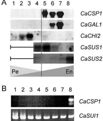
profiles showed that 11S proteins began to accumulate at 18 WAF, reaching a maximum at 27 WAF (figure 3). In parallel, the expression of the 11S-encoding gene began at 18 WAF reaching a peak between 22 and 27 WAF. Subsequently, the expression of the csp1 gene (hereafter renamed CaCSP1) ceased, becoming undetectable in the mature endosperm (35 WAF in that study). At the same time, a large amount of 11S storage proteins was shown to persist, despite slight protein degradation being noted in mature grains. The CaCSP1 gene expression overlapped the accumulation of 11S storage proteins and showed tight coupling between transcriptional and translational processes. Although perisperm and endosperm tissues were not separated in this study, the expression profiles obtained strongly suggest endosperm-specific expression of storage proteins (figure 8A). This tissue-specificity of CaCSP1 gene expression was also confirmed by RT-PCR experiments (figure 8B), and by the large accumulation of CaCSP1 homologous ESTs found only in fruit cDNA libraries from Arabica (table 1) and Robusta (Lin et al., 2005). This endosperm-specific expression of the CaCSP1 gene is under the transcriptional control of a promoter region able to direct the expression of the GUS reporter gene only in endosperm of transgenic tobacco seeds (Marraccini et al., 1999).
Lin et al. (2005) also identified a unigene (# 125230) coding for putative 2S storage proteins formed by the clusterization of 1219 ESTs mainly expressed during the middle stages (≈ 30 WAF) of Robusta seed development. However, a lower level of 2S-encoding ESTs was detected in the last stage of seed development (42-46 WAF), at the time when ESTs for 11S storage proteins were strongly detected. Up to now, 2S proteins have not been identified by 2D-gel electrophoresis in protein extracts of mature seeds of Arabica and Robusta (J.W. Rogers, personal communication), suggesting that they could be transiently synthesized during seed development in these two species. However, they could correspond to the low molecular weight (<10 kDa) proteins strongly detected in mature seeds of C. racemosa (Baú et al., 2001, P. Mazzafera, personal communication).
Analysis of α-D-galactosidase expression: α-D-galactosidase (α-Gal; EC 3.2.1.22) is a very abundant protein in mature coffee beans (see Sigma-Aldrich catalog ref G8507) and was one of the first plant α-D-galactosidases to be characterized (Courtois and Petek, 1966; Carchon and De Bruyne, 1975; Haibach et al., 1991). It acts on galactomannans by removing galactose residues at the O-6 position of the (1→4)-β-
man-Figure 8. Example of genes expressed during bean development in C. arabica. A: Total RNAs were extracted from whole seeds (perisperm and endosperm) and probed by 32 P-labelled cDNA of 11S storage proteins (CaCSP1, Y16976: Rogers et al., 1999a), α-galactosidase (CaGAL1, AJ877911: Marraccini et al., 2005) and class III chitinase (CaCHI2, AM159173). For CaSUS1 (AM087674) and CaSUS2 (AM087675) expression analysis, total RNAs were extracted from separated perisperm and endosperm tissues (Geromel et al., submitted). Fruits were harvested at 28 DAF (lane 1); 49 DAF (lane 2); 63 DAF (lane 3); 98 DAF (lane 4); 126 DAF (lane 5); 154 DAF (lane 6); 189 DAF (lane 7) and 245 DAF WAF (lane 8). The broken vertical line indicates the virtual separation between “perisperm-phase (Pe)” (0-120 DAF) and “endosperm phase (En)” (≈ 120 DAF up to the harvest). B: Analysis of CaCSP1 gene expression in different coffee tissues by RT-PCR. For CaSUI1, experimental conditions were previously described (Gaborit et al., 2003). RT-PCR conditions used for amplification of CaCSP1 were identical to those for used CaSUI1, except that SO11 and SO2-1 primers were used (Marraccini et al., 1999). Lanes: 1, young leaves (< 1 cm); 2, intermediate leaves (length 2-5 cm); 3, old leaves (> 5 cm); 4, flowers buds; 5, open flowers; 6, stems; 7, roots 8, beans at 189 DAF.
nan chain to give insoluble mannans. For this reason, it was proposed to be one of the main enzymes involved in the dep-osition of mannans in cell walls (Redgwell et al., 2003). This enzyme also attracted attention in the field of biotechnology due to its capacity to remove the terminal galactose units (α
carbohy-CYT
OLOGY
, BIOCHEMISTR
Y
AND MOLECULAR CHANGES DURING COFFEE FRUIT
DEVELOPMENT
Braz. J. Plant Physiol., 18(1):175-199, 2006
Class of genes GB accession number (protein mnemonics)
Number of
clusters Total ESTs
cDNA libraries
AR BP* CA CB CL CS EA FB* FR* IC LV* LP PA RM RT* RX SH* SI* SS O
Sugar metabolism
CaVAC1 CaINV1 CaRBCS1 CaMANA, CaMANB CaGAL1
CaSUS1 CaSUS2
CGA metabolism
CcPAL1, CcPAL2 CaCYP73 CaCYP98 Ca4CL1 CaHCT1
Storage proteins and proteases
CaCSP1
Aspartic protease Aspartyl protease Cystein protease
CaCHI1 (AR-52)
CaCHI2 CcCHI3
Caffeine metabolism
CmXRS11 CTS12, CTS22 CCS13
CAE01318 CAE01317 CAD11991
CAC08208, CAC08442 CAI47559
AM087674 AM087675
AAN32866, AAN32867 CAJ41419
CAJ41417 CAJ41420 CAJ40778
CAA76573 CAI45845 CAI45844 CAI45842 AAD26856 CAJ43737
DQ124036(4)
BAC43755
BAC43756, BAC43757 BAC43760
1 1 4 4 3 3 2
1 2 1 1 1
2 4 2 2 3 3 7
1 3
10 14 539 33 11 212 18
34 63 16 48 4
141 122 9 10 571 75 1122
19 78
2 1 1 1 3 2
1 3 2 5 2 1
84 24 2 1 1 1 32 24 20 227 15 20 72 3 5 8 1 3 2 4 6 1 12 1 2 1
2 4 1 3 1
1 3 58 20 19 7 14 3 16 24 1 6 4 12 18 4 2
1 2 3 1 1 1 1 4 1 2 1
1 2 5 5 11 8 1 1
7 1 1 1 16 6 1 24 1 2 1 2
3 1 1 1 5 3 1 1
8 1 7 12 2 12 1 2 1 2 2 1 1 1 134 6
3 8 16 6 17 1 15 8 1 18 1 1 4 8 4 5 6
3 1 2 2 1
2 8
1 5 2 6 1 24 46 8 193 60 80 2 140 3 1 1 2 14 32 1 1 6 15 1 1
38 1 17 28 57 48 199 182 6 108 92 9 4 7 11 21 1 3 290 1 3 1 5 4 3 1 1
1 6 2 3 6 3 16 17 14 1 1 1 3 4
in vitro with BION™ and brassinosteroides; CL: hypocotyl of coffee plantlets (± BION™); CS: coffee cells cultivated in vitro with mannose, NaCl and KCl;
drate moiety of glycoprotein complexes, thus generating type O red blood cells (Yatsiv and Flowers, 1971).
Coffee tissues (perisperm and endosperm) were separated during the maturation of fruits of C. arabica var. Caturra to analyze α-Gal enzymatic activity and gene expression (Marraccini et al., 2005). α-Gal activity was not detected in perisperm tissue but increased gradually with endosperm development, to reach a peak at 30 WAF that coincided with the maximal of α-Gal activity in whole coffee berries at the semi-ripe (yellow exocarp) developmental stage (Golden et al., 1993). Northern-blot experiments detected α-Gal transcripts at 22 and 27 WAF, overlapping α-Gal enzymatic activity detected at the same time in the endosperm. In mature beans, α-Gal gene expression was undetectable, despite the high activity persisting at this stage.
As for endo-β-mannanase (Marraccini et al., 2001b), high
α-Gal activity was also detected during in vitro germination of coffee beans of C. arabica var. Caturra, with a continuous increase from 0 to 40 days after imbibition (Marraccini et al., 2005). Because expression analysis was not performed, it is not yet known whether this high α-Gal activity detected during germination arose from the expression of the CaGAL1 gene or of other α-D-galactosidase-encoding genes.
Genes of the CGA pathway: The biosynthetic pathway of CGA in coffee should involve at least five different enzymes: PAL (phenylalanine ammonia-lyase), C4H (cinnamate-4-hydroxylase), 4CL (4-[hydroxy]cinnamoyl-CoA ligase), C3H (p-coumaroyl-shikimate/quinate 3’-hydroxylase) and HCT (hydroxycinnamoyl CoA: quinate hydroxycinnamoyl transferases) (Campa et al., 2003; Hoffmann et al., 2003).
In coffee, PAL enzymatic activity was light-stimulated in cell suspensions of C. arabica (Baumann et al., 1993) and also strongly detected in young leaflets but decreased during further leaf expansion (Aerts and Baumann, 1994). To the authors’ knowledge, no work has been published concerning measurements of enzymatic activities of the CGA pathway, either in whole or separated tissues of coffee fruits.
However, the availability of EST coffee sequences (see Vieira et al., in this issue) permits the search of genes coding for these enzymes. Consequently, preliminary results of EST clusterization led us to conclude that there are at least three clusters coding for phenylalanine ammonia-lyase in coffee (named CaPAL1-3) as well as for the 4CL enzyme (named Ca4CL1-3). A unique cluster should code for the C3H enzyme (CaCYP98), whereas at least two clusters were found for the C4H enzyme (CaCYP73) (table 1). The highest
degree of complexity was encountered for the HCT enzyme that seems to be coded by at least 8 different clusters, named from CaHCT1 to 8 (G. Melo and E.K. Takahashi, personal communication).
Specific primer sets were designed from clusters formed by ESTs preferentially encountered in fruit cDNA libraries, and used in RT-PCR experiments to analyze gene expression during endosperm and leaf development in C. arabica (Melo, 2005; Melo and Mazzafera, 2005). For each cluster tested (PAL, C4H, C3H, 4CL and HCT), expression declined as the endosperm and leaf aged, concomitantly with the decrease in 5CQA content. The same kind of approach was followed to study gene expression in whole fruits of C. canephora under development (Campa et al., 2005). In that case, two cDNAs were identified for PAL and C3H enzymes (respectively called PAL-1 and 2 and C3’H-C1 and 2, according to the authors’ nomenclature), and used to design primer sets for RT-PCR experiments. This showed that PAL-1 and C3’H-C1 genes were expressed independently of fruit maturation (from green to red exocarp), whereas the PAL-2 and C3’H-C2 genes were only expressed in the “red pulp” stage of fruit development. It is difficult to compare these data with those obtained in C. arabica due to the absence of tissue separation and the limited number of development stages analyzed. Other analyses are underway to obtain a better overview of gene expression of CGA biosynthetic enzymes in C. canephora (A. De Kochko, personal communication).
Recently, a full-length cDNA sequence from C. arabica homologous to the HCT enzyme from Nicotiana tabacum (Hoffmann et al., 2003) was cloned and named CaHCT1 (G. Melo, L.P. Ferreira, L.G. Vieira, P. Marraccini, P. Mazzafera, unpublished observations). Northern-blot analysis revealed that this gene was expressed in endosperm of C. arabica, with a pattern identical to that observed for 11S- and α -D-galactosidase-encoding genes. To confirm the implication of CaHCT1 gene in CGA synthesis, it would be interesting to test its expression in fruits of C. pseudozanguebariae since CGA metabolism was shown to be oriented towards CQA accumulation in this coffee species (Bertrand et al., 2003).
Northern-blots showed that the CaSUS1 gene was expressed at early stages of endosperm development, at the time of rapid cellular expansion (figure 8). However, high expression of CaSUS2 was only observed during the latest stages of endosperm development, which coincides with maximal of SUS activity (Geromel et al., 2005). Because CaSUS2 expression also overlapped the peak of sucrose in these tissues (figures 7A and 7B), the CaSUS2 isoform of SUS protein was proposed to be the main enzyme responsible for sucrose accumulation in coffee seeds.
The CaSUS1 isoform of SUS could function in the sense of sucrose degradation since its expression was never accompanied by sucrose accumulation (Geromel, 2002; Geromel et al., 2005). In addition, preliminary studies showed that CaSUS1 gene expression was higher in perisperm and endosperm tissues of beans grown in shade conditions compared to fruits kept in “full sun” conditions (L.P. Ferreira and P. Marraccini, unpublished observations). Taken together with the maintenance of high hexose to sucrose ratio for a longer period in the shade plant, favoring cell division and elongation in young perisperm and endosperm tissues, this could explain the larger bean size observed for coffee plants cultivated under this condition (Muschler, 2001; Vaast et al, 2006). Additional experiments are underway to analyze sugar content, SUSY activity and gene expression in fruits of Coffea species of different bean size. In addition, CcSUS1 and CaSUS2 genes have been cloned, respectively from C. canephora (Leroy et al., 2005)and C. arabica (Geromel et al., submitted). Both are presently under analysis, particularly to study their nucleic polymorphism in various coffee species differing by their sucrose content in mature beans (Bouchet et al., 2005).
Genes expressed during pericarp maturation: In coffee, the lack of uniform ripening has serious effects on coffee quality when immature fruits are harvested together with mature fruit. It has been demonstrated that coffee fruit ripening could be synchronized by the exogenous application of ethephon during early (10-20 % of fruit colored) fruit development (Crisosto et al., 1990, 1992; Winston et al., 1992). This demonstrates that ethylene controls the final ripening stages of coffee fruit development and has motivated the search of genes coding for ethylene biosynthesis with the aim of reducing ethylene formation in the pericarp of genetically modified coffee plants (Neupane et al., 1999; Stiles, 2001).
cDNA sequences coding for ACC (1-aminocyclopropane-1-carboxylic acid) oxidase and ACC synthase have been
cloned (Neupane et al., 1999; Galvão et al., 2001; Pereira et al., 2005). In C. arabica, the ACC oxidase (CaACO) gene was expressed in the final stages of pericarp maturation, when the exocarp turned from green to red. Interestingly, highest levels of CaACO transcripts were detected simultaneously with the peak of ethylene emission by the fruits (Pereira et al., 2005). In comparison with other studies showing that maximum rates of respiration during fruit maturation coincide with ripening time (Puschmann, 1975; Marín-López et al., 2003), these results argue for the climacteric nature of coffee fruit ripening. Attempts to reduce the ACC oxidase expression in transgenic coffee plants employing an antisense strategy are now underway (Stiles, 2001; Ribas et al., 2001, see Pereira et al., in this issue).
In addition to CaACO expression studies, higt enzymatic activities and gene expression of pectin methylesterase (EC 3.1.1.11) and polygalacturonase (EC 3.2.1.15) have also been reported, respectively at the intermediate and final stages of pericarp maturation (Pimenta et al., 2000; Pimenta and Vilela, 2002; Cação et al., 2003; Budzinski et al., 2005). These carbohydrate-degrading enzymes should be responsible for the increase of soluble material during the maturation of the pericarp (Marín-López et al., 2003), and favor other enzymatic pathways, like sucrose synthesis detected in later pericarp development (figure 7B).
Electronic expression profile: Generally, the frequency of EST correlates with transcript accumulation in the tissues from which the cDNA library originated. In table 1, we presented the number of ESTs found in cDNA libraries of the Brazilian Coffee Genome Project for some cDNAs already cloned and discussed above.