2020
UNIVERSIDADE DE LISBOA
FACULDADE DE MEDICINA VETERINÁRIA
THE EFFECT OF PREDNISOLONE THERAPY ON CANINE SERUM LEVELS OF
1,2-O-DILAURYL-RAC-GLYCERO GLUTARIC ACID-(6′-METHYLRESORUFIN) ESTER
(DGGR) LIPASE
BEATRIZ COSTA GAGO MENDOZA
ORIENTADOR:
2020
UNIVERSIDADE DE LISBOA
FACULDADE DE MEDICINA VETERINÁRIA
THE EFFECT OF PREDNISOLONE THERAPY ON CANINE SERUM LEVELS OF
1,2-O-DILAURYL-RAC-GLYCERO GLUTARIC ACID-(6′-METHYLRESORUFIN) ESTER
(DGGR) LIPASE
BEATRIZ COSTA GAGO MENDOZA
DISSERTAÇÃO DE MESTRADO INTEGRADO EM MEDICINA VETERINÁRIA
JÚRI: ORIENTADOR:
PRESIDENTE:
Doutora Maria Teresa da Costa Mendes Vítor Villa de Brito
Doutor Rodolfo Assis Oliveira Leal
VOGAIS:
Doutora Berta Maria Fernandes Ferreira São Braz
ii
DECLARAÇÃO RELATIVA ÀS CONDIÇÕES DE REPRODUÇÃO DA TESE OU DISSERTAÇÃO
Nome: Beatriz Costa Gago Mendoza
Título da Tese ou Dissertação: THE EFFECT OF PREDNISOLONE THERAPY ON CANINE SERUM LEVELS
OF 1,2-O-DILAURYL-RAC-GLYCERO GLUTARIC ACID-(6′-METHYLRESORUFIN) ESTER (DGGR) LIPASE
Designação do curso de Mestrado ou de Doutoramento: Mestrado Integrado em Medicina Veterinária Área científica em que melhor se enquadra (assinale uma):
☒ Clínica ☐ Produção Animal e Segurança Alimentar ☐ Morfologia e Função ☐ Sanidade Animal
Declaro sob compromisso de honra que a tese ou dissertação agora entregue corresponde à que foi aprovada pelo júri constituído pela Faculdade de Medicina Veterinária da ULISBOA.
Declaro que concedo à Faculdade de Medicina Veterinária e aos seus agentes uma licença não-exclusiva para arquivar e tornar acessível, nomeadamente através do seu repositório institucional, nas condições abaixo indicadas, a minha tese ou dissertação, no todo ou em parte, em suporte digital.
Declaro que autorizo a Faculdade de Medicina Veterinária a arquivar mais de uma cópia da tese ou dissertação e a, sem alterar o seu conteúdo, converter o documento entregue, para qualquer formato de ficheiro, meio ou suporte, para efeitos de preservação e acesso.
Retenho todos os direitos de autor relativos à tese ou dissertação, e o direito de a usar em trabalhos futuros (como artigos ou livros).
Concordo que a minha tese ou dissertação seja colocada no repositório da Faculdade de Medicina Veterinária com o seguinte estatuto (assinale um):
1. ☒ Disponibilização imediata do conjunto do trabalho para acesso mundial;
2. ☐ Disponibilização do conjunto do trabalho para acesso exclusivo na Faculdade de Medicina Veterinária durante o período de ☐ 6 meses, ☐ 12 meses, sendo que após o tempo assinalado autorizo o acesso mundial*;
*Indique o motivo do embargo (OBRIGATÓRIO)
Nos exemplares das dissertações de mestrado ou teses de doutoramento entregues para a prestação de provas na Universidade e dos quais é obrigatoriamente enviado um exemplar para depósito na Biblioteca da Faculdade de Medicina Veterinária da Universidade de Lisboa deve constar uma das seguintes declarações (incluir apenas uma das três:
1. É AUTORIZADA A REPRODUÇÃO INTEGRAL DESTA TESE/TRABALHO APENAS PARA EFEITOS DE INVESTIGAÇÃO, MEDIANTE DECLARAÇÃO ESCRITA DO INTERESSADO, QUE A TAL SE COMPROMETE.
Faculdade de Medicina Veterinária da Universidade de Lisboa, 28 de setembro de 2020 Assinatura:
iii
Acknowledgements
Os meus agradecimentos teriam de começar por quem me apoiou incondicionalmente até hoje: os meus queridos pais. Ao meu pai, que desde muito cedo trouxe a medicina veterinária para a minha vida. Que desde muito cedo me inspirou e mostrou uma vida de dedicação, curiosidade e interesse, não só pela medicina veterinária, como também por tudo o que nos rodeia. À minha linda e talentosa mãe, por todo o brilho que me continua a transmitir, todo o amor e apoio inigualável. Por todas as conversas e conselhos sem fim.
De seguida, por quem mais tempo passou comigo nos últimos 6 anos: César, Sara, Catarina, Bea e Ruanita. Só nós sabemos pelo que passámos. Todo o nosso apoio e entreajuda para que, juntos, chegássemos ao fim foi inacreditável. Obrigada amigos, do fundo do coração. E obrigada a todos os outros bons amigos que a faculdade me trouxe. À Joana, Sofia, Stefania, Carla, Rui, Daniela, Patty e Miguel. À minha AEFMV, por tudo o que me deu durante os 3 anos que lá passei. E a quem me esperava em casa ao fim de todos os dias dos últimos anos. À Diana, Miguel, Patrick, Filipe, Inês e James. O meu conforto de todos os dias.
Ao meu professor e orientador Rodolfo Leal, que acabou por se tornar também num grande amigo. À inspiração que é para se ser feliz enquanto se é um excelente profissional. Quem me deu ferramentas pessoais e profissionais que levo para o resto da vida. E quem me deu certezas da felicidade e realização que a profissão que escolhi poderá trazer. Um obrigada muito grande não chega.
Às minhas amigas do coração que, por muitas ausências, sempre continuarão a receber-me de braços abertos. A minha Mimi, Nadine, Catrina, Marta, Cátia, Diana, Inês e Ana.
Aos meus tios e Manelinho, a minha família sempre presente e disponível em Lisboa. À minha querida irmã, por toda a paciência e amor sem nunca desistir de mim. Ao meu querido avô Vilo, que por pouco não me acompanhou até ao fim e pelo qual irei cumprir tudo o que prometi. À minha grande avó Badete, uma verdadeira segunda mãe. Ao Tintim e Stooky que me recebem com todo o amor do mundo, mesmo depois de tantos anos longe de casa.
A todo o HEV. À incrível equipa que lá trabalha e que tanto me ensinou. Aos amigos que fiz. Ao laboratório Braço Forte pela colaboração neste projeto e à paciente e simpática equipa que sempre me recebeu tão bem nas minhas infinitas visitas. Ao sempre disponível Professor Telmo pelas inúmeras reuniões e opiniões estatísticas imprescindíveis para a realização deste trabalho. Por fim, agradeço também ao Peter Kook que sem hesitação respondeu ao e-mail de uma desconhecida, contribuindo também para este trabalho.
iv
Agradeço ao CIISA, a entidade responsável pelo financiamento do presente projeto (UID/CVT/276/2020).
v
O EFEITO DA PREDNISOLONA NO DOSEAMENTO DA 1,2-O-DILAURYL-RAC-
-GLYCERO GLUTARIC ACID-(6′-METHYLRESORUFIN) ESTER (DGGR) LIPASE
EM CÃES
Resumo
1,2-o-dilauryl-rac-glycero glutaric acid-(6'-methylresorufin) ester (DGGR) lipase é um biomarcador recentemente disponível, que tem vindo a ser cada vez mais utilizado na exploração clínica de pancreatite em cães, sobretudo pelo seu custo acessível face à lipase pancreática específica (cPLI). Foi demonstrada uma boa concordância entre a cPLI e a DGGR lipase. Estudos prévios avaliaram a influência da corticoterapia no doseamento de cPLI. Contudo, a influência na DGGR lipase ainda não é conhecida. Este estudo visa avaliar o efeito da prednisolona nos níveis sérios de DGGR lipase em cães.
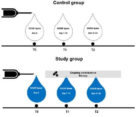
Foi efetuado um estudo prospetivo de coorte, que incluiu a medição da DGGR lipase em dois grupos: o grupo de estudo (GE) constituído por cães aos quais foi administrada prednisolona por via oral com justificação médica na dose inicial de 0.5-1.7mg/kg/dia durante pelo menos 3 semanas e o grupo controlo (GC) composto por cães saudáveis sem tratamento concomitante. Como critério de inclusão consideraram-se cães com valores de DGGR lipase abaixo do valor de referência (<80 U/L). A DGGR lipase foi quantificada em três pontos temporais (Dia 0 (T0), Dia 7-10 (T1) e Dia 21-30 (T2)). A análise foi efetuada com recurso a um kit previamente validado (Randox® DGGR lipase). Foram incluídos 34 cães (17 cães em
cada grupo, emparelhados relativamente ao género e idade). Em T0 não se observou diferença estatisticamente significativa entre grupos (p=0.868). A dose inicial média de prednisolona foi de 0.94 (±0.85) mg/kg/dia, tendo decrescido para 0.45 (±0.05) mg/kg/dia após T1. A concentração mediana de DGGR lipase no GE em cada ponto temporal (T0, T1 e T2) foi: 24.74 (14.45-31.48) U/L, 36.82 (23.8-80.16) U/L e 29.52 (15.91-48.48) U/L, respetivamente. Observou-se um efeito estatisticamente significativo da prednisolona nos valores de DGGR lipase ao longo de T0, T1 e T2 (p=0.007). Foi verificada uma baixa correlação entre as variações de DGGR lipase e a dose de prednisolona correspondente em T0-T1 e T1-T2 (rs=0.371 e rs=0.121, respetivamente). Em relação ao GC não se observaram
diferenças estatisticamente significativas ao longo de T0, T1 e T2 (p=0.926).
Sugere-se que a DGGR lipase seja afetada pela administração oral de prednisolona por justificação médica. No entanto, como os valores permanecem abaixo do limite máximo considerado (160 U/L), esta variação não aparenta ser clinicamente relevante.
vi
THE EFFECT OF PREDNISOLONE THERAPY ON CANINE SERUM LEVELS OF
1,2-O-DILAURYL-RAC-GLYCERO GLUTARIC ACID-(6′-METHYLRESORUFIN)
ESTER (DGGR) LIPASE
Abstract
1,2-o-dilauryl-rac-glycero glutaric acid-(6'-methylresorufin) ester (DGGR) lipase is a widely available biomarker, increasingly used in the investigation of canine pancreatitis mainly due to its low cost compared to pancreatic lipase immunoreactivity (cPLI). A previous study showed a good agreement between cPLI and DGGR lipase concentration. While the effect of corticotherapy on cPLI quantification has been studied, its influence on DGGR lipase is unknown. This study aims to evaluate the effect of prednisolone therapy in canine DGGR lipase serum levels.
A prospective cohort study was conducted, including the measurement of DGGR lipase in two groups: the study group (SG) composed of dogs treated with oral prednisolone for a medical reason, at the initial dosage of 0.5-1.7 mg/kg/day for at least 3 weeks, and the control group (CG) composed of healthy untreated dogs. As an inclusion criterion, animals had basal DGGR lipase within the reference range (<80 U/L). DGGR lipase was measured at three time points (Day 0(T0), Day 7-10(T1), and Day 21-30(T2)) in both groups. The analysis was performed using a previously validated kit (Randox® DGGR lipase). Thirty-four dogs were
included (17 dogs for each group, which were age and sex-matched). At T0, there was no significant difference in DGGR lipase concentrations between groups (p=0.868). Mean starting dosage of prednisolone was 0.94 (±0.85) mg/kg/day, decreasing to 0.45 (±0.05) mg/kg/day after T1.
The median DGGR lipase concentration in SG at each time point (T0, T1, and T2) was: 24.74 (14.45-31.48) U/L, 36.82 (23.8-80.16) U/L and 29.52 (15.91-48.48) U/L, respectively. There was a statistically significant effect of prednisolone on DGGR lipase values (p=0.007) over T0, T1, and T2. A poor correlation was verified between the variations of DGGR lipase and the correspondent prednisolone dosage of T0-T1 and T1-T2 (rs=0.371 e rs=0.121,
respectively). In CG, DGGR lipase did not significantly change over the three time points (p=0.926).
This study suggests that DGGR lipase levels are affected by oral prednisolone therapy in dogs treated for a medical reason. However, as values remained below the considered significant upper limit (160 U/L), this variation does not seem to be clinically relevant.
vii
Table of contents
Acknowledgements ... iii
Resumo ... v
Abstract ... vi
Table of contents ... vii
List of figures ... viii
List of tables ... viii
List of graphs ... ix
List of abbreviations and symbols ... x
1. TRAINEESHIP REPORT ... 1
2. LITERATURE REVIEW ... 5
2.1. Pancreas ... 5
2.1.1. Exocrine pancreas ... 6
2.2. Pancreatitis and lipase ... 7
2.3. Pancreatic biomarkers: state of the art ... 9
2.3.1. Trypsin-like immunoreactivity (TLI) ...11
2.3.2. Amylase ...13
2.3.3. Lipase ...14
2.3.3.1. Serum lipase activity ...14
2.3.1.1.1. Colorimetric method ...15
2.3.3.2. Serum lipase immunoreactivity ...17
2.4. DGGR lipase ...21
2.4.1. DGGR lipase and Spec cPL: What do we know? ...22
2.5. Pancreatitis diagnosis: the importance of biomarkers ...23
2.6. Disease interference on lipase - clinical relevance ...24
2.7. Corticotherapy in veterinary medicine ...26
2.7.1. Corticotherapy and pancreatic biomarkers: Is there any interference? ...27
3. THE EFFECT OF PREDNISOLONE THERAPY ON CANINE SERUM LEVELS OF DGGR LIPASE ...28
3.1. Introduction and objectives ...28
3.2. Material and methods ...28
3.2.1. Sample population ...28
3.2.1.1. Study Group ...28
3.2.1.2. Control Group ...29
viii
3.2.3. Reference interval and grey zone ...30
3.2.4. Prednisolone treatment ...30 3.2.5. Abdominal Ultrasound ...30 3.2.6. Statistical analysis ...30 3.3. Results ...31 3.3.1. Sample characterisation ...31 3.3.1.1. SG ...31 3.3.1.2. CG ...34
3.3.2. DGGR lipase over time ...35
3.3.3. DGGR lipase comparison between SG and CG ...35
3.3.4. Correlation between DGGR lipase and prednisolone dosage variations ...36
3.3.5 Abdominal ultrasound ...37 3.4. Discussion ...37 3.5. Conclusion ...42 4. REFERENCES ...43 5. ANNEXES ...51
List of figures
Figure 1. Reaction sequence of 1,2 DiG lipase method (Adapted from Fossati et al. 1992). .16 Figure 2. Reaction sequence of DGGR lipase method (Panteghini et al. 2001). ...21Figure 3. Study design scheme. ...30
List of tables
Table 1. Comparison between the sensitivity and specificity of TLI, Amylase, 1,2 DiG lipase, cPLI, and specific canine pancreatic lipase (Spec cPL) reported in previous studies ...12Table 2. Comparison between the sensitivity and specificity of Lipase, 1,2 DiG, DGGR lipase, and Spec cPL reported in previous studies...16
Table 3. Comparison between the sensitivity and specificity of SNAP® cPL and Spec cPL, reported in previous studies ...20
Table 4. Breed characterisation of the SG ...33
Table 5. Disease characterisation of the SG ...33
ix
List of graphs
Graph 1. Percentage of total time spent in each department. ... 1
Graph 2. Gender characterisation of the SG and CG ...32
Graph 3. Ages of the SG and CG ...32
Graph 4. Prednisolone dosage on T1 and T2 of the SG ...34
Graph 5. DGGR lipase over T0, T1 and T2 of the CG and SG ...35
Graph 6. Correlation of DGGR lipase and prednisolone variation on T0-T1 ...36
x
List of abbreviations and symbols
% - percentage L - litter
µg - microgram MCS - mean cumulative severity
1,2 DiG - 1,2 diglyceride mg - milligram
AI - activity index min. - minutes
ALT - alanine aminotransferase mRNA - messenger ribonucleic acid
A.m. - ante meridiem nm - nanometre
CEIE - Comissão de Ética para a Investigação e Ensino
p.m. - post meridiem
CG - control group RIA - radioimmunoassay
CI - chronicity index rcf – relative centrifugal force
cPL - canine pancreatic lipase rs - Spearman’s rank correlation coefficient
cPLI - canine pancreatic lipase immunoreactivity
SD - standard deviation
CT - computed tomography SG - study group
DGGR - 1,2-o-dilauryl-rac-glycero glutaric acid-(6′-methylresorufin) ester
Spec cPL - specific canine pancreatic lipase
ELISA - enzyme-linked immunosorbent assay
Spec fPL - specific feline pancreatic lipase
ENS - enteric nervous system TAP - trypsinogen activation peptide EPI - exocrine pancreatic insufficiency TLI - trypsin-like immunoreactivity
FFAs - free fatty-acids U - units
FMV-UL - Faculty of Veterinary Medicine - University of Lisbon
к - Cohen's kappa coefficient HAC - hyperadrenocorticism
ICC - intraclass correlation coefficient IDIU - Infectious Diseases Isolation Unit IF – intrinsic factor
IQR - interquartile range Kcat - catalytic constant kDa – kilodaltons Km - Michelis constant
1
1. TRAINEESHIP REPORT
This report describes the work done during the curricular internship that took place from the 2nd September 2019 to the 28th February 2020 at the Veterinary Hospital of the Faculty of
Veterinary Medicine - University of Lisbon. The activities were performed in a rotation basis across multiple departments such as internal medicine, ophthalmology, imagiology, surgery, dermatology, inpatient care, oncology, general practice, and infectious diseases under the supervision of the Veterinary Surgeon or the Veterinary Nurse on duty. During this period, there was an opportunity to participate in weekly journal clubs and staff seminars.
The time spent in each department varied in terms of the period of the day (day or night) as well as duration (from 8 to 12 hours a day). The internship had a duration of 6 months, totalling 1198 hours. The time spent in each department is depicted in graph 1.
Internal Medicine
Of all rotations, the one in internal medicine was the longest. This rotation was under the supervision of Professor Doctor Rodolfo Leal, the responsible for the department and board-certified specialist, who also supervised the project. This rotation was essential for the accomplishment of the current work since it allowed the recruitment of most animals enrolled in the study. 0 5 10 15 20 25 30 35 Internal Medicine General Practice Inpatient care Imagiology Ophthalmology Oncology Dermatology Surgery Infectious Diseases
Percentage of the internship total time (%)
Dep a rtm e n t
Graph 1. Percentage of total time spent in each department. Figure 1. Percentage of total time spent in each department.
Graph 2. Percentage of total time spent in each department. Figure 2. Percentage of total time spent in each department.
2
The daily routine started with medical rounds which usually took place around 8.30 a.m. focusing on the inpatient care animals in the presence of staff from several departments. At this stage, a detailed presentation of each animal was made by the night-shift clinician and the differential diagnosis was discussed. The entire group participated in the discussion of the complementary exams, surgeries, and treatments that needed to be performed during the day.
During the morning period, first-opinion, re-evaluation, referral, and second-opinion small-animal consultations were attended. Clinical cases were mostly from endocrinological, gastroenterological, nephrological, and respiratory diseases. Apart from Professor Rodolfo, once a week it was possible to attend consultations with the responsible intern of the internal medicine team (Dr. Joana Dias, Dr. Telmo Casimiro, or Dr. Sara Prata). Under supervision, it was possible to participate in the collection of prior clinical history and anamnesis of the patients as well as to perform a thorough physical examination. There was also the opportunity to participate in medical procedures such as blood sample collection, blood pressure measurements, and cystocentesis. Moreover, there was a chance to discuss the different possible approaches, differential diagnosis, and treatment options, which took place mainly in the afternoon. During this period, the results from the performed complementary exams were discussed and communicated to the owners. Also, it was possible to develop the skills required for the elaboration of a medical report, by actively taking place in this process. Complementary exams such as upper and lower gastrointestinal endoscopy, rhinoscopy, bronchoscopy with bronchoalveolar lavage, and bone marrow aspiration were performed about once a week.
Ophthalmology
The time spent with the ophthalmology team allowed the attendance of consultations and surgeries. The consultations were attended in both morning and afternoon periods over the entire week, while surgeries were attended during the morning periods over three defined days in each week.
During this rotation, the student had the opportunity to attend first-opinion, re-evaluation, referral, and second-opinion consultations of small animals, horses, and exotic animals as well. Moreover, it was possible to perform exams such as fundoscopic exam, Schirmer’s test, and fluorescein test. Also, peripheral intravenous catheterisations and blood collections were performed under supervision.
Additionally, several ophthalmological surgeries were attended, such as cataract surgery and enucleation.
3
Imagiology
The time spent in this rotation was divided into two periods: a week attending radiography and computed tomography (CT) exams and 2 weeks participating in ultrasonography exams. During this period, it was possible to improve skills related to exam positioning and image interpretation. The shifts had a duration of 8 hours and started at 9 a.m. or 2 p.m.
During the first week, which involved the attendance of radiography and CT exams, the student also assisted in various procedures such as myelography and contrast-CT. Moreover, all steps from anaesthetic induction to post-exam care were performed, under close supervision.
During the second period of 2 weeks, there was the opportunity to attend several ultrasonography exams as well as ultrasound guided percutaneous needle biopsies, cardiac and abdominal ultrasounds, and echo guided pericardiocentesis. Also, it was possible to perform ultrasound-guided cystocentesis and to train abdominal ultrasound under supervision.
General Practice
This was the second longest rotation of the internship. The shifts had a duration of 8 hours, starting at 8 a.m. or 1 p.m., depending on the week. During the morning shifts, it was possible to attend the medical round of the inpatient care animals. In this service, there was mainly the possibility of attending first-opinion and preventive-medicine consultations, which provided an excellent environment to improve communication skills and to perform medical procedures under supervision. During this period, it was possible to collect the prior clinical history, anamnesis, and to perform a good physical exam in most animals. During all the clinical cases there was the chance to discuss the different possible approaches with the several clinicians on duty, as well as the differential diagnosis, and treatment options. Some emergency procedures such as cardiorespiratory resuscitation were also assisted.
Infectious Diseases
During this rotation, the activities were developed in the Infectious Diseases Isolation Unit (IDIU) with afternoon shifts of 8 hours during a week. All the activities took place under the supervision of the responsible clinician on duty. The activities included clinical monitoring, medication preparation and administration, and the correct hygienization of contaminated infectious material. Moreover, it was possible to attend some infectious disease consultations and to participate in the clinical discussion of the differential diagnosis and treatment options.
4
Surgery
During this rotation, the scheduled surgeries took place from Monday to Friday during an 8 hour shift and emergency surgery took place whenever needed.
At the beginning of the shifts, which started at 8 a.m., the animals were admitted by the student, and a pre-surgery checklist was filled by asking some questions to the owners. During the surgery service, there was the opportunity to perform procedures such as peripheral venous catheterisation, pre-anaesthetic drugs preparation and administration, trichotomy and surgical asepsis, anaesthetic induction, animal intubation, animal transference onto the surgical table, monitoring, assistance in the surgical procedures, and post-surgical monitoring. These were performed under the supervision of the responsible surgeon or nurse.
In some cases, direct involvement during the surgery was not possible. Nonetheless, it was still feasible to observe and discuss with the surgeons the surgical techniques and other aspects regarding pre- and post-operative care. During this period, the attended surgeries included ovariohysterectomies, orchiectomies, caesarean sections with the respective neonatal reanimation, correction of brachycephalic syndrome, cystotomies, removal of mammary chains, among others.
Oncology
This rotation lasted two weeks, with shifts of 8 hours starting at 8 a.m. or 2 p.m. These weeks were focused on oncology consultations and chemotherapy sessions. Additionally, it was possible to attend first-opinion, re-evaluation, referral, and second-opinion small-animal consultations. During this period, prior clinical history and anamnesis were collected and the physical exam technique was improved. Under supervision, there was the opportunity of performing blood sample collections and some other procedures.
Dermatology
The period of 2 weeks spent in the dermatology department was mostly focused on attending first-opinion, re-evaluation, and second-opinion small-animal consultations. In addition to consultations, it was also possible to attend dermatology lectures. The 8 hour shift varied between morning and afternoon periods.
During this rotation, there was the opportunity to discuss the different possible approaches, differential diagnosis, and treatment options with the clinician on duty, performing some procedures such as blood collection, anamnesis, and physical exams. Also, skin biopsies and video otoscopies were attended.
5
Inpatient care
The time spent in this rotation was organised over the 6 month period including weekends with 12 hour day and night shifts. In this service, the shift started with a medical round followed by animal related activities.
During this period, there was the chance to follow the progress of the different clinical cases, participating in the animals’ clinical monitorization, drug preparation, and administration according to the indications of the clinician and nurse on duty.
The evolution of the clinical condition was discussed with the clinician as well as the discharge plan for each animal. Concerning medical procedures and similarly to the remaining services, there was the possibility of performing blood sample collection, cystocentesis, and peripheral venous catheterisations. Some of the cases offered the opportunity to attend some emergency procedures such as cardiorespiratory resuscitation.
Scientific communications
An abstract with the preliminary results was submitted and presented as an oral communication at Congresso Internacional Hospital Veterinário Montenegro, in February 2020 (Annexe 1).
A poster with the results will be presented at the European College of Veterinary Internal Medicine – Companion Animals Congress, in September 2020 (Annexe 2).
A paper with the results has been accepted to
the Journal of Veterinary Internal Medicine.
Apart from this study, a clinical case followed during the internal medicine rotation and concerning Shar-Pei fever was presented as a poster at Congresso Internacional Hospital Veterinário Montenegro, in February 2020 (Annexe 3).
2. LITERATURE REVIEW
2.1. Pancreas
The pancreas is located in the cranial abdomen, caudally to the stomach. It is composed of a left lobe positioned between the transverse colon and the greater curvature of the stomach, a right lobe adjacent to the proximal duodenum, and, at last, the body which is between the two lobes (Watson 2014, Evans 1993). It is composed of two different functional types of glandular tissue – the endocrine and exocrine. The endocrine portion is organised into discrete islets called islets of Langerhans, within the parenchyma, and makes up 2% (Evans 1993; Jubb and Stent 2015) of the pancreatic tissue. The exocrine portion comprises 98% of
6
the pancreatic tissue and its detailed anatomy and physiology are discussed in the following section (Evans 1993; Herdt and Sayegh 2013).
2.1.1. Exocrine pancreas
The pancreas is mostly composed of acinar tissue lacking a conventional capsule (Jubb and Stent 2015). This structure is characteristic of a typical acinar gland in which the acini are connected by a system of ducts (Herdt and Sayegh 2013). In most dogs, the smaller ducts of the pancreatic lobules coalesce in a structure to form two larger pancreatic ducts existing in the duodenum (Watson 2015). The exocrine pancreas is responsible for digestive secretions, bicarbonate, and intrinsic factor (IF) which are delivered into the intestinal lumen. The acinar cells produce the digestive enzymes, whereas centroacinar cells and duct cells secrete electrolyte solution rich in sodium bicarbonate (Herdt and Sayegh 2013) and intrinsic factor (Vaillant et al. 1990).
Some digestive enzymes can induce lesions to the pancreatic cells, and for this reason, they are synthesised as inactive precursors called zymogens (Herdt and Sayegh 2013). The zymogens are only activated in the intestinal lumen and are separated intracellularly from lysosomes (Herdt and Sayegh 2013). The zymogens of digestive enzymes are trypsinogen, chymotrypsinogen, proelastase, prophospholipase, kallikreinogen, and procarboxypeptidase (Steiner 2008). By contrast, other enzymes such as lipase, amylase, carboxylesterase, desoxyribonuclease, and ribonuclease are secreted in their active form (Steiner 2008).
Pancreatic secretion is, mainly, triggered by the presence of fat and proteins in the small intestinal lumen and involves the release of acetylcholine and hormones such as secretin and cholecystokinin (Watson 2014). There are nerve fibres from the enteric nervous system (ENS) ending close to the pancreatic acinar glands (Herdt and Sayegh 2013). These neurons will release acetylcholine by impulses arriving from other neurons of the ENS or by parasympathetic fibres from the vagus nerve (Herdt and Sayegh 2013). Cholecystokinin is the primary hormonal stimulus for acinar cells (Herdt and Sayegh 2013) and is synthesised by I cells located in the duodenum and jejunum (Rehfeld 2004). On the other hand, secretin is the primary hormonal stimulus for centroacinar and duct cells and is synthesised by S cells mostly located in the duodenum (Afroze et al. 2013). When these hormones interact with binding sites on the surfaces of pancreatic cells, secretion is stimulated (Herdt and Sayegh 2013).
All the digestive enzymes produced by the exocrine portion of the pancreas are crucial for the digestion of most nutrients. Although there is a complex structural and functional interrelationship between both endocrine and exocrine tissues, there is also an important interdependence between them (Jubb and Stent 2015).
7
The exocrine pancreas is considered a labile organ. It synthesises a large amount of protein and consumes the corresponding amount of precursor substrate (Jubb and Stent 2015). However, the homeostatic regulation of the pancreatic tissue continues to be poorly understood (Jubb and Stent 2015). It is known that there is evidence of trophic hormones from islets and that the response of exocrine pancreas to changes in nutrients intake is rapid and produces remarkable alterations in the composition of pancreatic secretion (Jubb and Stent 2015).
2.2. Pancreatitis and lipase
The pancreas is considered the only significant source of lipase (Watson 2014) and it was shown by Brobst et al. (1970)1 (cited by Hoffmann 2008) that experimentally induced
pancreatitis in dogs increases the serum activity of this enzyme (Hoffmann 2008). Thus, serum lipase has been a subject of discussion since it is considered a powerful tool to evaluate pancreatic lesions (Dröes and Tappin 2017). Considering pancreatitis as the most common exocrine pancreatic disease in both dogs and cats (Steiner 2008), it has become relevant to study its underlying pathophysiology and which risk factors should be considered relevant in the context of possible pancreatitis. Furthermore, this information integrated with the previous clinical history will help deciding the need of measuring a serum pancreatic biomarker such as lipase, which is secreted in its active form (Steiner 2008).
Trypsin is the major protease secreted by the pancreas. It is secreted as the zymogen trypsinogen and is central to the pathogenesis of pancreatitis. Inappropriate and prematurely intracellular activation of trypsinogen to trypsin, leads to activation of other zymogens such as proelastase, prophospholipase A2, and chymotrypsinogen, resulting in autodigestion and inflammation (Dröes and Tappin 2017). Trypsin activation within the acinar cells appears to be caused by blockage of the acinar cell apex, oxidative stress, or hypotension (Spillmann 2017).
Thus, under normal circumstances, self-defence mechanisms are in place to stop premature activation of trypsin (Jubb and Stent 2015; Watson 2015; Spillmann 2017). Therefore, the disruption of these mechanisms might underlie the causes of pancreatitis (Watson 2015). However, the causes are usually unknown, and several risk factors have been proposed (Spillmann 2017). It is documented that factors such as hypoxia, hypercalcemia, hyperlipidaemia, and other cellular injuries could affect trypsin activation control, making them
1 Brobst D, Ferguson AB, Carter JM. 1970. Evaluation of serum amylase and lipase activity in experimentally
induced pancreatitis in the dog. J Am Vet Med Assoc. 157(11):1697–702. url: https://pubmed.ncbi.nlm.nih.gov/5530372/
8
risk factors for pancreatitis (Jubb and Stent 2015). Other factors such as dietary indiscretion and overweight (Lem et al. 2008), zinc toxicosis (Mikszewski et al. 2003; Blundell and Adam 2012), and drugs are also documented. Some drugs have been reported to be associated with pancreatitis such as azathioprine (Moriello et al. 1987), potassium bromide with phenobarbitone (Gaskill and Cribb 2000), clomipramine (Kook et al. 2009) and sulfonamides (Trepanier et al. 2003), among others. Additionally, it is described that some breeds have a higher risk of developing pancreatitis. Cook et al. (1993)1 and Hess et al. (1999)2 (cited by Lem
et al. 2008) identified miniature schnauzers, Yorkshires terriers, and other terriers as a group and Watson et al. (2007) documented Cavalier King Charles spaniels, cocker spaniels, collies, and boxers. Also, Pápa et al. (2011) reported that pancreatitis is common in poodles, Dachshunds, laika, and Alaskan malamute. Moreover, dogs with concurrent endocrine diseases such as diabetes mellitus or hypothyroidism may be at increased risk of acute pancreatic necrosis (Jubb and Stent 2015). On the other hand, it is discussable whether hyperadrenocorticism (HAC) is a predisposing factor for pancreatitis (Behrend 2015). At last, other factors such as acute enteritis or gastroenteritis (Rallis et al. 1996), abdominal trauma, infections such as Babesia, and any condition that causes hypotension such as anaesthesia have also been identified as potential risk factors for pancreatitis in dogs (Xenoulis and Steiner 2013b). However, it is important to note that some of the associations considered above were not based on studies using histopathology, the gold standard for pancreatitis diagnosis (Xenoulis 2015).
“Functional and structural changes in the normal pancreas are not self-initiated, but rather occur in response to systemic metabolic activity, although in practice the relationship between cause and effect is often obscure” (Jubb and Stent 2015, p.353). Therefore, the aetiology of acute and chronic pancreatitis remains idiopathic (Spillmann 2017), but the risk factors aforementioned together with a compatible clinical investigation should raise the suspicion of pancreatitis.
Despite the fact that the pancreas is the main significant source of lipase, it is nowadays recognised that its production is not limited to this organ. For instance, Simpson et al. (1991) evaluated the effect of pancreatectomy on plasma lipase activity and other biomarkers
1 Cook AK, Breitschwerdt EB, Levine JF, Bunch SE, Linn LO. 1993. Risk factors associated with acute pancreatitis
in dogs: 101 cases (1985-1990). J Am Vet Med Assoc. 203(5):673–9. url: https://pubmed.ncbi.nlm.nih.gov/8407536/
2 Hess RS, Kass PH, Shofer FS, Winkle TJV, Washabau RJ. 1999. Evaluation of risk factors for fatal acute
9
(Simpson et al. 1991). The authors observed that lipase values decreased after pancreatectomy, but these values were not significantly different from the pre-operative ones. Therefore, this study reinforced the fact that the pancreas is not the only source of serum lipase activity. Also, Steiner et al. (2006) observed no significant differences in the serum lipase activity between dogs with and without exocrine pancreatic insufficiency (EPI), further supporting the hypothesis that there is a significant portion of lipase from non-pancreatic origin. Apart from the pancreatic lipase, many other lipases have been described, such as gastric, hepatic, lipoprotein, hormone-sensitive, and other less prominent lipases (Steiner et al. 2002). Nonetheless, pancreatic lipase is still considered a useful biomarker, being the research focused on directing the measurement exclusively to a pancreatic origin.
2.3.
Pancreatic biomarkers: state of the art
As pancreatic tissue is the source of several enzymes, their measurement is a logical approach to assess pancreatic disease (Simpson et al. 1991). These tests are based on the principle that different lesions will affect the levels of pancreatic enzymes leaking into the bloodstream. While acute lesions cause an increase in the levels of pancreatic enzymes, atrophy, which is associated with a chronic process, causes its decrease (Simpson et al. 1991). As pancreatitis can be present in both scenarios, it is difficult to distinguish in between these two possibilities by using this approach. Thus, it is important to note that such a distinction is only possible using histopathology (Xenoulis 2015). Acute pancreatitis is defined as a neutrophilic inflammation without fibrosis and exocrine atrophy (Newman et al. 2006). By contrast, the permanent histopathological changes such as fibrosis and acinar atrophy with mononuclear, often lymphocytic, inflammation are suggestive of chronic pancreatitis (Newman et al. 2004; Watson et al. 2007; Bostrom et al. 2012). The decreased leakage of pancreatic enzymes into the bloodstream is reflected by the lower correlation between some pancreatic serological markers and histopathologic features of chronic pancreatitis compared with histopathologic features of acute pancreatitis (Trivedi et al. 2011). However, the presence of atrophy, fibrosis, and hyperplastic nodules with acute lesions such as neutrophilic infiltration, pancreatic, and peripancreatic fat necrosis were also described. This suggests that some dogs presenting acute pancreatitis likely had previous episodes of pancreatitis or ongoing chronic pancreatitis (Neilson-Carley et al. 2011). Also, the presence of active pancreatic disease, such as pancreatic and peripancreatic fat necrosis, possibly contributes to the occurrence of clinical signs in chronic pancreatitis. On the other hand, the absence of active disease is less likely to develop clinical signs and might escape clinical diagnosis (Bostrom et al. 2012). While it has been traditionally accepted that acute pancreatitis is the most common manifestation in dogs, some results support that chronic pancreatitis is also common (Watson and Herrtage 2006;
10
Watson et al. 2007) and clinically significant in a referral population (Watson and Herrtage 2006), suggesting that it may be under-recognised or subclinical in some dogs (Xenoulis et al. 2008).
Despite the fact that there are no studies describing the pathophysiology of naturally occurring acute or chronic pancreatitis in dogs and cats, some authors discussed it based on findings from human and experimental animal work (Watson 2015). The relationship between acute and chronic pancreatitis is important since some chronic cases result from acute disease. However, it is unclear how many cases progress from acute to chronic disease or how many started due to chronic inflammation. This is the case in autoimmune chronic pancreatitis, which is caused by an unclear trigger and the role of an acute episode is still a hypothesis (Watson 2015).
The gold standard for the confirmation of pancreatitis and its definition as an acute or chronic disease is pancreatic histopathology (Xenoulis and Steiner 2013a). Nevertheless, it is still not routinely used since the sampling procedure presents several medical risks that should not be neglected (Dröes and Tappin 2017). Moreover, a single biopsy can be insufficient because the inflammation is usually non-uniform (Newman et al. 2004). For those reasons, results such as sensitivity and specificity should be interpreted with caution since they rely upon an imperfect gold standard (Xenoulis 2015). Therefore, in cases where there is not a reasonable reason to do a biopsy, other less invasive diagnostic tests may be considered. The digestive enzymes are the most commonly used biomarkers of pancreatic disease, namely pancreatitis (Ruaux 2003). However, several factors should be taken into consideration when considering the performance of these tests, since it relies upon different criteria used for diagnosis, type of pancreatitis and/or used cut-off values among studies (Xenoulis 2015). Table 1 provides an overview of selected studies evaluating the sensitivity and/or specificity of different laboratory tests for the diagnosis of pancreatitis in dogs, considering the different gold standards and the pancreatitis classifications.
Considering the pancreatic secretion and the pathophysiology of pancreatitis, trypsinogen, amylase, and lipase had been historically evoked as potential indicators of pancreatic inflammation. Based on the studies of Steiner et al. (2008), Watson et al. (2010), Trivedi et al. (2011) and McCord et al. (2012), the specificity and sensitivity of these tests are summarised in Table 1. As a result of the influence of histopathological lesions in enzyme leakage, it becomes important to refer to how these lesions have been classified in each study. The histopathologic characterisation, when considered, is based on a grading scheme for exocrine pancreatic lesions (Newman et al. 2006). This grading scheme comprises the
11
quantification and severity of lesions through the mean cumulative severity (MCS), which then allows calculating the activity index (AI) and the chronicity index (CI). The AI comprises processes involving necrosis or inflammation and the CI comprises processes more indicative of a chronic ongoing or previously resolved pancreatic lesion (Newman et al. 2006).
Regarding sensitivity, the results from a study with dogs that presented macroscopic evidence of pancreatitis documented a histologically acute mild to moderate pancreatitis in all the animals (Steiner et al. 2008). It is possible that the sensitivity would increase if there was a histological characterisation of more severe cases of pancreatitis. In another study, Watson et al. (2010) included dogs with histologically confirmed chronic pancreatitis, with severe and mild to moderate lesions associated, and the sensitivity for some serum markers was evaluated. Trivedi et al. (2011) encountered a group of dogs presenting, on the majority, histopathologic features of acute and chronic pancreatitis, concurrently. Therefore, the groups of acute and chronic pancreatitis were combined. The two formed groups were based on the MCS, and thus, the mild and moderate to severe pancreatitis groups were considered. The sensitivity and specificity were evaluated for several serum markers in dogs. At last, McCord et al. (2012) characterised a study group for acute pancreatitis not based on histopathology, but considering the medical record, laboratory findings, and ultrasound interpretation. However, a more recent analysis method has been used to evaluate unbiased parameters and calculate specificity and sensitivity.
2.3.1. Trypsin-like immunoreactivity (TLI)
TLI is an immunoassay that measures trypsinogen and, to a lesser degree, trypsin concentration in serum (Xenoulis 2015). Since serum TLI concentration in dogs increases after the experimental induction of pancreatitis but decreases more rapidly than amylase and lipase activities and present low sensitivity, it is considered a poor indicator of pancreatitis (Simpson et al. 1989; Mansfield and Jones 2000; Xenoulis 2015). The blood half-life of TLI has not been reported; however, it is likely short due to the rapid action of endopeptidases, whose main role is to inactivate pancreatic enzymes (Hoffmann and Solter 2008). Steiner et al. (2008) described a low sensitivity of 36.4% (Table 1) and no correlation with the AI for acute mild to moderate pancreatitis. On the other hand, Watson et al. (2010) described a sensitivity of 17% for chronic pancreatitis (Table 1). Trivedi et al. (2011) documented a sensitivity of 30% and 29%, for mild and moderate to severe pancreatitis, respectively (Table 1). In the same study, the reported specificity was 100%. However, this could be explained by the relatively low number of dogs without histopathologic evidence of pancreatitis.
12
Table 1. Comparison between the sensitivity and specificity of TLI, Amylase, 1,2 DiG lipase, cPLI, and specific canine pancreatic lipase (Spec cPL) reported in previous studies
Gold standard and pancreatitis
classification
TLI Amylase 1,2 DiG lipase cPLI Spec cPL
Sens. (%) Spec. (%) Sens. (%) Spec. (%) Sens. (%) Spec. (%) Sens. (%) Sens. (%) Spec. (%) Clinical criteria Acute pancreatitis - - - - ≥400 µg/l 70 77 Haworth et al. (2014) Histopathology Pancreatitis - - - - >200 µg/L ≥400 µg/L >200 µg/L ≥400 µg/L 58 33 80 90 Mansfield et al. (2012) Clinical criteria Acute pancreatitis - - >1240 U/L >750 U/L - >200 µg/L >400 µg/L >200 µg/L >400 µg/L McCord et al. (2012) 52.7 79.1 47.2 91.4 86.5- 93.6 71.7-77.8 66.3-77.0 80.5- 88.0 Histopathology - - - - >200 µg/L >400 µg/L Neilson- Carley et al. (2011) 95.0 97.5 Histopathology Mild pancreatitis >35 µg/L >1240 U/L >750 U/L - - >200 µg/L >400 µg/L > 200 µg/L >400 µg/L 30.0 100 7.0 100 54.0 43.0 43.0 21.0 86.0 100 Moderate to severe pancreatitis 29.0 14.0 71.0 71.0 71.0 Trivedi et al. (2011) Histopathology Chronic pancreatitis >35 µg/l - >1126 U/L >3378 U/L - >250 U/L >750U /L - >102 µg/L >200 µg/L - - Watson et al. (2010) 17 67 14 44 28 58 26 Histopathology Acute mild-moderate pancreatitis >35 µg/L - >1380 U/L - >691 U/L - >102 µg/L >200 µg/L >200 µg/L >400 µg/L - Steiner et al. (2008) 36.4 18.2 13.6 77.3 63.6 72.7 63.6
Legend: 1,2 DiG, 1,2 diglyceride; cPLI, canine pancreatic lipase immunoreactivity; Sens., sensitivity; Spec., specificity; Spec cPL, specific canine pancreatic lipase; TLI, Trypsin-like immunoreactivity. .
Legend: 1,2 DiG, 1,2 diglyceride; cPLI, canine pancreatic lipase immunoreactivity; Sens, sensitivity; Spec cPL, specific canine pancreatic lipase; TLI, Trypsin-like immunoreactivity. .
13
Nonetheless, it is used as a specific indicator of pancreatic exocrine insufficiency, as it is accepted that TLI is exclusive of pancreatic origin (Batt 1993). In agreement with these results, also Simpson et al. (1991) demonstrated that pancreatectomised dogs have markedly lower TLI concentrations. Thus, 100% of both sensitivity and specificity have been documented for EPI diagnosis (Williams and Batt 1988). The low sensitivity and the longer turnaround time for TLI compared to serum lipase and amylase have become TLI of little interest for pancreatitis evaluation in dogs (Hoffmann and Solter 2008).
Also, some investigators evaluated an enzyme linked immunosorbent assay for trypsinogen activation peptide (TAP). However, healthy dogs presented an unexpectedly broad range of TAP concentration detectable in the urine. Furthermore, plasma and urinary TAP concentrations were significantly increased only in dogs with severe and necrotizing pancreatitis (Mansfield and Jones 2000).
2.3.2. Amylase
Amylase is not exclusively of pancreatic origin, as it is also produced in the small intestine and liver (Hoffmann and Solter 2008). These findings are in agreement with previous studies conducted in pancreatectomised dogs from Simpson et al. (1991), where serum amylase activity results were consistent with non-pancreatic sources for amylase. Steiner et al. (2008) described a sensitivity of 18.2% (Steiner et al. 2008) (Table 1) and observed no correlation between AI and serum amylase activity. Concerning chronic pancreatitis, Watson et al. (2010) documented a sensitivity of 67% considering the reference interval of 167-1126 U/L, while a higher cut-off of 3378 U/L resulted in a sensitivity of 14% (Table 1). Trivedi et al. (2011) documented serum amylase as the least sensitive serological assay for the diagnosis of pancreatitis with a sensitivity of 7% for mild pancreatitis and 14% for moderate to severe pancreatitis (Table 1). The specificity described was 100% (Trivedi et al. 2011), considering the low number of dogs without histopathologic evidence of pancreatitis in this study (Table 1). McCord et al. (2012) documented 52.7% for sensitivity and 79.1% for specificity, emphasising the low performance of amylase (Table 1). Moreover, an older study reported that an increase in serum amylase activity could support a diagnosis of pancreatitis, but this was a poor indicator without the knowledge of lipase levels (Strombeck et al. 1981). Furthermore, it is described that amylase has the lowest sensitivity for mild or moderate to severe pancreatitis among pancreatic markers (Trivedi et al. 2011). For all reasons stated above, it should not be the first choice as a screening test (Kook 2017).
14
2.3.3. Lipase
As previously stated, lipase is mainly produced in the pancreas and its function is centred on triglycerides hydrolysis. Its sensitivity and specificity have also been described in the studies of Steiner et al. (2008), Watson et al. (2010), Trivedi et al. (2011), and McCord et al. (2012). These studies are all detailed in the following section. For a long time, amylase and lipase serum activities have been important tools for the diagnosis of pancreatitis.
Despite all extra-pancreatic sources, there is a specific pancreatic lipase that is of interest in the diagnosis of pancreatic disease. This molecular protein has 42 kDa and hydrolyses triglycerides at positions 1 and 3, releasing a monoglyceride. This lipase binds at the lipid-water interface emulsified in the presence of bile salts, calcium, and colipase (Hoffmann and Solter 2008).
Therefore, many studies have focused on developing more selective and accessible methods for the detection of lipase from a pancreatic origin. These assays are based on serum lipase activity and serum lipase immunoreactivity.
2.3.3.1. Serum lipase activity
Determination of serum lipase activity has been used for the diagnosis of pancreatitis in dogs for several decades (Steiner et al. 2006). Lipases hydrolyse lipids, such as triglycerides, which are apolar. As these lipids are important for long-term energy storage, they are converted into polar products, allowing their transport in and out of cells. As a result, there are many different lipases in the organism responsible for hydrolysing apolar triglycerides, which share functional and structural features (Steiner 2017).
Although the variety of existing methodologies rely on the detection of different substrates, many of these present low specificities. Thus, other enzymes from non-pancreatic origin may contribute to the detected lipolytic activity (Ruaux 2003).
Several methods have been developed for the measurement of lipolytic activity such as the titrimetric (Tietz and Fiereck 1966), turbidimetric (Burlina and Galzigna 1973; Orda et al. 1984), nephelometric (Wagner and Macy 1982) and colorimetric (Fossati et al. 1992) methods. The titrimetric method measures the neutralised free fatty-acids (FFAs), while the colorimetric method evaluates the coloured end product after hydrolysis. The nephelometric and turbidimetric methods measure, respectively, the light scattered and transmitted (Hasan et al. 2009; Encyclopaedia Britannica 2017). However, titrimetric, turbidimetric, and nephelometric methods presented enough limitations to be later replaced by other methods
15
(Fossati et al. 1992). Therefore, the colorimetric method is the current most utilised serum lipase activity quantification technique.
2.3.1.1.1. Colorimetric method
Around thirty years ago, a rapid and easy colorimetric method, based on the principle of Imamura et al. (1989)1 (cited by Fossati et al. 1992), was validated for assaying lipase
activity in human serum.
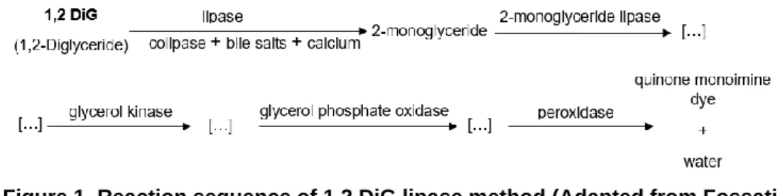
In veterinary medicine, concerning canine serum lipase assessment, a modification of the colorimetric method for automated analysers was compared to titrimetric method (Walter et al. 1992). The authors have shown that this is a reliable indicator of serum lipase activity in dogs, making it an attractive alternative to the titrimetric procedure (Walter et al. 1992). The determined reference range was 90-527 U/L (Walter et al. 1992). A few years later, Mackenzie et al. (1996) achieved similar results using a different automated analyser and determined a similar reference range (0-561 U/L). The results supported the utility of the colorimetric method as a reliable indicator of serum lipase activity in dogs. This methodology uses the natural long-chain fatty acid 1,diglyceride (1,2 DiG), a clear substrate that is hydrolysed into 2-monoglyceride by lipase in the presence of colipase, deoxycholate, and calcium ions (Figure 1) (Fossati et al. 1992; Walter et al. 1992). A sequence of enzymatic reactions with the 2-monoglyceride lipase, glycerol kinase, glycerol phosphate oxidase, and peroxidase, produce a violet quinone monoimine dye (Figure 1) with peak absorption at 550 nm (Fossati et al. 1992; Walter et al. 1992). The rate of increase in light absorbance is directly proportional to the lipase activity in the serum (Walter et al. 1992). The bile salts (deoxycholate) and colipase are responsible for preventing the rapid and irreversible inactivation of lipase. The calcium ions are added to increase the interaction of lipase and the substrate surface and to stabilize the enzyme’s active site (Fossati et al. 1992).
1lmamura S, Hirayama T, Arai T, et al. 1989. An Enzymatic Method Using 1,2-Diglyceride for Pancreatic Lipase in Serum. Clin Chem. 35(6): 1126,1989.
16
The specificity of the colorimetric assay for pancreatic lipase in human serum was attributed to 1,2 DiG and both colipase and deoxycholate, which initiate the conversion of the diglyceride to monoglyceride while inhibiting the hepatic and lipoprotein lipases (Fossati et al. 1992; Mackenzie et al. 1996).
Several studies have evaluated the specificity and sensitivity of these assays (Table 1 and 2). In studies where the clinical criteria were considered the gold standard, specificity achieved the highest values ranging from 73% to 91.4% (Graca et al. 2005; McCord et al. 2012), while Trivedi et al. (2011) who considered histopathology as the gold standard, reported a specificity of 43% (Table 1).
Table 2. Comparison between the sensitivity and specificity of Lipase, 1,2 DiG, DGGR lipase, and Spec cPL reported in previous studies
Gold standard and pancreatitis
classification
1,2 DiG lipase DGGR lipase Spec cPL
Sens. (%) Spec. (%) Sens. (%) Spec. (%) Sens. (%) Spec. (%) Clinical criteria Acute pancreatitis - - > 216 U/L ≥400 µg/L 85.7 – 90.9 64.0 – 74.3 81.0- 90.9 74.1-81.1 Cridge et al. (2018) Histopathology Chronic pancreatitis - - > 245 U/L >400 µg/L 57 100 42 100 Goodband et al. (2018) Acute pancreatitis 0 33 Clinical criteria Acute pancreatitis >500 U/L >699 U/L >500 U/L >699 U/L >120 U/L >180 U/L >120 U/L >180 U/L - - 73.3 60.0 73.0 73.0 93.3 73.3 53.3 66.6 Graca et al. (2005) Figure 1. Reaction sequence of 1,2 DiG lipase method (Adapted from Fossati
et al. 1992).
Figure 1. Reaction sequence of 1,2 DiG lipase method (Adapted from Fossati et al. 1992).
Legend: […], chemical reaction steps not included in the figure.
Figure 3.Reaction sequence of DGGR lipase method
Figure 1. Reaction sequence of 1,2 DiG lipase method (Adapted from Fossati et al. 1992).Legend: […], chemical reaction steps not included in the figure.
Figure 4.Reaction sequence of DGGR lipase method
Legend: 1,2 DiG, 1,2 diglyceride; DGGR lipase, 1,2-o-dilauryl-rac-glycero glutaric acid-(6′-methylresorufin) ester lipase; Sens., sensitivity; Spec., specificity; Spec cPL, specific canine pancreatic lipase.
Figure 5.Reaction sequence of DGGR lipase method
Legend: DGGR lipase, 1,2-o-dilauryl-rac-glycero glutaric acid-(6′-methylresorufin) ester lipase; Spec cPL, canine pancreas-specific lipase; Sens, sensitivity; Spec, specificity.
17
Concerning lipase sensitivity, the lowest values were documented in studies using animals with histopathologic chronic pancreatitis (Watson et al. 2010) and mild to moderate pancreatitis diagnosed (Steiner et al. 2008) (Table 1). Watson et al. (2010) documented a sensitivity of 44% and 28%, considering the different cut-offs of 250 U/L and 750 U/L, respectively (Table 1). Steiner et al. (2008) described a lower sensitivity of 13.6% but described a significant correlation between AI and serum lipase activity (Table 1).
Graca et al. (2005), who diagnosed pancreatitis based on clinical criteria, reported different sensitivities depending on the cut-off used, which was determined to be 73.3% for 500 U/L and 60% for 699 U/L (Table 1). Trivedi et al. (2011) found that 1,2 DiG lipase had the highest sensitivity when using a cut-off of 750 U/L, followed by Spec cPL (Kook 2017) (Table 1). However, the high sensitivity range of 54-71% should be interpreted with caution due to the nature of the study and the presence of dogs with extra-pancreatic disease. On the other hand, McCord et al. (2012), which used the clinical criteria as the gold standard, estimated a lower sensitivity of 47.2%.
However, it is recognised that human and canine patients can have increased serum lipase activity due to extra-pancreatic illness. On the other hand, this parameter can be normal in dogs that do have pancreatitis (Steiner 2003). Despite the fact that many assays have relied on the measurement of serum lipase activity, it is believed that “the 1,2-diglyceride assay is not useful for diagnosing pancreatitis in dogs and that usage of this assay most likely has contributed to the generally poor perception of traditional catalytic lipase assays” (Kook et al. 2014, p.867).
1,2 DiG was the most used substrate, at least until 2005, when a new and stable assay based on the use of 1,2-o-dilauryl-rac-glycero glutaric acid-(6′-methylresorufin) ester (DGGR) lipase was described (Graca et al. 2005). The findings from this work suggest that the DGGR lipase method is more specific than the 1,2 DiG method for the detection of pancreatic lipase activity in dogs (Graca et al. 2005). So, over the last 15 years, this substrate has been used.
More recently, a point of care colorimetric assay has been described, the FUJI DRI-CHEM SLIDE LIP-P. It uses triolein as a substrate and negatively charged detergent as an auxiliary agent (Ishioka et al. 2011). Currently, there are different available substrates for lipase assays, but 1,2 DiG and DGGR are the most utilised ones (Dröes and Tappin 2017).
2.3.3.2. Serum lipase immunoreactivity
The lipases in circulation share the same substrate specificity, but it is known that the lipase produced in the pancreas is antigenically and structurally distinct from the others (Ruaux
18
2003). In dogs, a single gene encoding for a pancreatic lipase has been identified (Mickel et al. 19891 cited by Hoffmann and Solter 2008). Apart from this, only one protein of 50.7 kDa
showed homology with classical pancreatic lipases from other species, which was recovered from dog pancreas by affinity purification (Steiner and Williams 2002). Also, it was shown through immunolocalization that canine pancreatic lipase (cPL) is exclusively expressed in pancreatic acinar cells, suggesting that cPL is a specific marker for pancreatic acinar cells (Steiner et al. 2002).
A radioimmunoassay (RIA) was developed for measuring canine pancreatic lipase immunoreactivity (cPLI) (Steiner and Williams 2003). RIA allows the measurement of the concentration of a specific analyte, while lipase activity measures the function of an analyte. Thus, theoretically, cPLI should only be increased during pancreatic inflammation. This method is well suited for this purpose but presented some disadvantages for clinical use (Steiner et al. 2003). Therefore, it was replaced by a quantitative enzyme-linked immunosorbent assay (ELISA) for the measurement of cPLI, and a reference interval from 2.2 to 102.1 µg/L was established in clinically healthy dogs (Steiner et al. 2003). Steiner et al. (2006) showed that cPLI is derived only from the exocrine pancreas. Using dual monoclonal antibodies for capture and detection, the cPLI assay was then developed into specific canine pancreatic lipase (Spec cPL) assay, which is suitable for commercial application and provides rapid results (Huth et al. 2010). Spec cPL values below 200 µg/L are considered normal, while values above 400 µg/L are considered highly suggestive of pancreatitis. The range from 200 to 400 µg/L is considered a grey zone (Steiner et al. 2008; Dröes and Tappin 2017).
The sensitivity of cPLI in dogs with macroscopic evidence of pancreatitis was documented as 77.3% for values higher than the upper limit of the reference range (102 µg/L) and 63.6% for the suggested cut-off of pancreatitis (200 µg/L) (Steiner et al. 2008) (Table 1). Concerning the Spec cPL, the sensitivity for values above the upper limit of the reference range (200 µg/L) was 72.7% and 63.6% for the suggested cut-off of pancreatitis (400 µg/L) (Table 1). However, in this study, most of the considered animals had mild pancreatitis as judged by histologic AI scores. Concerning the influence of chronic histopathological features, Watson et al. (2010) also described the sensitivity for cPLI in dogs, which ranged from 58% (>102 µg/L) to 26% (>200 µg/L) (Table 1). The higher correlation with the AI score described by Steiner et
1 Mickel FS, Weidenbach F, Swarovsky B, LaForge KS, Scheele GA. 1989. Structure of the canine pancreatic lipase
19
al. (2008) combined with these findings from Watson et al. (2010) suggests that the Spec cPL may be more useful for the identification of dogs with acute pancreatitis than with chronic pancreatitis (Trivedi et al. 2011).
According to Trivedi et al. (2011), Spec cPL presented a good overall performance as a pancreatic biomarker. The sensitivity for mild to severe pancreatitis ranged from 43.0-71.0% (>200 µg/L) to 21.0-71.0% (>400 µg/L) (Table 1) and the specificity ranged from 86.0% (>200 µg/L) to 100% (>400 µg/L) (Table 1). Still, the specificity in this study might be partially attributable to a small population of healthy animals (n=7). However, these findings are in agreement with the results from Neilson-Carley et al. (2011) where the documented specificity was 97.5% using a cut-off of 400 µg/L (Table 1). This high specificity documented in both studies suggests that this test has a low rate of false positives (Trivedi et al. 2011).
Mansfield et al. (2012) also described the specificity and sensitivity of the Spec cPL assay in a sample population of sick dogs but using histopathology as the gold standard for diagnosis. Although the nature of the study has preselected dogs with severe disease, the sensitivity was low and ranged from 33% (≥400 µg/l) to 58% (>200 µg/l), which is less than the other studies (Table 1). However, this aspect could also be due to the very small number of animals with true disease. The specificity identified was similar to previous studies with histopathologic based diagnosis (Neilson-Carley et al. 2011) (Table 1).
With the objective of developing an in-clinic rapid test, a point-of-care semiquantitative assay (SNAP® cPL) was developed using the same antibodies for pancreatic lipase (Beall et
al. 2011). A negative result corresponds to Spec cPL below 200 µg/L, while a positive result corresponds to Spec cPL above 200 µg/L. The results from this test are well correlated to Spec cPL results (Beall et al. 2011). The colour intensity of a spot is determined by the observer and compared to a reference spot, being the results lighter than (200 µg/L), equal to (200-400 µg/L) or darker than (400 µg/L) the reference spot (Dröes and Tappin 2017). McCord et al. (2012) reported a sensitivity of 91.5-94.1% and a specificity of 71.1-77.5% for SNAP® cPL (Table 3).
Regarding the Spec cPL, the sensitivity was 86.5-93.6% (>200 µg/L) and 71.7-77.8% (>400 µg/L), while the specificity was 66.3-77.0% (>200 µg/L) and 80.5-88.0% (>400 µg/L) (McCord et al. 2012). In another study, lower specificities were described, probably due to an exclusive population of sick dogs with similar signals and without a histologic diagnosis (Haworth et al. 2014) (Table 3). However, the sensitivity of 70% for Spec cPL was consistent with other studies (Table 1) and SNAP® presented a sensitivity of 82% (Haworth et al. 2014). Recently, Cridge
71.1-20
77.8%, respectively (Table 3). Concerning the Spec cPL, it registered a sensitivity of 81.0-90.9% and a specificity of 74.1-81.1% (Cridge et al. 2018) (Table 3).
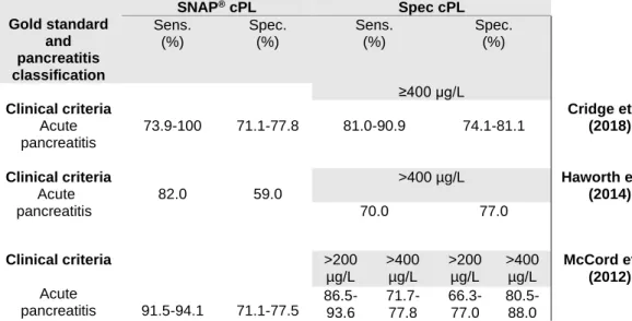
Table 3. Comparison between the sensitivity and specificity of SNAP® cPL and Spec cPL,
reported in previous studies
Therefore, the studies mentioned above are in agreement regarding the performance of the SNAP® cPL tests (McCord et al. 2012; Haworth et al. 2014; Cridge et al. 2018). Also, it
is known that SNAP® and Spec cPL have a higher sensitivity than measurement of total lipase
for the diagnosis of clinical acute pancreatitis (McCord et al. 2012). Although there is a good agreement between SNAP® cPL and Spec cPL (Haworth et al. 2014; Cridge et al. 2018), there
is also some degree of discordance in dogs without clinical acute pancreatitis presenting positive SNAP® results with Spec cPL lower than 200 µg/L (McCord et al. 2012; Haworth et al.
2014; Cridge et al. 2018).
A negative SNAP® cPL result is useful to rule out pancreatitis when there are
suggestive clinical signs, while a positive result is highly suggestive of pancreatitis (Steiner 2017). Still, the collection of a serum sample to quantitatively measure the cPL is recommended in order the allow monitoring disease progression (Steiner 2017).
Recently, an in-house semiquantitaive assay, the VetScan cPL Rapid Test, was developed, giving a rapid point-of-care numerical result rather than a binary one (Cridge et al. 2018). However, this test failed basic analytical validation when performed in a research laboratory under non-clinical circumstances (Steiner and Lidbury 2018; Steiner et al. 2019). Moreover, lack of linearity, precision, and reproducibility was reported for another in-house
Gold standard and pancreatitis classification SNAP® cPL Spec cPL Sens. (%) Spec. (%) Sens. (%) Spec. (%) Clinical criteria Acute pancreatitis 73.9-100 71.1-77.8 ≥400 µg/L 81.0-90.9 74.1-81.1 Cridge et al. (2018) Clinical criteria Acute pancreatitis 82.0 59.0 >400 µg/L Haworth et al. (2014) 70.0 77.0 Clinical criteria Acute pancreatitis 91.5-94.1 71.1-77.5 >200 µg/L >400 µg/L >200 µg/L >400 µg/L McCord et al. (2012) 86.5- 93.6 71.7-77.8 66.3-77.0 80.5-88.0
Legend: SNAP® cPL, SNAP® canine Pancreatic Lipase;Sens., sensitivity; Spec., specificity; Spec cPL,
specific canine pancreatic lipase.
Figure 7.Reaction sequence of DGGR lipase method
Legend: SNAP® cPL, SNAP® canine Pancreatic Lipase; Spec cPL, Specific canine Pancreatic Lipase; Sens,
sensitivity; Spec, specificity.