Interleukin-1 Receptor Antagonist Gene VNTR Polymorphism is
Associated with Coronary Artery Disease
Ahmet Arman, Ozer Soylu, Ahmet Yildirim, Andrzej Furman, Nesrin Ercelen, Hakki Aydogan, Ajda Coker,Tuna Tezel
Departamento de Cardiologia da Faculdade de Engenharia da Universidade Marmara, Hospital Dr. Siyami Ersek; Instituto de Ciências Ambientais, Universidade Bogazici; Departamento de Genética Médica, Hospital Americano; Departamento de Biologia, Artes e Ciências da Universidade Marmara, Istambul, Turquia.Summary
Background:Coronary Artery Disease (CAD) is the atherosclerosis of coronary arteries that carry blood to the heart muscle. Atherosclerosis is an inflammatory disease. Cytokine gene variations such as those associated with the IL1 family are involved in the pathogenesis of atherosclerosis.
Objective: The purpose of this study was to determine the relationship between IL1 family polymorphisms (IL1RN VNTR, IL1B positions -511 and +3953) and CAD in Turkish population.
Methods:427 individuals were submitted to coronary angiography and were grouped as 170 control subjects and 257 CAD patients. The CAD subjects were divided into two subgroups: 91 Single Vessel Disease (SVD) and 166 Multiple Vessel Disease (MVD) subjects. The genotypes of IL1RN and of IL1B (-511, +3953) were determined by polymerase chain reaction (PCR) followed by restriction digestion analysis.
Results:No significant difference was found in IL1RN and IL1B (-511 and +3953) genotype distributions between CAD and control subjects or MVD and control subjects. However, significant association was seen in IL1RN 2/2 genotype between SVD and control subjects (P= 0.016, x2: 10.289, OR: 2.94, 95% CI: 1.183-7.229). Similarly, no statistically significant difference was found in IL1RN and IL1B (-511 and +3953) allele frequencies between CAD and control subjects, MVD and control subjects or SVD and control subjects.
Conclusion: No association was found in either allele frequency or genotype distribution of IL1RN and IL1B polymorphisms between CAD and the control groups. However; IL1RN 2/2 genotype may be a risk factor for SVD in the Turkish population. (Arq Bras Cardiol 2008; 91(5) : 268-273)
Key words:Interleukin 1; mini-satellite repeats; coronary arteriosclerosis; population; Turkey / epidemiology.
Mailing address: Ahmet Arman •
The department of Engineering, Marmara University, Goztepe Campus, 34722, Istanbul,Turkey.
E-mail: [email protected]
Manuscript received August 28, 2007; revised manuscript received January 24, 2008; accepted January 14, 2008.
Introduction
Atherosclerosis is an inflammatory disease that affects medium or large arteries causing gangrene, stroke and heart diseases. Coronary Artery Disease (CAD) is a multifactorial heart disease caused by atherosclerosis of coronary arteries1. Growth and
clotting factors, cytokines, adhesion molecules and their effects on vascular Endothelial Cells (EC) and Smooth Muscle Cells (SMC) were studied for insight into atherosclerotic processes2.
Inflammation in the artery wall triggers the production of primary proinflammatory cytokines such as Interleukin-1β (IL-1β), Tissue Necrosis Factor α (TNFα) which can induce the secondary proinflammatory cytokine (IL-6), the chemokine (IL-8), and adhesion molecule (E-selectin) production3.
The IL-1 family has three well-studied members, two agonists, IL-1α and IL-1β, and the antagonist 1Ra. IL-1Ra inhibits IL-1-induced inflammation action by blocking the binding of IL-1 to IL-1 Type I Receptor (IL-1RI)4.
IL-1Ra is expressing from IL1RN gene which has a length variation within intron 25 caused by 86 bp Variable Number
of Tandem Repeats (VNTR)6. According to the number of 86
bp repeats, there are six alleles corresponding to 1, 2, 3, 4, 5, 6 repeats6-7. Single nucleotide polymorphisms (SNPs) have
been determined at promoter position -511 C/T8 and in exon
5 at position +3953 C/T of IL1B gene9. These polymorphisms
in IL1RN and IL1B genes are thought to influence gene expression8-9.
There are various reports showing a relationship between VNTR and SNPs on the IL1 gene family and diseases. IL1RN genotype 2/2 has been found to be significantly associated with Single Vessel Disease (SVD) in Sheffield English population10.
Significant relevance was found between IL1RN allele 2 and CAD with Type 2 diabetes11. Also IL1RN allele 2 has a
Coronary Angioplasty (PTCA)12. But no association was found
between IL1B SNPs and CAD10,13.
The purpose of this study was to investigate whether polymorphisms in IL1RN (VNTR) and IL1B (-511, +3953) genes are associated with CAD in Turkey. Our results showed that neither IL1B (+3953) nor IL1B (-511) SNP genotype distributions and allele frequencies are susceptible to CAD, but people carrying IL1RN 2/2 genotype may be susceptible to SVD in the Turkish population.
Methods
Subjects
The study population was selected from patients undergoing coronary angiography at Dr. Siyami Ersek Hospital between 2003-2006. Indications for coronary arteriography in our clinic are based on guidelines for coronary angiography14.
According to coronary angiography results12,15 participants
who presented ≥ 70 % stenoses in at least one of the major coronary arteries were considered as CAD (n= 257) and the remainders who presented ≤ 30 % stenosis were accepted in the normal group (n= 170). CAD group was divided into two subgroups: SVD (only one coronary artery stenosis; n= 91) and Multiple Vessel Disease (MVD; more than one coronary artery stenosis; n= 166).
Risk factor assessment
Age, gender, family history of CAD, smoking habit, history of hypertension were obtained by filling a questionnaire form. Weight, height, Systolic (SBP) and Diastolic Blood Pressures (DBP) were measured. Body Mass Index (BMI) was calculated from height and weight measurements. The presence of
Diabetes Mellitus (DM) was defined by repeated fasting glucose level > glucose 126 mg/dl, the use of antidiabetic drugs or both. Total Cholesterol (TC) and High Density Lipoprotein (HDL), Low Density Lipoprotein (LDL) cholesterol levels were determined enzymatically, and also measured enzymatically after dextrane sulfate magnesium precipitation16.Information
about the study was given to all patients and control group, the informed consent was received from each of them.
Genotyping
Blood samples were taken from 427 participants and kept in tubes with EDTA. Genomic DNA isolation was done using GENTRA DNA isolation kit. We have amplified VNTR region in intron 2 of IL1RN gene, IL1B (-511) and IL1B (+3953 exon 5 regions as described previously17. Genotypes of IL1RN gene
were determined by repeating unit size of PCR products. IL1B (-511) genotype was done based on the size of digested PCR products with AvaI, and IL1B (+3953) genotype was determined based on the digested PCR products with Taq I.
Statistical analysis
Mann-Whitney U and x2 tests were used for the comparison
of two groups of individuals according to CAD risk factors. Allele and genotype frequencies among cases and controls were compared with Hardy-Weinberg predictions using x2
–analysis. Multivariate logistic regression analysis was done
to assess the distribution of IL1RN, IL1B genotypes in the control groups and cases. The results were expressed as odds ratio (OR) and 95% Confidence Interval (CI). The final model was adjusted for age, gender, family history of CAD, smoking habit, history of hypertension. Allele frequencies and genotype distribution among cases and controls were compared and values were calculated by x2 test. Probability for p<0.05 values
(2-sided) were considered statistically significant. SPSS 11.5 software program was used for all statistical analysis.
Ethics approval
This research was approved by the ethics committee of Marmara University in Turkey. All human subjects’ rights in this research are protected and any necessary approval was secured from the ethics committee.
Results
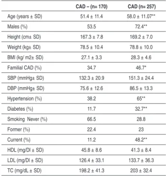
Baseline characteristics of control and CAD groups can be found in Table I. Significant difference between CAD and control subjects for gender (P= <0.001), family history of CAD (P= 0.014), Diabetes (P<0.001) History of Hypertension (P= <0.001) and current smoking (P= <0.001) can be found in Table 1.
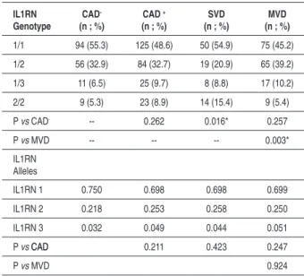
Genotype distribution and allele frequency of IL1RN VNTR are shown in Table 2. Four of six genotypes at IL1RN gene were observed in CAD and control subjects. Among all these genotypes, only IL1RN 2/2 is significantly different
Table 1 - Baseline characteristics of control and CAD groups in Turkish Population
CAD – (n= 170) CAD (n= 257)
Age (years ± SD) 51.4 ± 11.4 58.0 ± 11.07**
Males (%) 53.5 72.4**
Height (cm± SD) 167.3 ± 7.8 169.2 ± 7.0
Weight (kg± SD) 78.5 ± 10.4 78.8 ± 10.0
BMI (kg/ m2± SD) 27.1 ± 3.3 28.3 ± 4.6
Familial CAD (%) 34.7 46.7*
SBP (mmHg± SD) 132.3 ± 20.9 151.3 ± 24.4
DBP (mmHg± SD) 75.6 ± 12.6 86.5 ± 13.3
Hypertension (%) 38.2 65**
Diabetes (%) 11.7 32.7**
Smoking Never (%) 66.5 28.8
Former (%) 22.4 23
Current (%) 11.2 48.2**
HDL (mg/Dl ± SD) 45.8 ± 8.6 41.3 ± 8.4
LDL (mg/Dl ± SD) 126.4 ± 33.1 133.7 ± 36.3
Table 2 - Genotype distribution and allele frequency of IL-1RN VNTR polymorphism in Turkish Population
IL1RN Genotype
CAD
-(n ; %)
CAD +
(n ; %)
SVD (n ; %)
MVD (n ; %)
1/1 94 (55.3) 125 (48.6) 50 (54.9) 75 (45.2)
1/2 56 (32.9) 84 (32.7) 19 (20.9) 65 (39.2)
1/3 11 (6.5) 25 (9.7) 8 (8.8) 17 (10.2)
2/2 9 (5.3) 23 (8.9) 14 (15.4) 9 (5.4)
P vs CAD- -- 0.262 0.016* 0.257
P vs MVD -- -- -- 0.003*
IL1RN Alleles
IL1RN 1 0.750 0.698 0.698 0.699
IL1RN 2 0.218 0.253 0.258 0.250
IL1RN 3 0.032 0.049 0.044 0.051
P vs CADCAD 0.211 0.423 0.247
P vs MVD 0.924
Genotypes are expressed as number of patients (proportion in % within brackets), p values are from chi square test.
Table 3 - Genotype distribution and allele frequency of the IL1B (-511) SNP polymorphisms in CAD, SVD, MVD and the control groups in Turkish Population
IL1B (-511) Genotype
DAC
-(n ; %)
DAC +
(n ; %)
SVD (n ; %)
MVD (n ; %)
1/1 51 (30.0) 75 (29.2) 30 (33) 45 (27.1)
1/2 74 (43.5) 130 (50.6) 41 (45.1) 89 (53.6)
2/2 45 (26.5) 52 (20.2) 20 (22) 32 (19.3)
P vs CAD- 0.242 0.712 0.142
P vs MVD 0.416
IL1B (-511) Alleles
IL1B 1 0.518 0.545 0.555 0.539
IL1B 2 0.482 0.455 0.445 0.461
P vs CADCAD- 0.442 0.462 0.589
P vs MVD 0.781
Genotypes are expressed as number of patients (proportion in % within brackets), p values are from chi square test.
Table 4 - Genotype distribution and allele frequency of IL-1B +3953 SNP polymorphisms in CAD, SVD, MVD and control groups.
IL1B (+3953) Genotype
DAC
-(n ; %)
DAC +
(n ; %)
SVD (n ; %)
MVD (n ; %)
1/1 93 (54.7) 151 (58.8) 52 (57.1) 99 (59.6)
1/2 68 (40) 91 (35.4) 35 (38.5) 56 (33.7)
2/2 9 (5.3) 15 (5.8) 4 (4.4) 11 (6.6)
P vs CAD- 0.629 0.907 0.472
P vs MVD 0.627
IL1B (+3953) Alleles
IL1B 1 0.747 0.765 0.764 0.765
IL1B 2 0.253 0.235 0.236 0.235
P vs CAD- 0.569 0.750 0.591
P vs MVD 0.973
Genotypes are expressed as number of patients (proportion in % within brackets), p values are from chi square test.
between SVD and healthy controls (P= 0.016, x2: 10.289, 3
df, OR: 2.94, 95% CI: 1.183-7.229). However, no significant difference was found between both controls and CAD (P= 0.262) and MVD vs control subjects (P= 0.257). Also, the genotype distribution between SVD and MVD was found to be statistically significant (P= 0.003, x2: 13.806, 3 df, OR:
5.22, 95% CI: 1.995-14.195). For IL1RN allele frequencies, no association was detected between control and cases: CAD (P= 0.211), MVD (P= 0.247), SVD (P= 0.423) (Table 2).
Genotype distribution and allele frequency of IL1B (-511) in CAD, SVD, MVD and control groups are shown in Table 3. The IL1B -511 genotype showed no significant difference between CAD and controls (P= 0.242), SVD and control (P= 0.712) and MVD and control (P= 0.142). Similarly, no significant differences were observed in allelic distribution between controls and CAD (P= 0.442), SVD (P= 0.098), and MVD (P= 0.142) (Table 3).
The allelic frequency and genotype distribution of IL1B +3953 in CAD, SVD and MVD and control groups are shown in Table 4. When genotype distribution of IL1B +3953 was compared among groups, no significant difference was observed between control groups and CAD (P= 0.629), SVD (P= 0.907), and MVD (P= 0.472). Also, no significant difference was observed in allelic distribution between controls and CAD (P= 0.569), SVD (P= 0.750) and MVD (P= 0.591) (Table 4).
Genotypes are expressed as number of patients (proportion in % within brackets), p values are from chi square test.
Homozygosity for IL1RN 2 allele was found significant between SVD and control groups (x2: 7.510, P= 0.010, OR:
3.253, % 95 CI: 1.349-7.844), However, this association can not be seen in heterozygosity for IL1RN 2 allele (P=0.757) (Table 5). Also, significant association was found in homozygosity for IL1RN 2 allele between MVD and SVD
groups (x2: 7.160, P= 0.011, OR: 2.838, % 95 CI:
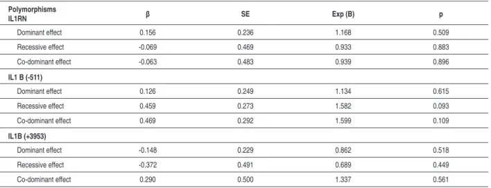
1.278-6.299); (Table 5). Additionally, the relative effect of the IL1 family polymorphism on the risk of CAD was not seen in a multiple logistic regression model including dominant, recessive and co-dominant effects (Table 6).
Discussion
Table 5 - Gene dosage-dependent association of IL-1RN 2 allele and CAD
IL1RN 2 Carrier p Homozygote % 95 CI p Heterozygote % 95 CI
CAD-vs CAD+ 0.160 (0.793-3.999) 0.545 (0.775-1.713)
CAD-vs MVD 0.959 (0.397-2.651) 0.268 (0.841-2.008)
CAD-vs SVD 0.010 * (1.349-7.844) 0.757 (0.542-1.558)
MVD vs SVD 0.011 * (1.278-6.299) 0.234 (0.836- 2.391)
P < 0.05 is taken as statistically signiicant.
Table 6 - A multiple logistic regression model for evaluating the relative effects of the polymorphisms on the risk of CAD
Polymorphisms
IL1RN β SE Exp (B) p
Dominant effect 0.156 0.236 1.168 0.509
Recessive effect -0.069 0.469 0.933 0.883
Co-dominant effect -0.063 0.483 0.939 0.896
IL1 B (-511)
Dominant effect 0.126 0.249 1.134 0.615
Recessive effect 0.459 0.273 1.582 0.093
Co-dominant effect 0.469 0.292 1.599 0.109
IL1B (+3953)
Dominant effect -0.148 0.229 0.862 0.518
Recessive effect -0.372 0.491 0.689 0.449
Co-dominant effect 0.290 0.500 1.337 0.561
Model adjusted for age, gender, familial CAD, smoking habit, history of hypertension.
etiology and development processes of CAD are not well understood, but it has been shown that both inflammation and genetics play an important role in the pathogenesis of atherosclerosis2. Many epidemiological studies have
investigated the association between CAD and inflammatory cytokine gene polymorphisms19.One of the candidate
proinflammatory cytokines associated with CAD susceptibility is IL-1β, which affects the majority of cells and is involved in immunity, sepsis, infection and inflammation. In contrast, IL-1Ra acts as an anti-inflammatory agent that inhibits the action of IL-120.
Francis et al10 found the association between IL-1RN 2/2
genotype and SVD in the UK Caucasian Sheffield population, but not in the London population. Also, no significant difference was found for MVD in either population group10. Other reports
did not find any statistical significance between IL1RN 2 allele and SVD or IL1RN 2 allele and CAD21,13.Furthermore, it has
been reported that IL1RN 2 allele is significantly associated with CAD in the Type 2 diabetes patients11.Also, IL1RN 2
allele has a protective effect on restenosis after PTCA12 and
an association between IL1RN 2 and carotid atherosclerosis was reported22. It was also reported that future Myocardial
Infarction risk was increased with IL1RN 2 allele and high CRP levels in young patients23. In this study, we found an association
between SVD and IL1RN 2/2 genotype (P= 0.016, OR: 2.94, 95% CI: 1.183-7.229), but no statistical association between IL1RN alleles and CAD (P= 0.211) or MVD (P= 0.247) or SVD (P=0.423). In addition, Genotype distribution analysis indicated that homozygous IL1RN 2 carrier was significantly different between SVD and control groups (x2: 7.510, P=
0.010, OR: 3.253, % 95 CI: 1.349-7.844). People withPeople with homozygous IL1RN allele 2 are 3.25 times more likely to have had SVD than people carrying the other genotypes. However,However, heterozygosity for IL1RN 2 carrier did not show this association (P=0.757). Also homozygous IL1RN 2 carrier was significantly different between MVD and SVD groups (x2: 7.160, P=
0.011, OR: 2.838, % 95 CI: 1.278-6.299). People carrying homozygous IL1RN allele 2 are 2.83 times more likely to have SVD than MVD. However, heterozygosity for IL1RN 2 carrier did not show that association (P=0.234; Table 5). Futhermore, we could not find any statistically significant influence of IL1 family polymorphisms (dominant, recessive and co-dominant effects) on the risk of CAD (Table 6). Even though IL1 family polymorphisms did not show any direct relative effect on risks of CAD, those polymorphisms may modulate some of processes for the development of CAD.
are controversial results for the function of IL1RN 2 allele in IL-1Ra expression. It has been reported that IL1RN 2 allele is associated with increased IL-1Ra levels in vitro25-27, decreased
levels in Ulcerative colitis27 but at similar levels for South
African patients with inflammatory bowel disease28. The
results above imply that IL1RN 2 polymorphism function in affecting IL-1Ra protein expression depends on cell type and ethnic origin.
It has been suggested that IL-1β may play an important role in the pathogenesis of atherosclerosis by the stimulation of vascular SMC. Also, increased levels of IL-1β mRNA was detected in atherosclerotic plaques29. The polymorphisms
in IL-1B gene can affect severity or susceptibility to different diseases. Two important SNPs at -511 C/T in the promoter and +3953 C/T in exon 5 of IL1B gene are shown respectively8,9.
Different controversial results have been reported for the effect of IL1B (-511) polymorphism on the production of IL-1Ra and IL-1β. It has been reported that individuals who have IL1B (-511) allele 2 show higher levels of IL-1Ra26. LPS-induced
IL-1β production was increased 2-3 fold by a T allele at -511 position30. No significant relationship between -511 alleles of
IL1B and CAD has been determined10,13. However, Iacoviello
et al31 indicated that the IL1B (-511) 1/1 (C/C) genotype was
associated with MI at young age31. Similarly, Zhang et al32
reported that IL1B (-511) polymorphism was associated to the severity of coronary heart disease in the Chinese population32.
However, we did not find any risk for CAD related to IL1B (-511) polymorphism in the Turkish population. No significant differences were found in the frequencies of allele 1 and 2 or the genotypes of 1/1 and 2/2 of IL1B (-511) between SVD and control or MVD and control or CAD and control groups (Table 3).
Another polymorphism in the IL1B gene is located at position +3953 in exon 5 and thought to influence IL-1β expression. Different results were reported on the effect
of IL1B (+3953) allele on IL-1β protein production. Some studies showed that an association was seen between IL1B (+3953) and increased plasma levels of IL-1β9,30, but others
found no affect on IL-1 levels33-35. No studies are available
on the relationship between CAD and polymorphism in the IL1B gene (+3953). No association was seen between either IL1B (+3953) allele frequency or genotype distribution and CAD in the British population10. Similarly, no relationship was
found between CAD and IL1B (+3953) polymorphism in the Turkish population (Table 4).
The present study has shown that the homozygote carrier of IL-1RN allele 2 may be a risk factor for SVD. Controversial results were obtained for the association between IL1RN 2 and CAD from the different studies and different locations. This showed that ethnic background and different geographical locations play an important role for that association.
Acknowledgement
This research was supported by the FEN-YLS-100105-0068 grant by Marmara University Research Fund and by a partial grant from American Hospital in Turkey. Professor Philip E. Auron, Duquesne University Department of Biological Sciences, are acknowledged for their critical review of the manuscript.
Potential Conflict of Interest
No potential conflict of interest relevant to this article was reported.
Sources of Funding
There were no external funding sources for this study.
Study Association
This study is not associated with any graduation program.
References
1. Ross R. Atherosclerosis: an inflammatory disease. N Engl J Med. 1999; 320: 115-26.
2. Russell R. The pathogenesis of atherosclerosis: a perspective for the 1990s. Nature. 1993; 362: 801-8.
3. Saadeddin SM, Habbab MA, Ferns GA. Markers of inflammation and coronary artery disease. Med Sci Monit. 2002; 8: RA5-12.
4. Dinarello CA. Interleukin-1. Cytokine & Growth Factors Rev. 1997; 8: 253-65.
5. Steinkasserer A, Koelble K, Sim RB. Length variation within intron 2 of the human IL-1 receptor antagonist gene (IL-1 RN). Nucleic Acid Res. 1991; 19: 5095.
6. Tarlow JK, Blakemore AI, Lennard A, Solari R, Huges HN, Steinkasserer A, et al. Polymorphism in human IL-1 receptor antagonist gene intron 2 is caused by variable numbers of an 86-bp tandem repeat. Hum Genet. 1993; 91: 403-4.
7. Vamvakopoulos JE, Taylor CJ, Morris-Stiff GJ, Green C, Metcalfe S. The interleukin-1 receptor antagonist gene: a single-copy variant of the intron 2 variable number tandem repeat (VNTR) polymorphism. Eur J Immunogenet.
2002; 29: 337-40.
8. di Giovine FS, Takhsh E, Blakemore AI, Duff GW. Single base polymorphism at -511 in the human interleukin-1 beta gene (IL1 beta). Hum Mol Genet. 1992; 1 (6): 450.
9. Pociot F, Molvig J, Wogensen L, Worsaae H, Nerup J. A Taq I polymorphism in the human interleukin-1 beta (IL-1 beta) gene correlates with IL-1 beta secretion in vitro. Eur J Clin Invest. 1992; 22 (6): 396-402.
10. Francis SE, Camp NJ, Dewberry RM, Gunn J, Syrris P, Carter ND, et al. Interleukin-1 receptor antagonist gene polymorphism and coronary artery disease. Circulation. 1999; 99: 861-6.
11. Marculescu R, Endler G, Schillinger M, Iordanova N, Exner M, Hayden E, et al. Interleukin-1 receptor antagonist genotype is associated with coronary atherosclerosis in patients with type 2 diabetes. Diabetes. 2002; 51: 3582-5.
12. Francis SE, Camp NJ, Burton AJ, Dewberry RM, Gunn J, Stephens-Lloyd A, et al. Interleukin 1 receptor antagonist gene polymorphism and restenosis after coronary angioplasty. Heart. 2001; 86: 336-40.
Haematologica. 2003; 88: 54-60.
14. Scanlon PJ, Faxon DP, Audet AM, Carabello B, Dehmer GJ, Eagle KA, et al. ACC/AHA guidelines for coronary angiography: a report if the American College of Cardiology / AHA. Task Force on practice guidelines (Committee on Coronary Angiography). Developed in collaboration with the Society for Cardiac Angiography and Interventions. J Am Coll Cardiol. 1999; 33: 1756-824.
15. Deligonul U. Coronary angiography as a prognostic tool. Anadolu Kardiyol Derg. 2001; 3: 189-96.
16. Friedewald WT, Levi RI, Fredrickson DJ. Estimation of the concentration of low density lipoprotein cholesterol in plasma without use of the ultracentrifuge. Clin Chem. 1972; 18: 499-502.
17. Arman A, Yılmaz B, Coker A, Inanc N, Direskeneli H. Interleukin-1 receptor antagonist (IL-1RN) and interleukin-1B gene polymorphisms in Turkish patients with rheumatoid arthritis. Clin Exp Rheumatol. 2006; 24 (6): 643-8.
18. Onat A. Risk factors and cardiovascular disease in Turkey. Atherosclerosis. 2001; 156: 1-10.
19. Andreotti F, Porto I, Crea F, Maseri A. Inflammatory gene polymorphisms and ischaemic heart disease: review of population association studies. Heart. 2002; 87: 107-12.
20. Dinarello CA. Proinflammatory cytokines. Chest. 2000; 118: 503-8.
21. Zee RY, Lunze K, Lindpaintner K, Ridker PM. A prospective evaluation of the interleukin-1 receptor antogonist intron 2 gene polymorphism and the risk of myocardial infarction. Thromb Haemost. 2001; 86: 1141-3.
22. Worrall BB, Azhar S, Nyquist PA, Ackerman RH, Hamm TL, DeGraba TJ. Interleukin-1 receptor antagonist gene polymorphisms in carotid atherosclerosis. Stroke. 2003; 34: 790-3.
23. Manzoli A, Androtti F, Varlotta C, Mollichelli N, Verde M, Van de Greed W, et al. Allelic polymorphism of the IL-1Ra gene in patients with acute or stable presentation of ischaemic heart disease. Cardiologia. 1999; 44: 825-30.
24. Arend WP. The balance between IL-1 and IL-1 Ra in disease. Cytokine Growth Factor Rev. 2002; 13: 323-40.
25. Danis VA, Millington M, Hyland VJ, Grennan D. Cytokine production by normal human monocytes: inter-subject variation and relationship to an IL-1
receptor antagonist (IL-1Ra) gene polymorphism. Clin Exp Immunol. 1995; 99: 303-10.
26. Hurme M, Santtila S. IL-1 receptor antagonist (IL-1Ra) plasma levels are co-ordinately regulated by both IL-1Ra and IL-1beta genes. Eur J Immunol. 1998; 28 (8): 2598-602.
27. Tountas NA, Casini-Raggi V, Yang H, di Giovine FS, Vecchi M, Melani L, et al. Functional and ethnic association of allel 2 of the interleukin-1 receptor antagonist gene in ulcerative colitis. Gastroenterology. 1999; 117: 806-13.
28. Mwantembe O, Gaillard MC, Barkhuizen M, Pillay V, Berry SD, Dewar JB, et al. Ethnic differences in allelic association of the interleukin-1 gene cluster in South African patients with inflammatory disease and in control individuals. Immunogenetics. 2001; 52: 249-54.
29. Offner FA, Feichtinger H, Stadlmann S, Obrist P, Marth C, Klingler P, et al. Transforming growth factor-beta synthesis by human peritoneal mesothelial cells. Am J Pathol. 1996; 148 (5): 1679-88.
30. Hall SK, Perregaux DG, Gabel CA, Woodworth T, Durham LK, Huızınga TW, et al. Correlation of polymorphic variation in the promoter region of the interleukin-1 beta gene with secretion of interleukin-1 beta protein. Arthritis Rheum. 2004; 50 (6): 1976-83.
31. Iacoviello L, Donati MB, Gattone M. Possible different involvement of interleukin-1 receptor antagonist gene polymorphism in coronary single vessel disease and myocardial infarction. Circulation. 2000; 101 (18): E193.
32. Zhang YM, Zhong LJ, He BX, Li WC, Nie J, Wang X, et al. The correlation between polymorphism at position -511C/T in the promoter region of interleukin 1B and the severity of coronary heart disease: Zhonghua Yi Xue Yi Chuan Xue Za Zhi. 2006; 23 (1): 86-8.
33. Camargo JF, Correa PA, CastIblanco J, Anaya JM. Interleukin-1beta polymorphisms in Colombian patients with autoimmune rheumatic diseases. Genes Immun. 2004; 5 (8): 609-14.
34. Dominici R, Malferrari G, Mariani C, Grimaldi L, Biunno I. The Interleukin 1-beta exonic (+3953) polymorphism does not alter in vitro protein secretion. Exp Mol Pathol. 2002; 73 (2): 139-41.