Key Words
Applied research; models, animals; coronary angiography; heart catheterization.
The Importance of Pre-Clinical Animal Testing in Interventional
Cardiology
Yoriyasu Suzuki, Alan C. Yeung, Fumiaki Ikeno
Division of Cardiovascular Medicine, School of Medicine, Stanford University, Stanford, California, USA
Mailing Address: Fumiaki Ikeno, M.D. •
Stanford University, Division of Cardiovascular Medicine, 300 Pasteur Drive, Falk CVRB007, Stanford, CA94305a
E-mail: [email protected]
Summary
The treatment of cardiovascular disease has changed dramatically over the past 2 decades, allowing patients to live longer and better quality lives. The introduction of new therapies has contributed much to this success. Nowhere has this been more evident than in interventional cardiology, where percutaneous cardiovascular intervention has evolved in the past 2 decades from a quirky experimental procedure to a therapeutic cornerstone for patients with symptomatic cardiovascular disease. The development of these technologies from the earliest stages requires preclinical experiments using animal models. Once introduced into the clinical arena, an understanding of therapeutic mechanisms of these devices can be ascertained through comparisons of animal model research findings with clinical pathological specimens.
This review provides an overview of the emerging role, results of preclinical studies and development, and evaluation of animal models for percutaneous cardiovascular intervention technologies for patients with symptomatic cardiovascular disease.
Introduction
To improve human health, scientific discoveries and technologies must be translated into practical applications. Such advances typically begin at “the bench” with basic research where scientists study disease at a molecular or cellular level and then progresses to the clinical level, or the patient’s “bedside.” Translational research is an interchangeable term that underscores the pressing need to translate the practical benefits for those affected by symptomatic cardiovascular disease with the extensive investments divested by the private and public sectors in biomedical research. Translational research should be viewed as a two-way road: bench to bedside, and bedside to bench. In particular, to facilitate a more effective translation process, a new road map should be implemented to foster interaction and cooperation between basic researchers, clinicians, laboratory professionals and device manufacturers.
Nowhere has this been more evident than in interventional cardiology, where percutaneous cardiovascular intervention has evolved from a quirky experimental procedure to a therapeutic cornerstone for patients with symptomatic cardiovascular disease. Inherent in the development of these technologies is the role of preclinical testing using animal models. Once these technologies enter the clinical arena (bench to bedside), a further understanding of their therapeutic mechanisms can be realized through comparative analysis of animal model research findings with those of clinical pathological specimens (bedside to bench).
This review will provide an overview of the clinical application status and limitations of current percutaneous cardiovascular intervention technologies, future technologies under development, and results of preclinical studies including animal models.
Experimental animal model for restenosis and coronary intervention technologies
A new era in the field of percutaneous coronary intervention has arrived: the drug-eluting stent (DES)1, 2. These stents are coated with antiproliferative drugs such as sirolimus, and have been shown to limit in-stent restenosis in discrete lesions3. Other drug coatings, such as paclitaxel analogs, have shown similar efficacy4. The success of these antiproliferative agents is founded not only in initial human clinical data but also on preclinical studies using the porcine coronary restenosis model5-8. Presently, it is unclear whether any single animal species is more predictive of the human response to such coated stents. As such, we maintain that most animal models currently available can still provide mechanistic insight into fundamental biological processes and response. These animal models can therefore help prove critical hypotheses regarding putative mechanisms of action of an intervention yet cannot be used to predict efficacy9. The following section will summarize an overview of current coronary stent technologies as well as the animal models used in their evaluation.
Brief over view of each animal model Small animal model
study molecular biology14-16. Conversely, this model assumed less importance after several early studies of angiotensin-converting enzyme inhibitors17-21, where failure of this model to predict negative clinical trial results caused it to lose favor among investigators.
The mouse arterial injury as a restenosis model developed from the availability of the mouse genome and molecular methods to study events after arterial injury22,23. Since this model has very small vessels, traditional injury methods by balloon or stent are not practical. Vascular injury may instead be performed by rotating a small guide wire in the vessel or electrical injury, and placement of a non-constrictive peri-vascular cuff around the mouse femoral artery24,25. Through the use of these methods, variable focal neointimal thickening forms at injury sites proportionate to the amount of injury with little thrombus formation. A thin neointima (roughly 0.03 mm2) forms by three weeks. The power of molecular biology and genetics in the mouse model will permit substantial advances in understanding the interactions between cell proliferation, cell migration, thrombus formation, and remodeling.
Large animal model
The rabbit iliac restenosis model has also been studied extensively to test restenosis therapies and to understand cellular and molecular mechanisms26-28. Blood cholesterol levels are typically >1,000 mg/dl and cause biochemical arterial injury which can be further supplemented by mechanical injury. These models can have initial injury induced by air desiccation to hypercholesterolemic diets and then balloon inflation to further injure the vessel. Histopathology in this model shows foam cells (macrophages that have ingested excessive lipid) and voluminous extracellular matrix. One criticism of this model is that foam cells are rare in human restenotic neointima. However, balloon angioplasty in this model does cause histopathologic injury comparable to that seen with human angioplasty, with medial dissection and plaque fracture. Platelet deposition occurs rapidly at sites of a balloon-induced plaque fracture. Thus, antiplatelet agents, as a potential anti-restenosis therapy, were studied early in the history of this model29, 30 and showed efficacy in reducing neointimal thickness.
The coronary arteries of domestic pigs after injury respond in similar fashion as human coronary arteries31, 32. Furthermore, when porcine coronary arteries are injured, thick neointima will be seen within 28 days and is identical to human restenotic neointima. When a balloon-only injury is performed, a typical medial laceration occurs and is filled at 28 days by neointima. In addition, the amount of neointimal thickening is directly proportional to injury thereby permitting the creation of an injury-response regression relationship that can further quantify the response to potential treatment therapies33,34.
Experience suggests that the coronary arteries in domestic swine and iliac arteries of rabbits are suitable in that their size, access, and injury response are similar to human vessels, and therefore allow assessment of devices that might be used in clinical evaluation35. Although armed with fewer collateral vessels, there are pertinent similarities between the pig and human cardiovascular system, including the distribution of epicardial coronary arteries.
Technical consideration of porcine coronary model
Porcine coronary models using vascular injury methods are now accepted standards by which potential restenosis therapies are studied35.
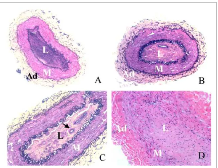
In the porcine model, the left main coronary artery (LMCA) generally bifurcates early into the left anterior descending (LAD) and circumflex (LCx) coronary arteries (Fig.1). These vessels are of similar diameter to those in the human (2.0–4.0 mm). Similarly, the LCx commonly has 1–3 marginal branches and the LAD supplies the septum (Fig.1). Furthermore, the porcine right coronary artery (RCA) is usually of similar diameter to the human RCA, although it is less often dominant
than in humans, where RCA dominance occurs in 80% of
cases (Fig.1).
Cardiac catheterization techniques
Cardiac catheterization techniques in the pig are similar in many ways to the techniques used in humans. Procedures are often performed under general anesthesia. Intubation can be achieved using human endotracheal tubes, although the pig’s longer pharynx may make this more technically challenging. Arterial access can be achieved via the carotid or femoral arteries, using a cut-down approach or by direct percutaneous puncture using a modified Seldinger technique, respectively.
Standard human diagnostic and interventional equipment may be used, with catheter choice dependent on the approach. In general, via the carotid approach, our group has usually used a Judkins left 3.5 or 4.0 guide catheter36-39. Via the femoral approach, the hockey stick guide catheter may be used for both left main and RCA ostia8, or Judkins right 4.0 guide catheter for the RCA (Fig.1).
Antiplatelet therapy is recommended, using aspirin (300 mg) started the day before the procedure and continuing until sacrifice6,8. Clopidogrel (75 mg) has also been used in addition to aspirin36,40. Heparin (100–300 U/kg) is often used during the procedure as an antithrombotic although stent thrombosis may still occur with this regimen, as in the human41.
Stent implantation
Only one stent should be implanted per artery except when issues of stent overlap or multiple stent dosing are considered. The stent may be placed in multiple different arteries in the same animal including bare metal stents such as copper and/or gold stents for development of restenosis model, carrier-only such as polymeric material, and carrier plus DES for evaluation of DES. Regarding DES evaluation, recommendations from a preclinical studies consensus group suggest that the stent should be appropriately sized by visual or quantitative coronary
artery measurement using a stent:artery ratio ≤1:1 , as using a
higher stent:artery ratio could induce severe arterial injury and considerable coronary artery stenosis9.
Stent overlap frequently occurs during clinical implant and overlapping DES present the possibility of a combined effect from drug released from the 2 stents.
Evaluation of stent performance
1) Injury and inflammation score: Inflammation by histopathologic evaluation can include an injury score at each stent strut site (Fig.2), an inflammation description (absent, or cell types and location), and an inflammation score for the overall vessel as well as for the adventitia, media, neointima, and at stent strut sites. When possible, cell density in tissue compartments should be recorded as number of cells per area32,42.
2) Stent strut position and adjacent tissue: Other observational data should include stent strut apposition to the vessel wall and stent struts covered by tissue or endothelium. A subjective description should also be rendered for adjacent tissue, including medial thinning, loss of cellularity, and hyalinization.
3)Quantitative histomorphometry: histomorphometry of histopathologic sections is essential for stent evaluation. Measurements at all sections should include medial area, IEL area (area within the internal elastic lamina), EEL area (area within the external elastic lamina), lumen area, and stent area (area within the stent itself). Neointimal measurement is important for efficacy assessment and should include thickness at each stent strut site and total neointimal area9.
4) Vascular response, and healing: Drug choice and release kinetics are the most important components of DES technology because they determine the type of vascular response and time-course of healing. From numerous studies,
considerable data exist on how sirolimus and paclitaxel differ in terms of their effects on the arterial wall43,46.
Endothelialization after stent implantation should be recorded as absent, partial, or complete in all sections and the time of re-endothelialization should be estimated. In the porcine coronary stent model, a thick neointima was reliably induced by 28 days (Fig. 3) and several reports have investigated the phasic, time-dependent cellular response after stenting47-49.
Preclinical studies of both sirolimus-eluting stents (SES) and paclitaxel-eluting stents (PES) have demonstrated the efficacy compared with BMS6,8. However, enthusiasm for this technology has recently dampened by concerns of late stent thrombosis. A major criticism of earlier preclinical studies leading to the United States Food and Drug Administration (FDA) approval of both stents was their failure to detect significant differences in the healing response of the arterial wall when compared with BMS at 28 days while human angioscopic and autopsy data clearly demonstrated significant differences in healing50,51. Most preclinical studies have failed to show any significant differences between DES and BMS in the extent of endothelial coverage when a 1.1:1 balloon artery ratio was chosen. It was not until the results of a study using overlapping commercially available SES and PES stents in the rabbit iliac artery model showed incomplete endothelialization Figure 1 - Porcine and human epicardial coronary anatomy; Porcine (A and B). A, Left coronary system. B, Right coronary artery. Human (C and D). C, Left coronary system. D, Right coronary artery. Coronary angiography was undertaken via the right femoral approach in both pig and human. Similar anatomy and coronary distribution is shown of the left anterior descending, left circumlex, and right coronary arteries40
compared with matched BMS controls that these differences were recongnized52. Recently, Nakazawa et al.53 have reviewed the comparison of preclinical data from SES, PES, and the phosphorylcholine-coated Endeavor zotarolimus-eluting stent (ZES; Medtronic Vascular, Santa Rosa, CA). In this review, incomplete endothelial coverage was seen in non-overlapping and overlapping sites of both SES and PES compared with both ZES and BMS, though the differences were more pronounced in overlapping segments (Fig.4). Accompanying these findings were more increases in fibrin and inflammatory cells in SES than in either ZES or BMS, which persisted out to 90 days after stent implantation53. Two studies using human autopsy samples suggested that incomplete endothelial coverage of stent struts played one of the very important roles as the morphometric predictor of late stent thrombosis although the cause of late stent thrombosis is likely multifactorial, with delayed re-endothelialization in combination with other clinical and/or procedural risk factors51,52.
Two time points should be used for the evaluation of stent performance, the first at 28 days to observe for neointimal hyperplasia, and at least one later time point to examine long term effects. The later time point (3 or 6 months) depends on when “healing” and drug release are both complete. Of note, the FDA has typically recommended 6 months follow up as the interval to acquire preclinical stent data.
Porcine heat-injury restenosis model
The porcine coronary stent restenosis model is a well-accepted standard. However, the fundamental drawback
of this model is that there is no stenosis lesion in coronary arteries and that the stent itself is foreign material. As a result, this model may not be suitable to evaluate the performance of bifurcation or bioabsorbable stents due to lack of a true stenosis lesion. Also, results of coronary artery imaging such as computed tomography (CT), magnetic resonance imaging (MRI), intravascular ultrasound (IVUS), and optical coherence tomography (OCT) may be hampered as the stent can produce artifact.
Radiofrequency thermal balloon angioplasty was introduced as a new technique for percutaneous arterial dilatation in the 1990’s54-56, yet due to increased restenosis rates observed in patients receiving this therapy57, it was soon abandoned as a percutaneous interventional treatment option. Using this system, Staab et al.58 and our laboratory39 have investigated a porcine heat-injury restenosis model.
In our study using 22 swine with a total of 54 coronary arteries, coronary artery stenoses were consistently developed at 4 weeks after heat-injury (Fig.5). In light of these results, this porcine coronary restenosis model might be useful for the evaluation of bifurcation stents and bioabsorbable stents, coronary imaging studies such as MRI, CT, IVUS, and OCT, and for the technical training of complex percutaneous coronary interventions such as bifurcation stenting and atherectomy39.
Experimental animal model for chronic total occlusion (CTO)
Recent advances of DES technology has shifted focus within interventional cardiology from restenosis prevention Figure 2 -Scoring systems for the porcine response to stent injury; A - Anatomic (Schwartz score); 0 - endothelium denuded; IEL intact; Media compressed, not lacerated; 1 - IEL lacerated; Media compressed, not lacerated; 2 - IEL lacerated; Media lacerated; EEL compressed, not lacerated; 3 - Media severely lacerated; EEL lacerated; Adventitia may contain stent struts; B - Inlammatory; 0 - no inlammatory cells surrounding stent struts; 1 - light, non-circumferential cellular iniltrate, localized to stent strut; 2 - moderate, non-circumferential cellular iniltrate, localized to stent strut. 3 - dense, circumferential cellular iniltrate; M - media; A - adventitia; Dots denote inlammatory cellular iniltrate; Adapted from Schwartz et al.32 and Kornowski et al42
Figure 3 -A - Photomicrographic section shows gross neointimal proliferation causing a signiicant stenosis (Elastic-van Gieson stain, x30); The gross proliferation and luminal compromise by neointima is obvious; B - Microscopic section in a case in which, fortuitously, not all coil wires penetrated into the vessel media; In this section, the two coils farthest left penetrated the media (arrows) and resulted in substantial proliferation; A short segment of vessel media at the lowermost portion of the igure is entirely normal, without any proliferation, although this segment was stretched by the balloon; Elastic- van Gieson stain was used (x30); L - lumen; M - media; NI - neointima;*, holes from coil wires 31.
to the treatment of CTO. It is often stated that successful chronic total occlusion (CTO) recanalization represents the “last frontier” of percutaneous coronary intervention. This interest has stimulated the development of specialized devices such as the Frontrunner (Lumend Inc., Redwood City, CA) that performs blunt micro-dissection59, and the Safecross (Intraluminal Therapeutics Inc., Carlsbad, CA) that utilizes optical coherence reflectometry for traversing the CTO60. Despite its common occurrence, there is surprisingly little information about the pathophysiology of CTO, and why some CTO can be crossed while in others, crossing is unsuccessful. For the past several years, researchers have developed CTO models to guide therapeutic investigations, including image-guidance intervention and device development.
Overview of animal model for CTO
Spontaneous atherosclerotic plaque rupture and subsequent arterial occlusion do not occur naturally in any animal model, even among those models that have been genetically engineered to have increased atheroma formation. For the past several years, researchers have tried to develop several kinds of animal CTO models. The initial method of producing a total occlusion utilized external ligature or ameroid constriction61,62. However, a fundamental drawback of this method is the
inability to facilitate the development of devices to recanalize CTO. Subsequent techniques for endoluminal formation of CTO in coronary and peripheral arteries have differed in their fundamental approach. Murphy et al.63 evaluated 4 methods in a rabbit iliac model for developing peripheral arterial thrombosis obliterans. Of these, gas-drying with thrombin and a high-cholesterol diet was deemed the most efficacious. Strauss et al. subsequently modified the thrombin injection model by infusing collagenase before wire passage64. Several characteristics of human CTO were evident in this model, including mature fibrous tissue, multiple small intraluminal vascular channels, occasional extracellular lipid deposits, and disruption of the internal elastic lamina (Fig.6). Their reports suggested that the microchannels may be a critical determinant of successful CTO guide wire crossing65. Other CTO models have included stents with occluded outflow and even direct alcohol injection to promote thrombosis66. Developing an accurate and reproducible human-like coronary CTO model has been a complex undertaking because 1) coronary vessels are less amenable to a direct surgical approach; 2) simulating luminal and medial pathology, including microcalcification, has been difficult; and 3) an inflammatory component must be present to mimic human CTO lesions67,68. Balloon angioplasty and stent implantation in animal coronary arteries, both standard Figure 5 -Representative Coronary Angiogram of the porcine treated with thermal balloon;Time course of coronary artery treated with thermal balloon; A severe tandem coronary artery stenosis is observed in the left anterior descending artery (LAD) at 4 weeks after thermal balloon injury even though the LAD remained overstretched until 2 weeks after the procedure39
Figure 6 - Chronic Total Occlusion (CTO) in the rabbit iliac model; Lumen (L) is occluded by ibrotic intimal lesion; Small vascular channels are evident (B, arrow in C); M indicates media; A - adventitia; A and B, Movat, x10; C, Movat, x20; D, H&E, x20. Adapted from the reference 64.
methods of denuding the vessel and engendering neointimal proliferation, rarely result in CTO development. More aggressive measures tried have involved the use of thermal injury and copper stent implantation as described above39. Polymers have also been used to invoke chronic coronary occlusions. Early polymeric implants were abandoned as stent platforms because they induced severe inflammatory responses and vessel occlusion69. Prosser et al.70 reported placement of a microporous poly L-lactic acid polymer into pig and dog coronary arteries. The polymer is absorbed by 28 days, resulting in a micro-channeled occlusion histologically similar to a human CTO70. These animal models may contribute to a deeper understanding of the biology of human CTO and enable new device and pharmacological investigation to improve recanalization success in these challenging lesions.
Percutaneous interventions for the treatment of valvular heart disease
Valve repair and replacement are common surgical procedures and are typically effective in eliminating or significantly reducing valve dysfunction. However, these procedures remain
controversial as sole treatments for patients with a low ejection fraction. The challenge in treating patients with congestive heart failure due to valvular disease is deciding the mode of repair to address multiple factors such as alignment of the leaflets, the size of annulus, and geometries of subvalvular apparatus. Coupled with the risk of morbidity and mortality due to open heart surgery, these reconstructive procedures have proven to be a challenge to the surgeon and a risk to the patient, thereby motivating scientists to design devices that can treat valvular dysfunction in a minimally invasive manner. Based on the experience gained from the development of surgical valve prostheses, the FDA has established detailed guidelines for the assessment of fatigue, flow dynamics, and hydrodynamics of valve implants as well as processes for in vitro and in vivo preclinical testing of heart valve prostheses. In these preclinical studies, not only device development and durability testing but also optimal imaging and deployment protocols should be established and comprehensive user training should be initiated in the latter stages of the preclinical evaluation71.
Aortic valve replacement
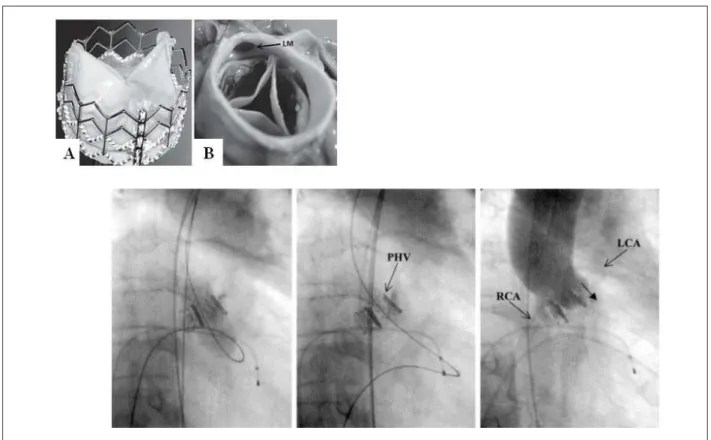
Age-related aortic valve disease contributes to continuously increasing morbidity and mortality worldwide and is associated with an increase in aortic valve replacement procedures over the past decade. Degenerative aortic stenosis, a common adult valvular abnormality72, has been the focus of percutaneous treatments since the mid 1980s . To date, 2 percutaneous aortic valve procedures have been introduced in clinical setting73,74 (Fig.7).
The ovine model is preferred for in vivo assessment of percutaneous aortic valve devices. Currently there is no chronic animal model representative of aortic stenosis. While the healthy ovine model has provided validation of catheter function, prosthesis anchoring, device function post-implantation, and unimpaired coronary blood flow, this model has several limitations:
1) the size of femoral arteries (typically ≤5mm);
2) the angulation of the aortic arch (the cause of kinking of the delivery system);
3) the length of the aortic arch (shorter than that of human); and
4) the location of coronary ostia (closer to the aortic valve than in the human)71. Newer valve technologies may provide solutions to access issues and other limitations of first generation devices.
Mitral Valve Repair
The mitral valve is a complex anatomic and physiologic structure. Its proper function depends on coordinated interaction of the mitral annulus, leaflets, chordae, papillary muscles, and the left ventricle. Improved understanding of the mechanisms of mitral valve dysfunction coupled with advances in catheter-based technology has resulted in several potential percutaneous approaches to mitral valve repair75. Innovations attempt to duplicate techniques of surgical mitral repair. Similar to aortic valve devices, the ovine model is preferred for in vivo assessment of percutaneous mitral valve devices. To date, 2 types of diseased animal models were used in published data76-79. One type is the rapid-pacing heart failure model and the other type is the ischemic mitral regurgitation (MR) model. By using progressive rapid ventricular pacing for 5 to 8 weeks (180-240 beats/minute), the increase in left ventricular dimension results in congestive hear failure and MR. In some publications, left ventricular ejection
fraction (LVEF) reduced up to 24-28% and moderate to severe
MR was developed after rapid pacing77,78,80. One drawback of this model is that after recovery from rapid pacing, LV function return to levels seen in the healthy animals81. In light of this observation, researchers should pay attention to this fact with regard to evaluation of device efficacy. Ischemic MR is induced by coronary arterial occlusion, however, variable anatomy of the coronary artery tree poses a challenge. Gorman et al.82 has investigated the relationship between the coronary arterial
Figure 8 -A - The MitraClip device (Evalve) with the arms of the clip in the open position; B - The left atrium is accessed with the device via transseptal catheterization and the clip engages the mitral lealets from the left ventricular surface; C - A suction port (arrow) is used to draw the lealets into proximity of the catheter (Edwards Lifesciences) in order to suture (arrow, d ) the central portion together; D - A double oriice (arrowheads) mitral valve is the result. Adapted from the reference 87.
Table 1– Transcatheter Mitral valve treatment
Repair category/Name Description status
Edge-to-Edge Repair
MitraClip (Evalve) Clip for edge-to-edge repair Phase II clinical trial
MOBIUS (Edwards) Suture-based edge-to-edge repair Phase I clinical trial
Medtronic Edge-to-edge repair Preclinical
St. Jude Edge-to-edge repair Preclinical
Annuloplasty
MONARCH (Edwards) Coronary-sinus based with anchors and tensioning element Phase I clinical trial
Carillon (Cardiac Dimensions) Coronary-sinus based with anchors and cinching element Phase I clinical trial
PTMA (Viacor) Coronary-sinus based with reversible and adjustable treatment effect Phase I clinical trial
Implant (Extensia) Coronary-sinus based with anchors and tensioning element Preclinical
Mitralign Transventricular suture-based system using coronary sinus as anatomic guide Preclinical
Accucinch (Guided
Delivery Systems) Transventricular annular cinching Preclinical
PS3 (Ample) Transventricular and transseptal approach to shorten septal-lateral mitral dimension Preclinical
Other
anatomy and development of ischemic MR and concluded that only posterior infarction by occlusion of the left circumflex could induce acute or chronic ischemic MR. The primary concern of this model is the mortality and consistence of MR development.
Mortality related to myocardial infarction is about 30-40% and consistence of MR development is about 20-30%82. A “diseased” model might not be necessary for device development or for durability testing of optimal deployment protocols. Two percutaneous approaches – edge to edge repair and annuloplasty – have been subjected to extensive preclinical testing and to preliminary clinical investigation as shown in the table: 1) Edge-to-Edge repair; The first phase I feasibility trial on a percutaneous mitral device has been completed83. A transcatheter mitral clip (MitraClipTM , EValve, Menlo Park, CA) placed on the free edges of the mitral leaflets through a transseptal puncture, mimics the Alfieri surgical procedure (Fig.8).
Another edge-to-edge repair technology, the MIOBIUS leaflet repair system (Edwards Lifesciences, Irvine, CA) uses a suture-based technology to complete an Alfieri-type repair. The preclinical results have confirmed the feasibility of this approach for creation of an edge-to-edge repair84. 2) Annuloplasy: A variety of percutaneous technologies have been developed to alter mitral annular geometry such as coronary sinus-based annuloplasty, direct intracavitary annuloplasty, and other novel cinching devices76-79, 85. Shortening or reshaping the annulus by insertion of a device into the coronary sinus has the potential to mimic surgical annuloplasty. Proof of concept has been demonstrated
experimentally with recent publication of an initial human feasibility study using the MONARCHTM (Edwards Lifesciences, Irvine, CA) which consists of a self-expanding nitinol implant with distal and proximal stent-like anchors86.
A variety of other percutaneous valve devices are under development. Over the next decade, clinical trials will clarify the roles of these new approaches in relation to each other and to current surgical and medical therapies. These percutaneous technologies will be carefully studied and subjected to a level of scrutiny far beyond those applied to new surgical therapies.
Conclusion
The field of percutaneous cardiovascular intervention technology is developing quickly and reflects the time sensitivity of the information contained within this chapter. The basic concepts, however, will be important to understand as all further advances will be generated by the early beginnings including preclinical studies. The success of this interventional subspecialty will be driven by the results of a collaborative relationship between the cardiologist, cardiac surgeon, and the medical device industry. A new era is coming yet again for the discipline of cardiovascular diseases, with new implications not only involving patient care but also in the area of multidisciplinary cooperation.
Acknowledgment
In the preparation of this review, we thank Heidi Bonneau, RN, MS, CCA for her editorial review.
References
1. Serruys PW, Degertekin M, Tanabe K, Abizaid A, Sousa JE, Colombo A, et al. Intravascular ultrasound findings in the multicenter, randomized, double-blind RAVEL (RAndomized study with the sirolimus-eluting VElocity balloon-expandable stent in the treatment of patients with de novo native coronary artery Lesions) trial. Circulation. 2002; 106 (7): 798-803.
2. Sousa JE, Costa MA, Abizaid A, Sousa AG, Feres F, Mattos LA, et al. Sirolimus-eluting stent for the treatment of in-stent restenosis: a quantitative coronary angiography and three-dimensional intravascular ultrasound study. Circulation. 2003; 107 (1): 24-7.
3. Morice MC, Serruys PW, Sousa JE, Fajadet J, Ban Hayashi E, Perin M, et al. A randomized comparison of a sirolimus-eluting stent with a standard stent for coronary revascularization. N Engl J Med. 2002; 346 (23): 1773-80.
4. Park SJ, Shim WH, Ho DS, Raizner AE, Park SW, Hong MK, et al. A paclitaxel-eluting stent for the prevention of coronary restenosis. N Engl J Med. 2003; 348 (16): 1537-45.
5. Gallo R, Padurean A, Jayaraman T, Marx S, Roque M, Adelman S, et al. Inhibition of intimal thickening after balloon angioplasty in porcine coronary arteries by targeting regulators of the cell cycle. Circulation. 1999; 99 (16): 2164-70.
6. Suzuki T, Kopia G, Hayashi S, Bailey LR, Llanos G, Wilensky R, et al. Stent-based delivery of sirolimus reduces neointimal formation in a porcine coronary model. Circulation. 2001; 104 (10): 1188-93.
7. Teirstein PS. Living the dream of no restenosis. Circulation. 2001; 104 (17): 1996-8.
8. Heldman AW, Cheng L, Jenkins GM, Heller PF, Kim DW, Ware M Jr, et al. Paclitaxel stent coating inhibits neointimal hyperplasia at 4 weeks in a porcine
model of coronary restenosis. Circulation. 2001; 103 (18): 2289-95.
9. Schwartz RS, Edelman ER, Carter A, Chronos N, Roger C, Robinson KA, et al. Drug-eluting stents in preclinical studies: recommended evaluation from a consensus group. Circulation. 2002; 106 (14): 1867-73.
10. Clowes AW, Schwartz SM. Significance of quiescent smooth muscle migration in the injured rat carotid artery. Circ Res. 1985; 56 (1): 139-45.
11. Lindner V, Reidy MA, Fingerle J. Regrowth of arterial endothelium. Denudation with minimal trauma leads to complete endothelial cell regrowth. Lab Invest. 1989; 61 (5): 556-63.
12. Zempo N, Koyama N, Kenagy RD, Lea HJ, Clowes AW. Regulation of vascular smooth muscle cell migration and proliferation in vitro and in injured rat arteries by a synthetic matrix metalloproteinase inhibitor. Arterioscler Thromb Vasc Biol. 1996;16 (1): 28-33.
13. Jackson CL, Pettersson KS. Effects of probucol on rat carotid artery responses to balloon catheter injury. Atherosclerosis. 2001; 154 (2): 407-14.
14. Perlman H, Luo Z, Krasinski K, Le Roux A, Mahfoudi A, Smith RC, et al. Adenovirus-mediated delivery of the Gax transcription factor to rat carotid arteries inhibits smooth muscle proliferation and induces apoptosis. Gene Ther. 1999; 6 (5): 758-63.
15. Ascher E, Scheinman M, Hingorani A, Seth P, Marella VK, Jacob T, et al. Effect of p53 gene therapy combined with CTLA4Ig selective immunosuppression on prolonged neointima formation reduction in a rat model. Ann Vasc Surg. 2000; 14 (4): 385-92.
by adenovirus-mediated gene transfer of the cell surface-directed plasmin inhibitor ATF.BPTI. Gene Ther. 2001;8 (7): 534-41.
17. Powell JS, Muller RK, Rouge M, Ku hn H, Hefti F, Baumgartner HR. The proliferative response to vascular injury is suppressed by angiotensin-converting enzyme inhibition. J Cardiovasc Pharmacol. 1990; 16 (Suppl 4): S42-49.
18. Berger PB, Holmes DR Jr, Ohman EM, O’Hanesian MA, Murphy JG, Schwartz RS, et al. Restenosis, reocclusion and adverse cardiovascular events after successful balloon angioplasty of occluded versus nonoccluded coronary arteries: results from the Multicenter American Research Trial With Cilazapril After Angioplasty to Prevent Transluminal Coronary Obstruction and Restenosis (MARCATOR). J Am Coll Cardiol. 1996; 27 (1): 1-7.
19. Peters S, Gotting B, Trummel M, Rust H, Brattströn A. Valsartan for prevention of restenosis after stenting of type B2/C lesions: the VAL-PREST trial. J Invasive Cardiol. 2001;13 (2): 93-7.
20. Matsumoto K, Morishita R, Moriguchi A, Tomita N, Aoki M, Sakonjo H, et al. Inhibition of neointima by angiotensin-converting enzyme inhibitor in porcine coronary artery balloon-injury model. Hypertension. 2001; 37 (2): 270-4.
21. Ellis SG. Do ACE inhibitors or ARBs limit restenosis after stenting? --assimilating the data. J Invasive Cardiol. 2001; 13 (2): 98-9.
22. Horiba M, Kadomatsu K, Nakamura E, Muramatsu H, Ikematsu S, Sakuma S, et al. Neointima formation in a restenosis model is suppressed in midkine-deficient mice. J Clin Invest. 2000; 105 (4): 489-95.
23. Sata M, Maejima Y, Adachi F, Fukino K, Saiura A, Sugiura S, et al. A mouse model of vascular injury that induces rapid onset of medial cell apoptosis followed by reproducible neointimal hyperplasia. J Mol Cell Cardiol. 2000; 32 (11): 2097-104.
24. Moroi M, Zhang L, Yasuda T, Virmani R, Gold HK, Fishman MC, et al. Interaction of genetic deficiency of endothelial nitric oxide, gender, and pregnancy in vascular response to injury in mice. J Clin Invest. 1998; 101 (6): 1225-32.
25. Lindner V. Vascular repair processes mediated by transforming growth factor-beta. Z Kardiol. 2001; 90 (Suppl 3):17-22.
26. Kalinowski M, Alfke H, Bergen S, Klose KJ, Bary JJ, Wagner HJ. Comparative trial of local pharmacotherapy with L-arginine, r-hirudin, and molsidomine to reduce restenosis after balloon angioplasty of stenotic rabbit iliac arteries. Radiology. 2001; 219 (3): 716-23.
27. Nagae T, Aizawa K, Uchimura N, Tani D, Abe M, Fujishima K, et al. Endovascular photodynamic therapy using mono-L-aspartyl-chlorin e6 to inhibit Intimal hyperplasia in balloon-injured rabbit arteries. Lasers Surg Med. 2001; 28 (4): 381-8.
28. Kanamasa K, Otani N, Ishida N, Inoue Y, Morü H, Ishikawa K, et al. A 7-day administration of tPA or heparin in the prevention of intimal hyperplasia following vascular injury in atherosclerotic rabbits. J Interv Cardiol. 2002;15 (3): 191-5.
29. Steinhubl SR, Ellis SG, Wolski K, Lincoff AM, Topol EJ. Ticlopidine pretreatment before coronary stenting is associated with sustained decrease in adverse cardiac events: data from the Evaluation of Platelet IIb/IIIa Inhibitor for Stenting (EPISTENT) Trial. Circulation. 2001; 103 (10): 1403-9.
30. Nagaoka N, Matsubara T, Okazaki K, Masuda N, Shikaura K, Hotta A, et al. Comparison of ticlopidine and cilostazol for the prevention of restenosis after percutaneous transluminal coronary angioplasty. Jpn Heart J. 2001; 42 (1): 43-54.
31. Schwartz RS, Murphy JG, Edwards WD, Camrud AR, Vliestra RE, Holmes DR, et al. Restenosis after balloon angioplasty: a pr actical proliferative model in porcine coronary arteries. Circulation. 1990; 82 (6): 2190-200.
32. Schwartz RS, Huber KC, Murphy JG, Edwards WD, Camrud AR, Vliestra RE, et al. Restenosis and the proportional neointimal response to coronary artery injury: results in a porcine model. J Am Coll Cardiol. 1992; 19 (2): 267-74.
33. Schwartz RS, Holder DJ, Holmes DR, Veinot JP, Camrud AR, Jorgenson MA, et al. Neointimal thickening after severe coronary artery injury is limited by a short-term administration of a factor Xa inhibitor: results in a porcine model. Circulation. 1996; 93 (8): 1542-8.
34. Huber KC, Schwartz RS, Edwards WD, Camrud AR, Bailey KR, Jorgenson MA, et al. Effects of angiotensin converting enzyme inhibition on neointimal proliferation in a porcine coronary injury model. Am Heart J. 1993; 125 (3): 695-701.
35. Schwartz RS, Edelman ER, Carter A, Chronos NA, Rogers C, Robinson KA, et al. Preclinical evaluation of drug-eluting stents for peripheral applications: recommendations from an expert consensus group. Circulation. 2004; 110 (16): 2498-505.
36. Ikeno F, Buchbinder M, Yeung AC. Novel stent and delivery systems for the treatment of bifurcation lesions: porcine coronary artery model. Cardiovasc Revasc Med. 2007; 8 (1): 38-42.
37. Ikeno F, Inagaki K, Rezaee M, Mochly-Rosen D. Impaired perfusion after myocardial infarction is due to reperfusion-induced deltaPKC-mediated myocardial damage. Cardiovasc Res. 2007; 73 (4): 699-709.
38. Suzuki Y, Lyons JK, Yeung AC, Ikeno F. In vivo porcine model of reperfused myocardial infarction: in situ double staining to measure precise infarct area/ area at risk. Catheter Cardiovasc Interv. 2008; 71 (1): 100-7.
39. Suzuki Y, Lyons JK, Yeung AC, Ikeno F. The porcine restenosis model using Thermal Balloon Injury: comparison with the model by coronary stenting. J Invasive Cardiol. 2008; 20 (3): 142-6.
40. Lowe HC, Schwartz RS, Mac Neill BD, Jang JK, Hayase M, Rogers C, et al. The porcine coronary model of in-stent restenosis: current status in the era of drug-eluting stents. Catheter Cardiovasc Interv. 2003; 60 (4): 515-23.
41. Lowe HC, Kumar RK, Chesterman CN, Fahmy RG, Khachigian LM. Coronary stent thrombosis: insights from the porcine coronary stent model. Thromb Haemost. 2001; 86 (3): 937-8.
42. Kornowski R, Hong MK, Tio FO, Bramwell O, Wu H, Leon MB, et al. In-stent restenosis: contributions of inflammatory responses and arterial injury to neointimal hyperplasia. J Am Coll Cardiol. 1998; 31 (1): 224-30.
43. Tanner FC, Boehm M, Akyurek LM, San H, Yang ZY, Tashiro J, et al. Differential effects of the cyclin-dependent kinase inhibitors p27(Kip1), p21(Cip1), and p16(Ink4) on vascular smooth muscle cell proliferation. Circulation. 2000; 101 (17): 2022-5.
44. Sun J, Marx SO, Chen HJ, Poon M, Marks AR, Rabbani LE. Role for p27(Kip1) in Vascular Smooth Muscle Cell Migration. Circulation. 2001; 103 (24): 2967-72.
45. Viñals F, Chambard JC, Pouyssegur J. p70 S6 kinase-mediated protein synthesis is a critical step for vascular endothelial cell proliferation. J Biol Chem. 1999; 274 (38): 26776-82.
46. Abal M, Andreu JM, Barasoain I. Taxanes: microtubule and centrosome targets, and cell cycle dependent mechanisms of action. Curr Cancer Drug Targets. 2003; 3 (3): 193-203.
47. Carter AJ, Laird JR, Farb A, Kufs W, Wortham DC, Virmani R. Morphologic characteristics of lesion formation and time course of smooth muscle cell proliferation in a porcine proliferative restenosis model. J Am Coll Cardiol. 1994; 24 (5): 1398-405.
48. Edelman ER, Rogers C. Pathobiologic responses to stenting. Am J Cardiol. 1998; 81 (7A): 4E-6E.
49. Virmani R, Kolodgie FD, Farb A, Lafont A. Drug eluting stents: are human and animal studies comparable? Heart. 2003; 89 (2): 133-8.
50. Kotani J, Awata M, Nanto S, Uematsu M, Oshima F, Minamiguchi H, et al. Incomplete neointimal coverage of sirolimus-eluting stents: angioscopic findings. J Am Coll Cardiol. 2006; 47 (10): 2108-11.
51. Joner M, Finn AV, Farb A, Mont EK, Kolodgie FD, Ladich E, et al. Pathology of drug-eluting stents in humans: delayed healing and late thrombotic risk. J Am Coll Cardiol. 2006; 48 (1): 193-202.
52. Finn AV, Kolodgie FD, Harnek J, Guerrero LJ, Acampado E, Tefera K, et al. Differential response of delayed healing and persistent inflammation at sites of overlapping sirolimus- or paclitaxel-eluting stents. Circulation. 2005; 112 (2): 270-8.
53. Nakazawa G, Finn AV, John MC, Kolodgie FD, Virmani R. The significance of preclinical evaluation of sirolimus-, paclitaxel-, and zotarolimus-eluting stents. Am J Cardiol. 2007; 100 (8B): 36M-44M.
54. Becker GJ, Lee BI, Waller BF, Barry KJ, Kaplan J, Connolly R, et al. Radiofrequency balloon angioplasty: rationale and proof of principle. Invest Radiol. 1988; 23 (11): 810-7.
compression and molding of atherosclerotic vascular tissue with use of radiofrequency energy: implications for radiofrequency balloon angioplasty. J Am Coll Cardiol. 1989; 13 (5): 1167-75.
56. Yamashita K, Satake S, Ohira H, Ohtomo K. Radiofrequency thermal balloon coronary angioplasty: a new device for successful percutaneous transluminal coronary angioplasty. J Am Coll Cardiol. 1994; 23 (2): 336-40.
57. Saito S, Arai H, Kim K, Aoki N. Initial clinical experiences with rescue unipolar radiofrequency thermal balloon angioplasty after abrupt or threatened vessel closure complicating elective conventional balloon coronary angioplasty. J Am Coll Cardiol. 1994; 24 (5): 1220-8.
58. Staab ME, Srivatsa SS, Lerman A, Sangiorgi G, Jeong MH, Edwards WD, et al. Arterial remodeling after experimental percutaneous injury is highly dependent on adventitial injury and histopathology. Int J Cardiol. 1997; 58 (1): 31-40.
59. Orlic D, Stankovic G, Sangiorgi G, Airoldi F, Chieffo A, Michev I, et al. Preliminary experience with the Frontrunner coronary catheter: novel device dedicated to mechanical revascularization of chronic total occlusions. Catheter Cardiovasc Interv. 2005; 64 (2): 146-52.
60. Ng W, Chen W-H, Lee PY, Lau CP. Initial experience and safety in the treatment of chronic total coronary occlusions with a new optical coherent reflectometry-guided radiofrequency ablation guidewire. Am J Cardiol. 2003; 92 (6): 732-4.
61. Elzinga WE. Ameroid constrictor: uniform closure rates and a calibration procedure. J Appl Physiol. 1969; 27 (3): 419-21.
62. Bredee JJ, Blickman JR, Holman van der Heide JN, Kootstra GJ, Zeelenberg HJ, Zijlstra WG. Standardized induction of myocardial ischaemia in the dog. Eur Surg Res. 1975; 7 (4-5): 269-86.
63. Murphy TP, Dorfman GS, Esparza AR, Duwaji MS, Smith WJ. Arteriosclerosis obliterans in a rabbit model. Invest Radiol. 1992 ; 27 (12): 1059-63.
64. Strauss BH, Goldman L, Qiang B, Nili N, Segev A, Butany J, et al. Collagenase plaque digestion for facilitating guide wire crossing in chronic total occlusions. Circulation. 2003; 108 (10): 1259-62.
65. Strauss BH, Segev A, Wright GA, Chiang B, Munce N, Anderson KJ, et al. Microvessels in chronic total occlusions: pathways for successful guidewire crossing? J Interv Cardiol. 2005; 18 (6): 425-36.
66. Nikol S, Armeanu S, Engelmann MG, Pelisek J, Fuchs A, Zahringer C, et al. Evaluation of endovascular techniques for creating a porcine femoral artery occlusion model. J Endovasc Ther. 2001 ;8 (4): 401-7.
67. Katsuragawa M, Fujiwara H, Miyamae M, Sassayama S. Histologic studies in percutaneous transluminal coronary angioplasty for chronic total occlusion: comparison of tapering and abrupt types of occlusion and short and long occluded segments. J Am Coll Cardiol. 1993; 21 (3): 604-11.
68. Srivatsa SS, Edwards WD, Boos CM, Grill DE, Sangiorgi GM, Garratt KN, et al. Histologic correlates of angiographic chronic total coronary artery occlusions: influence of occlusion duration on neovascular channel patterns and intimal plaque composition. J Am Coll of Cardiol. 1997; 29 (5): 955-63.
69. Tanguay JF, Zidar JP, Phillips HR 3rd, Stack RS. Current status of biodegradable stents. Cardiol Clin. 1994; 12 (4): 699-713.
70. Prosser L, Agrawal CM, Polan J, Elliott J, Adams DG, Bailey SR. Implantation of oxygen enhanced, three-dimensional microporous L-PLA polymers: a reproducible porcine model of chronic total coronary occlusion. Catheter Cardiovasc Interv. 2006; 67 (3): 412-6.
71. Fann JI, Chronos N, Rowe SJ, Michiels R, Lyons BE, Leon MB, et al. Evolving strategies for the treatment of valvular heart disease: Preclinical and clinical pathways for percutaneous aortic valve replacement. Catheter Cardiovasc Interv. 2008; 71 (3): 434-40.
72. Iung B, Baron G, Butchart EG, Delahaye F, Gohlke-Barwolf C, Levang OW, et al. A prospective survey of patients with valvular heart disease in Europe: The Euro Heart Survey on Valvular Heart Disease. Eur Heart J. 2003; 24 (13): 1231-43.
73. Cribier A, Eltchaninoff H, Bash A, Borenstein N, Tron C, Bauer F, et al. Percutaneous transcatheter implantation of an aortic valve prosthesis for calcific aortic stenosis: first human case description. Circulation. 2002; 106 (24): 3006-8.
74. Grube E, Laborde JC, Zickmann B, Gerckens U, Felderhoff T, Sauren B, et al. First report on a human percutaneous transluminal implantation of a self-expanding valve prosthesis for interventional treatment of aortic valve stenosis. Catheter Cardiovasc Interv. 2005; 66 (4): 465-9.
75. Gillinov AM, Liddicoat JR. Percutaneous mitral valve repair. Semin Thorac Cardiovasc Surg. 2006; 18 (2): 115-21.
76. Liddicoat JR, Mac Neill BD, Gillinov AM, Cohn WE, Chin CH, Prado AD, et al. Percutaneous mitral valve repair: a feasibility study in an ovine model of acute ischemic mitral regurgitation. Catheter Cardiovasc Interv. 2003; 60 (3): 410-6.
77. Kaye DM, Byrne M, Alferness C, Power J. Feasibility and short-term efficacy of percutaneous mitral annular reduction for the therapy of heart failure-induced mitral regurgitation. Circulation. 2003; 108 (15): 1795-7.
78. Maniu CV, Patel JB, Reuter DG, Mayer DM, Edwards WD, Rihal CS,. et al. Acute and chronic reduction of functional mitral regurgitation in experimental heart failure by percutaneous mitral annuloplasty. J Am Coll Cardiol. 2004; 44 (8): 1652-61.
79. Daimon M, Shiota T, Gillinov AM, Hayase M, Ruel M, Cohn WE, et al. Percutaneous mitral valve repair for chronic ischemic mitral regurgitation: a real-time three-dimensional echocardiographic study in an ovine model. Circulation. 2005; 111 (17): 2183-9.
80. Komamura K, Shannon RP, Pasipoularides A, Ihara T, Lader AS, Patrick TA, et al. Alterations in left ventricular diastolic function in conscious dogs with pacing-induced heart failure. J Clin Invest. 1992; 89 (6): 1825-38.
81. Yamamoto K, Burnett JC Jr, Meyer LM, Sinclair L, Stevens TL, Redfield MM. Ventricular remodeling during development and recovery from modified tachycardia-induced cardiomyopathy model. Am J Physiol. 1996; 271 (6 Pt 2): R1529-1534.
82. Gorman RC, McCaughan JS, Ratcliffe MB, Gupta KB, Streicher JT, Ferrari VA, et al. Pathogenesis of acute ischemic mitral regurgitation in three dimensions. J Thorac Cardiovasc Surg. 1995; 109 (4): 684-93.
83. Feldman T, Wasserman HS, Herrmann HC, Gray W, Block PC, Whitlow P, et al. Percutaneous mitral valve repair using the edge-to-edge technique: six-month results of the EVEREST Phase I Clinical Trial. J Am Coll Cardiol. 2005; 46 (11): 2134-40.
84. Alfieri O, Elefteriades JA, Chapolini RJ, Steckel R, Allen WJ, Reed SW, et al. Novel suture device for beating-heart mitral leaflet approximation. Ann Thorac Surg. 2002; 74 (5): 1488-93.
85. Feldman T. Percutaneous valve repair and replacement: challenges encountered, challenges met, challenges ahead. Circulation. 2006; 113 (6): 771-3.
86. Webb JG, Harnek J, Munt BI, Kimblad PO, Chandavimol M, Thompson CR, et al. Percutaneous transvenous mitral annuloplasty: initial human experience with device implantation in the coronary sinus. Circulation. 2006; 113 (6): 851-5.