Assessing Ankle-Brachial Index (ABI) by Using Automated
Oscillometric Devices
Takao Kawamura
Cardioclínica Araçatuba, Araçatuba, SP - Brazil
Mailing address: Takao Kawamura •
Rua Tiradentes, 1301 -16015-020 - Araçatuba, SP - Brazil E-mail: [email protected]
Manuscript received August 14, 2007; revised manuscript received Novem-ber 09, 2007; accepted January 15, 2008.
Summary
Background: Assessing Ankle-Brachial Index is an essential procedure in clinical settings, but since its measurement by the gold standard Doppler Ultrasonic (DU) technique is impaired by technical difficulties, it is underperformed.
Objective: The aim of this study was to assess the efficacy of an automated oscillometric device (AOD) by performing Ankle-Brachial Index (ABI) assessments and to suggest delta brachial-brachial (delta-BB) and delta-ABI as markers of cardiovascular risk.
Methods: In this observational and descriptive study, 247 patients (56.2% females, mean age 62.0 years) had their arterial blood pressure (ABP) measured for ABI calculation. Two AOD (OMRON-HEM705CP) devices were used for simultaneous measurements of the ABP, first of the two arms and then of the arm with higher systolic ABP and a leg, first the left and then the right one. When leg ABP measurements were not possible, ABI determination was performed by using the standard Doppler Ultrasonic (DU) technique. Patients were designated to Group N (normal ABI: 0.91 to 1.30) or Group A (abnormal ABI: <0.90 or >1.30). Other indexes were also calculated: delta-BB (absolute difference in mmHg of systolic ABP between arms) and delta-ABI (absolute difference of ABI between legs) and the results were compared.
Results: In most patients (90.7%), it was possible to determine the ABI. Group N data allowed calculation of the 95th percentile reference values (RV) of delta-BB (0 to 8 mmHg) and delta-ABI (0 to 0.13). When compared to Group N, Group A had a significantly higher prevalence of high values greater than the RVs of delta-ABI (30 of 52 and 10 of 195, respectively; Odds Ratio = 25.23; p<0.0001) and delta-BB (13 of 52 and 7 of 195, respectively; Odds Ratio = 8.95; p<0.0001).
Conclusion: In most patients, the ABI could be measured by AOD. Both indexes, delta-BB and delta-ABI greater than the RVs, were significantly more prevalent in patients with abnormal ABI values, and their usefulness as new markers of cardiovascular disease should be further appraised in epidemiological studies. (Arq Bras Cardiol 2008; 90(5): 294-298)
Key words: Predictive value of tests; brachial artery/physiopathology; blood pressure; peripheral vascular diseases.
not shared by Ramanathan et al7 who found no significant correlation among measurements made with VD when compared to those made by the DINAMAP (Device for Indirect Noninvasive Automatic Mean Arterial Pressure). Recently, a group from Harvard demonstrated the applicability of AOD in determining ABI, and identified 88% sensitivity and 85% specificity for the diagnosis of PAD8. In this study, the authors used merely an AOD, which did not eliminate the systematic error of temporal arterial blood pressure variability.
Methods
This study was designed to assess the applicability of ABI determination with the use of automated oscillometric blood pressure devices. For this, we used two AOD devices with BP measurements performed simultaneously, both on the upper limbs (ULs), in order to determine the limb with the higher systolic blood pressure (SBP) value, such as the higher arm SBP measurement, and on the ankle on each side. Compared to previous methods, this presents the innovation of describing new indices derived from this method: delta-BB (absolute SBP difference in mmHg of the ULs) and delta-ABI (absolute ABI difference of the LLs) and discussing possible future use
Introduction
of these as prognostic markers of cardiovascular risk.
ABI determination
In this descriptive and observational study, 247 consecutive outpatients from a cardiology clinic were included (56.3% of them females, with mean age 62.0±17.0 years). Excluded were obese individuals (who require special cuffs), those with contraindications for ankle BP measurements (painful inflammatory processes, wounds, phlebitis, or extreme edema), and patients with significant cardiac arrhythmias (atrial fibrillation and frequent extra-systoles). Our sample size was approximately 20% greater than the population analyzed in a previous study8.Pressure was measured on all four limbs during routine clinical examinations with two AODs (OMRON HEM 705 CP) duly validated by the British Hypertension Society (BHS)9 and the Association for Advancement of Medical Instrumentation (AAMI)10 as per the technique described below:
Description of the procedure
1) Patient is put in dorsal decubitus resting in a cool, calm environment (room temperature around 25oC) for at least 5 minutes.
2) Cuffs are then comfortably set in place, adjusted to the arms at the same distance above the cubital malleolus with the cuff directed towards the brachial artery trajectory on each side.
3) Simultaneous BP determination in the ULs. After data was recorded and annotated, the arm with the higher systolic arterial blood pressure (SBP) was selected in order to confront its result with the results of the LLs. When the SBP results of the LLs and ULs are identical, the right arm (RA) is chosen. If a difference equal to or greater than 10 mmHg is noted, a second measurement is made, and the latter data are adopted.
4) Simultaneous BP determination on the arm with the higher SBP and on the ankle, first on the left and then on the right side, with the cuff directed towards the trajectory of the posterior tibial artery. If the BP cannot be recorded in this position, the cuff is directed towards the trajectory of the dorsal artery of the foot. If it again is not possible to record the BP in this position, the ABI is determined by the conventional method using VD.
5) Calculation of ABI for each limb based on data obtained by using the formula: ABI = (SBPank / SBParm) [SBPank = SBP of the ankle; SBParm = SBP of the arm].
Group composition and determination of reference values After ABI determination, patients were divided into Group N (normal ABI: 0.91 to 1.30) and Group A (altered ABI: < 0.90 or > 1.30); using the same databank, the following indices may also be determined:
1) Delta-BB (Delta Brachial-Brachial: absolute difference of SBP in mmHg between the arms measured simultaneously).
2) Delta-ABI (absolute difference of ABI between the ankles).
3) PP (Pulse Pressure: difference between the SBP and PAD in mmHg of the arm with the higher SBP).
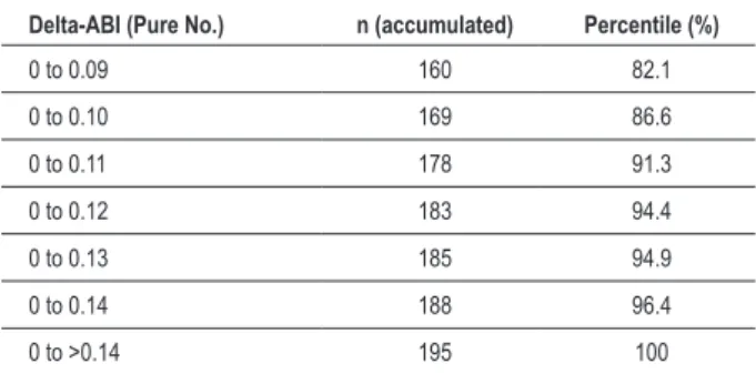
Since these are new indices not yet described in medical literature, we proceeded to determine Reference Values (RV) both for delta-BB and for delta-ABI. Using Group N, we distributed the members over a normal frequency curve, determined the cut-off level at the point closest to the 95th percentile, and therefore, found the RV = 0 to 8 mmHg for delta-BB and RV = 0 to 0.13 for delta-ABI (Tables 1 and 2).
Statistical analysis
Analysis of the parameters delta-BB and delta ABI in the groups with normal and altered ABI was performed by comparison of the 95% confidence intervals of the Odds Ratio (OR) Index of the respective groups. A comparison test of two proportions was applied to the results of the normal and altered ABI groups, with a 5% significance level. In the other quantitative variables, Student’s t test was used for comparison of the means. SBP/ULs data from both groups of patients were compared by Variance Analysis (ANOVA) with subdivided groupings and a 5% significance level. SAS (Statistical Analysis System) software was used for statistical analyses.
Results
It was possible to measure pressure levels with AOD in 224 out of the 247 patients (90.7%); only 23 (9.3%) cases
Table 1 - Determination of Reference Values (RV) in the 95th
Percentile of Delta-BB
Delta-BB (mmHg) n (accumulated) Percentile (%)
0 to 6 168 86.2
0 to 7 179 91.8
0 to 8 188 96.4
0 to 9 191 97.9
0 to 10 191 97.9
0 to >10 195 100
Data from Group N (normal ABI): the range of Delta-BB values closest to the 95th percentile was 0 to 8 mmHg.
Table 2 - Determination of Reference Values (RV) in the 95th
Percentile of Delta-ABI
Delta-ABI (Pure No.) n (accumulated) Percentile (%)
0 to 0.09 160 82.1
0 to 0.10 169 86.6
0 to 0.11 178 91.3
0 to 0.12 183 94.4
0 to 0.13 185 94.9
0 to 0.14 188 96.4
0 to >0.14 195 100
required the use of VD for ABI determination, which proved to be normal in 195 (78.9%) and altered in 52 (21.1%) of them. In all patients in whom data could not be measured by AOD and who were referred to undergo VD, there was confirmation of altered ABI levels (Table 3).
Clinical and demographic characteristics
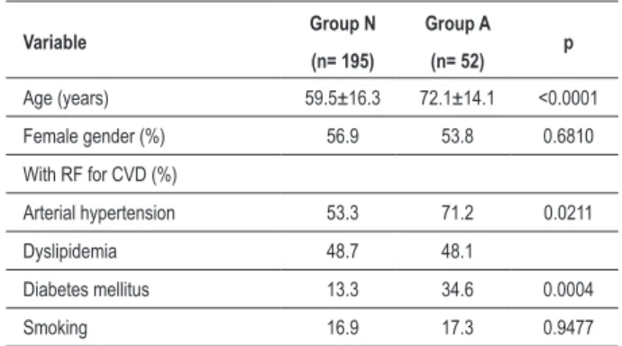
During clinical evaluation, we sought to detect the presence of classic cardiovascular risk factors such as smoking (the habit of smoking at least one cigarette a day) and arterial hypertension, dyslipidemia, and Type II diabetes (using diagnostic standards of the Brazilian Society of Cardiology
[Sociedade Brasileira de Cardiologia] and the Brazilian Society
of Diabetes [Sociedade Brasileira de Diabetes] (Table 4). In comparison with the normal ABI group (Group N), the individuals of the altered ABI group (Group A) tended to be older and had higher rates of cardiovascular risk factors, such as arterial hypertension and diabetes mellitus (Table 4). They also had significantly higher mean pulse pressures (PP) and a greater incidence of risk-related values (Table 3).
Curiously, in the normal ABI group, the incidence of higher SBP/ULs was twice as high in the right arm than in the left, a fact not noted in the group of altered ABI in which the incidence of this parameter was well balanced in both limbs (Table 5).
Results of ABI determination
The presence of delta-BB results greater than the RVs was significantly higher in Group A (Table 6) than in Group N (13
Table 3 - Results of ABI Determination in Groups of Normal ABI and Altered ABI (Groups N and A)
Variable Group N Group A p (n= 195) (N= 52)
Incidence of higher SBP/ULs (%)
Right 61.0 51.9 0.2355
Equal 9.7 3.8 0.2824
Left 29.2 44.2 0.0399
Mean SBP/ULs (mmHg)
Right 139.3 151.8 <0.0001
Left 138.1 148.7 <0.0001
Mean PP of arm with higher
SBP (mmHg) 58.2±16.4 73.6±22.6 <0.0001 Incidence of PP>63 mmHg (%) 32.3 63.5 <0.0001
ABI measurable by AOD (%) 100 55.8 <0.0001
Need to use Vascular Doppler
(%) 0 44.2 <0.0001
Mean Delta-BB (mmHg) 3.8 8.1 <0.0001
Delta-ABI 0.05 0.19 <0.0001 SBP - systolic blood pressure; ULs - upper limbs; PP - pulse pressure; ABI - ankle-brachial index; AOD - automated oscillometric device.
Table 4 - Clinical and Demographic Characteristics of Normal ABI and Altered ABI Groups (Groups N and A)
Variable Group N Group A p (n= 195) (n= 52)
Age (years) 59.5±16.3 72.1±14.1 <0.0001
Female gender (%) 56.9 53.8 0.6810
With RF for CVD (%)
Arterial hypertension 53.3 71.2 0.0211
Dyslipidemia 48.7 48.1
Diabetes mellitus 13.3 34.6 0.0004
Smoking 16.9 17.3 0.9477
RF - risk factors; CVD - cardiovascular disease.
Table 5 - SBP in ULs in Groups with Normal ABI and Altered ABI (Groups N and A)
Higher SBP/ULs RA LA p Group N (n= 195)
Incidence (%) 61.0 29.2
Mean (mmHg) 139.3±23.9 138.1±23.4 0.0614
Group A (n= 52)
Incidence (%) 51.9 44.2
Mean (mmHg) 151.8±29.3 148.7±30.3 0.0095
SBP - systolic blood pressure; ULs - upper limbs; RA - right arm; LA - left arm.
Table 6 - Delta-BB and Delta-ABI in Groups of Normal ABI and Altered ABI (Groups N and A)
Variable
Group A n = 52
Group N n = 195
OR 95% CI p
Delta-BB
> RV 13 7 8.95 3.08 to 26.79 < 0.0001
Delta-ABI
> RV 30 10 25.23 10.15 to 64.31 < 0.0001
OR - Odds Ratio; CI - Conidence Interval; Delta-BB > RV - Delta-BB above Reference Values; Delta-ABI > RV - Delta-ABI above Reference Values.
of 52 in Group A versus 7 of 195 in Group N; Odds Ratio: 8.95; 95% Confidence Interval: 3.08 to 26.79; p<0.0001). Additionally, the presence of delta-ABI results greater than the RVs was significantly higher in Group A (Table 6) than in Group N (30 of 52 in Group A versus 10 of 195 in Group N; Odds Ratio: 25.23; 95% Confidence Interval: 10.15 to 64.31; p <0.0001). In addition, the means of both delta-BB and delta-ABI values were significantly higher (p<0.0001) in Group A (8.1 mmHg and 0.19, respectively) than in Group N (3.8 mmHg and 0.05, respectively) (Table 3).
limit in patients with PAD: 65.6% versus 34.4% and means of 73 mmHg versus 58 mmHg, respectively, for Group N and Group A (Table 3).
On the other hand, in Group N, the limb with greatest prevalence for the higher SBP/ULs (Table 3) was the right arm (RA = 61.0%, Equal = 9.7% and LA = 29.2%; RA: right arm; LA: left arm). However, the SBP mean for both ULs was practically the same in both arms in Group N (RA = 139 mmHg; LA = 138 mmHg; p=0.0614) and a little higher on the right side (RA = 152 mmHg; LA = 149 mmHg; p=0.0095) in Group A (Table 5).
Discussion
ABI determination in clinical practice can be performed by simultaneously using duly validated simple AODs, except when the SBP in the LLs is extremely low. In this minority of cases, it is necessary to use conventional methodology with VD. The practicality of this method has advantages such as saving time, facilitating performance of the test, and decreasing errors. Consequently, it affords greater access to ABI determination in patients with CV risk, resulting in better clinical assessments and cost/benefit ratios since the financial expenditure is very low. Contrary to what might be supposed, this method is not opposed to the traditional method with VD. By making ABI more accessible in daily clinical practice, a greater number of patients with established PAD may be diagnosed, and among those, approximately half will require conventional ABI determination with VD (Table 3). Since it is practical, simple, and easy to perform, ABI with AOD can be repeated more often during clinical follow-up.
Rationale
The limbs of the human body are symmetrical and mirror identical images; therefore, it would be expected that pressure levels found in one limb would be very similar to those of the other, except in cases of abnormalities such as arterial stenosis. One limb serves as the perfect and unique control of its opposed limb on the same individual, and in the absence of an anatomical abnormality, it would be expected that the difference between the two limbs in any measurable parameter evaluated would be near zero. The greater the distance from this value the greater the chance of an abnormality.
The new parameters described above, delta-BB and delta-ABI, when situated outside of their reference values, also calculated here, have proved to be highly prevalent in individuals with PAD. Another interesting detail to be highlighted is that arterial stenosis in the upper limbs can be diagnosed when delta-BB is high. This is greatly significant since in this case, preventive and diagnostic steps should be taken and BP accompaniment should always be made on the limb with the higher levels of arterial pressure and not on its opposed limb, which could provide false impressions of normality. In individuals who always had similar BP levels in both upper limbs during routine clinical accompaniment and present sudden clinical symptoms of chest pain accompanied by an acutely modified delta-BB, the presumptive diagnosis of acute aorta dissection can lead to appropriate imaging tests. Future epidemiologic studies may determine the true
importance of these new parameters in the morbidity and mortality from cardiovascular and cerebrovascular diseases.
Curiously, it is noteworthy that the presence of higher SBPs in the ULs is more frequent in the right arm in the normal ABI group, a difference that is almost inexistent in the altered ABI group. As to the cause, could we assume that the first wave of cardiac systole directed towards the brachiocephalic trunk affords a greater pressure in the right arm, and this same wave would sequentially reach the root of the left subclavian artery with less impetus? Alternatively, is arterial blood pressure higher in the upper limb with greater strength and that is used more often? On the other hand, would arterial stenosis occur more frequently in the right arm than in the left?
Future perspectives
As is true with every new procedure, determination of ABI with AOD should be further tested against a gold standard method in order to be duly validated. Perhaps the best solution is not to confront it with conventional methodology carried out with VD, considering the inevitable systematic errors cited previously and especially because both are indirect methods. In this situation, the parameter to be calculated should be the level of agreement (or disagreement) between the two methods (kappa “k” value.) The ideal situation would be to analyze it with imaging tests as happened in the past with the conventional technique.
In the future, some ideas may be used to optimize ABI determination. Therefore, we might use four AOD measurements in the four limbs, especially in those cases of borderline ABI values (< 1.0) associated with symptoms suggestive of intermittent claudication or that show trophic modifications consistent with ischemia or even a decrease or absence of pulses in the extremities of the LLs. In this situation, four AOD measurements of SBP carried out simultaneously in the four limbs immediately after an exercise stress test may increase the sensitivity of ABI determination. Another improvement may come from the companies that manufacture AODs, such as cuffs with mirrored images specific for right and left limbs and other more conically shaped cuffs appropriate for ankles and arms of obese patients. A further solution for the obese would be to use normal cuffs and measure BP in the forearms where the shape and bone structure resemble that of the ankles.
The oscillometric method currently used in most cases of AOD provides great precision for mean BP determination, which is, ultimately the best parameter for quantifying flow. Therefore, the best strategy for an equally precise and accurate ABI in the future may be that one derived from mean BP values of the arms and ankles.
References
1. Newman AB, Siscovick DS, Manolio TA, Polak J, Fried LP, Borhani NO, et al. Ankle-arm index as a marker of atherosclerosis in the Cardiovascular Health Study. Cardiovascular Health Study (CHS) Collaborative Reserch Group. Circulation. 1993; 88 (3): 837-45.
2. Makdisse M: Índice tornozelo-braquial: importância e uso na prática clínica. São Paulo: Segmento Farma; 2004.
3. Yusuf S, Sleight P, Pogue J, Bosch J, Davies R, Dagenais G. Effects of an angiotensin-converting-enzyme inhibitor, ramipril, on cardiovascular events in high-risk patients.The Heart Outcomes Prevention Evaluation Study Investigators. N Engl J Med. 2000; 342 (3):145-53.
4. Carter SA. Indirect systolic pressures and pulses waves in arterial occlusive disease of the lower extremities. Circulation. 1968; 37: 624-37.
5. Ray SA, Srodon PD, Taylor RS, Dormandy JA. Reliability of ankle-brachial pressure index measurement by junior doctors. Br J Surg. 1990; 81: 188-90.
6. Adiseshiah M, Cross FW, Belsham PA. Ankle blood pressure measured by automatic oscillotonometry: a comparison with Doppler pressure measurements. Ann R Coll Surg Engl. 1987; 69: 271-3.
7. Ramanathan A, Conaghan PJ, Jenkinson AD, Bishop CR. Comparison of
ankle-brachial pressure index measurements using an automated oscillometric device with the standard Doppler ultrasound technique. ANZ J Surg. 2003; 73: 105-8.
8. Beckman JA, Higgins CO, Gerhard-Herman M. Automated oscillometric determination of the ankle-brachial index provides accuracy necessary for office practice. Hypertension. 2006; 47: 35-8.
9. O’Brien E, Petrie J, Littler WA, de Swiet M, Padfield PL, Altman DG, et al. The British Hypertension Society protocol for the evaluation of blood pressure measuring devices. J Hypertens. 1993; 11 (6): 677-9.
10. Association for Advancement of Medical Instrumentation. American National Standart. Electronic or automated sphygmomanometers. ANSI/AAMI SP 10-1992. Arlington: AAMI; 1993.
11. Madhavan S, Ooi WI, Cohen H, Alderman MH. Relation of pulse pressure reduction to the incidence of myocardial infarction. Hypertension. 1994; 23: 395-401.
12. Fang J, Madhavan S, Cohen H, Alderman MH. Measures of blood pressure and myocardial infarction in treated hypertensive patients. J Hypertens. 1995; 13: 413-9.
the humerus, which alone does not have the same protective capacity, but rather, facilitates artery constriction. We believe that this is the primary reason for the fact that BP measured at the ankles is higher than that measured at the arms.
Even though all these possibilities are welcome, we could not forget to point out that the main objective of this study is to present a simple, easy, and inexpensive method that can be used by any well-trained physician and provide extremely useful information in daily clinical practice. We will leave the other ideas as suggestions for those who are more knowledgeable and for Reference Centers.
Acknowledgements
I am grateful to Dra. Márcia Makdisse, Manager of the Cardiology Program of the Hospital Israelita Albert Einstein
and Coordinator of the “Corações do Brasil” Project of the Peripheral Artery Disease Committee [Comitê de Doença
Arterial Periférica – Projeto “Corações do Brasil”] for having
taught me to perform ABI measurements; to my business partner, Dr. Marco Antonio Goiato, for lending me his intelligent ideas; to Prof. Dra. Maria Lúcia M. Sundefeld, Chief of the Statistics discipline at the School of Dentistry of Araçatuba / UNESP for her careful statistical analysis; and finally, to my brother-in-law, Roberto Nakandakare, for having initiated me in the use of Excel software.
Potential Conflict of Interest
No potential conflict of interest relevant to this article was reported.
Sources of Funding
There were no external funding sources for this study.
Study Association