DOI: 10.1590/0004-282X20150008 ARTICLE
Upper airway dimensions in patients with
craniocervical junction malformations with and
without sleep apnea. A pilot case-control study
Dimensões da via aérea superior em pacientes com malformações da transição
craniocervical com e sem apnéia do sono. Um estudo piloto caso-controle
Ramon Barbalho Guerreiro1, Lia Bittencourt2, Rodolfo Casimiro Reis1, José Marcus Rotta3, Sérgio Tufik2,
Ricardo Vieira Botelho1
he craniocervical transition malformations (CCTM) comprise a spectrum of malformations frequently associ -ated with occipital bone hypoplasia and posterior fossa vol -ume depletion1,2,3,4,5.
Studies have shown that patients with CCTM tend to sufer more frequently from sleep respiratory disturbanc -es than the general population6. In CCTM cases, these
disturbances are more frequent and intense in patients with basilar invagination7.
he depleted airway dimensions have been associated with the pathophysiology of obstructive sleep apnea8. he ex
-perimental models of CCTM reveal that there is narrowing of airway dimensions associated with the posterior fossa reduc -tion in these patients9.
1Instituto de Assistência Médica ao Servidor Público Estadual, Pós-graduação em Ciências da Saúde, Sao Paulo SP, Brazil;
2Universidade Federal de São Paulo, Departamento de Psicobiologia, Sao Paulo SP, Brazil;
3Hospital do Servidor Público Estadual de São Paulo, Departamento de Neurocirurgia, Sao Paulo SP, Brazil.
Correspondence: Ricardo Vieira Botelho; Rua Tuim, 585 / apto 122A, Vila Uberabinha. 04514-102. São Paulo SP, Brazil. E-mail: [email protected] Conlict of interest: There is no conflict of interest to declare.
Received 06 July 2014; Received in final form 29 November 2014; Accepted 19 December 2014.
ABSTRACT
Objective: Patients with craniocervical junction malformations (CCJM) tend to suffer more frequently from sleep respiratory disturbances, which are more frequent and severe in patients with basilar invagination. Here we evaluate if patients with CCJM and sleep respiratory disorders (SRD) present smaller airway dimensions than patients without SRD. Method: Patients with CCCM with and without sleep respi-ratory disturbances were evaluated clinically by Bindal’s score, modified Mallampati classification, full-night polysomnography and upper airway cone beam tomography. Results: Eleven patients had sleep respiratory disorders (SRD), and nine patients performed control group without SRD. CCJM patients with SRD were predominantly female, older, had higher BMI, were more likely to have Mallampati grades 3 and 4 and had statistically significant smaller anteroposterior diameter of the upper airway than patients without SRD. Conclusion: Patients with CCJM and sleep respiratory disturbances have higher BMI, higher Mallampati score and smaller anterior posterior diameter of the upper airway.
Keywords: Chiari type I malformation, sleep apnea, polysomnography.
RESUMO
Objetivo: Pacientes com malformação da transição craniocervical (MTCC) tendem a apresentar mais frequentemente distúrbios respira-tórios do sono (DRS), os quais são mais intensos em pacientes com invaginação basilar. O objetivo desse estudo é avaliar se pacientes com MTCC e DRS apresentam dimensões das vias aéreas reduzidas em comparação a pacientes sem DRS. Método: Pacientes com MTCC com e sem apneia do sono foram avaliados com a escala de Bindal, classificação de Mallampati modificada, polissonografia de noite inteira e tomografia da via aérea superior. Resultados: Onze pacientes tinham DRS e nove não apresentaram esses distúrbios (grupo controle). Pacientes com MTCC e DRS foram principalmente mulheres, mais velhos, apresentaram maior IMC e maior gradação na escala de Mallam-pati, além de menor diâmetro anteroposterior de via aérea superior do que pacientes sem DRS. Conclusão: Pacientes com MTCC e DRS têm maior IMC, maior pontuação na escala de Mallampati e menor diâmtero anteroposterior da via aérea superior.
We evaluated the possibility that patients with Chiari type I and sleep respiratory disorders (SRD) may present smaller airway dimensions than Chiari type I patients without SRD.
METHOD
Patients with signs and symptoms suggestive of Chiari type I malformation and magnetic resonance images com -patible with Chiari type I malformation and/or basilar in -vagination were selected at two brazilian tertiary hospitals located in Sao Paulo from January to December of 2011. All patients signed a consent form and this clinical study was ap -proved by the Ethics Committee of both institutions.
Signs and symptoms speciic for CCTM were evaluated clinically with the scale developed by Bindal et al. Baseline full-night polysomnography (PSG) was performed by trained professionals using the EMBLA system (EMBLA® S7000, Inc., Broomield, CO, USA). he biological variables were mea -sured with electroencephalography (C3/A2, C4/A1, O1/A2 and O2/A1), electrooculography, submentonian and anterior tibial electromyography and electrocardiography (V2 modi -ied). Airlow was measured through a thermistor and a nasal cannula with a pressure transducer. he thoracic and abdom -inal movements were monitored with non-calibrated induc -tance plethysmography; snoring was recorded using a micro -phone; oxyhemoglobin was measured with pulse oximetry, and the body position was monitored with a sensor. Sleep scoring was performed by a professional who was trained in polysomnography and was based on the criteria set forth by Rechtschafen and Kales. he respiratory events and arousals were analyzed according to international criteria.
he patients were divided into two groups based on their PSG results: one with SRD and one without SRD (without central or obstructive sleep apnea). he patients with an ap -nea/hypopnea index (AHI) greater than or equal to 5 (with a predominance of obstructive events) were considered to have Obstructive Sleep Apnea Syndrome (OSAS) if they pre -sented with at least two of the following signs and symptoms: excessive daily drowsiness, snoring or episodes of respiratory pause observed by others.
he following anthropometric data were evaluated due to their importance in the genesis of SRD: weight, height and body mass index (BMI).
Both groups were evaluated according to the modi -ied Mallampati classiication (Figure 1), as proposed by Fredman et al., with the patient seated, the mouth opened maximally and the relaxed tongue inside the oral cavity. he patients were stratiied into four groups: (1) grade I: – all of the oropharynx was visible, including the soft palate, the ton -sil pillars, the palatine ton-sils and the tip of the uvula; (2) grade II: – the soft palate, the fauces and the uvula were vis -ible; (3) grade III: – the soft palate and the uvula base was vis -ible; and (4) grade IV: – the soft palate was not visible et al.10.
hepatients classiied as Mallampati 1 and 2 and Mallampati 3 and 4 were sub-grouped into two groups for analysis.
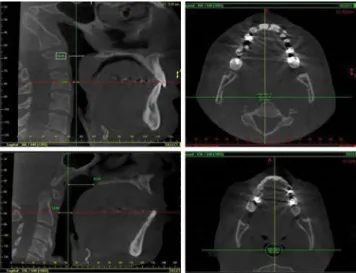
All of the patients were subjected to cone beam tomogra -phy of the upper airway (Figure 2). he images acquired by the tomography were analyzed in a specialized radiologic center with an I-CAT® (Imaging Sciences International, Hatield, PA, USA) instrument. All images were calibrated using 36.90 mA, 120 kVp, an exposure time of 40 s, an extended height ield of view (FOV) with a volume element of 0.3 mm, and a resolu -tion of 1024 X 1024 pixels and 12 bytes per pixel (4096 gray -scale). For image acquisition, the patient remained seated with eyes open and looking at a mirror in front of him. he patients were positioned using the laser beams of the machine with the Frankfurt plane parallel to the ground and the median sagit -tal plane perpendicular to the former. Image capture extended from 2 cm above the glabella to the inferior margin of the C4 vertebra. Axial images of 0.3 mm width were obtained and ex -ported in the DICOM (Digital Imaging and Communication in Medicine) format, and then recorded on a CD-ROM.
Dolphin Imaging® 3-D software, version 11.7 (Dolphin Imaging & Solutions, Chatsworth, CA) was used to process the volumetric data (DICOM iles) of the nasopharynx (NF), oropharynx (OF), and hypopharynx (HF) to calculate the total volume of these structures and the greatest constriction point.
Figure 1. Mallampati classification.
Figure 2. Upper airway Cone Beam Tomography. The
he anteroposterior and transverse diameters were mea -sured and the cross sectional area was calculated during normal respiration and maximal inspiration to delineate the maximally constricted point of the upper airway.
he maximum length of the upper airway was measured from the hard palate to the level of the epiglottal tip.
Statistical analysis
he anthropometric and image data from both groups were presented as the averages and standard deviations and compared using the Mann-Whitney test. Both genders were compared using the Chi-squared test. he signiicance level was determined as p < 0.05. he Mallampati data were ana -lyzed among both groups using the Chi-squared test.
RESULTS
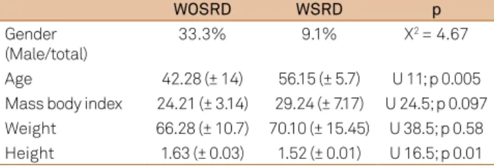
We studied twenty patients with type I Chiari malforma -tion during the pilot project. Eleven had SRD, and nine did not. hree patients without SRD (WOSRD group, n = 9) were male (33.3%) and 6 were female (66.7%). Among the eleven patients with SRD (WSRD group, n = 11), we studied one man (9.1%) and ten women (90.9%), (Chi-squared p = 0.47). he mean age was 42.28 (± 14) years old in the WOSRD group and 56.15 (± 5.7) years old in the WSRD patients (U 11, p = 0.005). he body mass index (MBI) was 24.21 (± 3.14) among pa -tients in the WOSRD group and 29.24 (± 7.17) in the WSRD patients (U 24.5, p = 0.097) (Table 1).
Concerning the modiied Mallampati grade (MMG) among the WOSRD group, three patients had an MMG of 1, three had an MMG of 2, two had an MMG of 3 and one had an MMG of 4. In the WRSD group, none had an MMG of 1, one had an MMG of 2, four had an MMG of 3 and six had an MMG of 4, with a total of 10 patients with an MMG of 3 or 4 in the WRSD group compared with 3 in the WOSRD group (Chi-squared p = 0.003) (Table 2).
Bindal score
he mean result for the Bindal clinical score for the WORSD group was 68.57 (± 41) points compared with 96.92 (± 39) in the WSRD patients (U 29, p = 0.11).
Polysomnography
All patients were subjected to polysomnography, and the following data were collected and studied: the exam length, the minutes of sleep, the number of respiratory events, the apnea-hypopnea index (AHI), the number of central, ob -structive and mixed apneas, and the lowest, mean and high -est oxygen saturation levels (Table 3).
he mean AHI was 3.14 among the WOSRD group com -pared with 28.75 in the WSRD patients (p = 0.001).
Computerized tomographic measurements of the pharyngeal airway
During normal breathing (Figure 3), the WOSRD patients had a pharyngeal anteroposterior diameter of 8.5 ± 1.31 mm compared with 4.73 ± 0.9 mm among the WSRD patients (U 21, p = 0.031). he laterolateral diameter difered between groups (22.6 ± 2.5 vs 18 ± 1.8 mm); however, this diference did not reach statistical signiicance (U 26.5, p = 0.08). he longi -tudinal length was 48.78 ± 1.77 mm in the WOSRD patients and 49.46 ± 2.38 mm in the WSRD group (U 49.5, p =1.0).
In regard to the maximum inspiration, the anteroposte -rior diameter was 7.29 ± 1.16 mm in the WOSRD patients and 5.89 ± 1.47 mm in the WSRD group (U 35.5, p = 0.29), the lat -erolateral diameter was 19.77 ± 2.72 mm in the irst group compared with 17.11 ± 2.52 mm in the second group (U 39, p = 0.45), and the longitudinal length was 49.66 ± 2.41 mm in the WOSRD patients compared with 49.22 ± 2.41 mm in the WSRD group (U 48, p = 0.94).
DISCUSSION
Cases of spontaneous respiratory collapse in adult and infant Chiari type I patients have been well documented, re -sulting in some deaths11.
During sleep, the voluntary respiratory control becomes attenuated; the maintenance of the respiratory system de -pends on the automatic ventilatory command, and the pa -tient is more susceptible to respiratory disturbances. his study of sleep respiratory disorders has the potential to re -veal SRD in the case of malformations.
With the advent of polysomnography, the respiratory dis -orders that occur during sleep can be better characterized, and the real importance of CCTM can be better elucidated.
Obstructive sleep apnea is the most prevalent respirato -ry disorder among CCTM patients12,13. Experimental studies
have associated CCTM with an abnormality of the occipital somites derived from the paraxial mesoderm13. here can be
a genetic predisposition to the condition14. Table 1. Anthropometric measurements of patients with and
without SRD (gender was analyzed with the Chi-squared test).
WOSRD WSRD p
Gender (Male/total)
33.3% 9.1% X2 = 4.67
Age 42.28 (± 14) 56.15 (± 5.7) U 11; p 0.005 Mass body index 24.21 (± 3.14) 29.24 (± 7.17) U 24.5; p 0.097 Weight 66.28 (± 10.7) 70.10 (± 15.45) U 38.5; p 0.58 Height 1.63 (± 0.03) 1.52 (± 0.01) U 16.5; p 0.01 U: Mann-Whitney U test.
Table 2. Modified Mallampati grade (MMG) of patients with (WSRD) and without (WOSRD) sleep respiratory disorders.
MMG1+2 MMG3+4 Total
WOSRD 6 3 9
*Difference between APNANB and APANB was statistically significant (U 21, p = 0.031). APNANB: Anteroposterior diameter of non-apneic patients during normal breathing; APANB: Anteroposterior diameter of apneic patients during normal breathing; LATNANB: Lateral diameter of non-apneic patients during normal breathing; LATANB: Lateral diameter of apneic patients during normal breathing; LONGNANB: Longitudinal length of non-apneic patients during normal breathing; LONGANB: Longitudinal length of apneic patients during normal breathing.
Figure 3. Computerized tomographic measurements of the pharyngeal airway during normal breathing.
Table 3. Polysomnnographic data.
Patient MOF EL AHI NCA NOA NMA HSaO2 MSaO2 LSaO2
Female, 63y 264 06:47 2 16 0 16 97.9 97.8 94
Female, 61y 304.5 06:34 5.7 61 0 61 97 95.9 88
Female, 61y 326 08:05 16 62 5 57 93.8 90.9 78
Female, 57y 396 08:15 3.8 13 0 13 95.3 94.7 90
Female, 61y 320.5 07:10 0 0 0 0 98 97.6 94
Male, 60y 437.5 07:46 1.5 11 2 9 97.9 97.7 93
Female, 37y 407.5 07:17 3.4 10 0 10 98.5 98.1 95
Female, 62y 404.5 07:40 22.4 201 18 46 97.4 96.8 89
Female, 50y 353 06:30 8.7 80 0 20 96.3 96 83
Female, 47y 218 07:23 6 6 0 1 97 96.8 90
Female, 47y 177.5 07:49 37.9 112 0 112 93.9 93.4 88
Female, 47y 429.5 07:57 15.6 111 0 111 96.6 96 83
Female, 45y 433.5 08:30 20.9 151 0 151 97 95.1 89
Male, 44y 360 06:51 1.3 8 1 7 97.1 96.1 93
Male, 49y 422.5 07:31 2 20 3 11 96.6 96.7 91
Male, 51y 337 07:13 27.6 153 21 153 95.5 94.9 78
Female, 51y 382 07:54 61.88 394 0 328 92 90 59
Female, 58a 392.1 06:58 9 59 6 53 95.4 95.2 87
Female, 15a 378.5 06:49 56.3 355 219 125 94.7 93.6 70
Female, 60a 390 07:42 46 299 4 183 95.5 94.5 84
Marin-Padilla’s experimental model revealed that, in ad -dition to occipital bone involvement, there is a narrowing of the airway dimensions associated with a posterior fossa vol -ume reduction in these patients15.
Alternatively, Lee RWW et al. have already demonstrated that other factors, such as the growth and development of the maxillomandibular relationships, the maxilla and the mandi -ble relationship to the cranial base, and the growth of local soft and adipose tissues may contribute to upper airway collapse16.
Our main results show that Chiari type I patients with SRD are predominantly female, older, more obese, more like -ly to have Mallampati grades 3 and 4 and a smaller antero -posterior diameter of the upper airway in comparison with Chiari type I patients without SRD.
Epidemiological studies have shown that the prevalence of OSAS in adults may vary from 1.2% to 7.5%, when considering the presence of EDS and AHI above 5 events per hour of sleep as the diagnostic criteria16. A recent epidemiological study, per
-formed in Sao Paulo, that used laboratory polysomnography (the gold standard test for diagnosis of this syndrome), ob -served that the OSAS prevalence reached the alarming rate of 32% of the analyzed population17. In a study of patients with
malformations, the OSAS prevalence reached 68%.
he main risk factors for OSAS are male gender, advanced age and being overweight. Important epidemiological stud -ies have demonstrated that male gender is a well-known risk factor for OSAS, with males being up to four times more af -fected than females18. he greater tendency of males to exhib
-it central fat depos-ition, the protective action of progester -one and the inducing action of testoster-one on the collapse of the upper airway and anatomical diferences in the airway are some of the possible reasons for this association19. In our
sample of Chiari malformation patients WSRD, the frequen -cy of females was greater than for those without WOSRD, even though this was not statistically signiicant.
Here, we found a higher proportion of older patients among the WSRD group than the WOSRD group, which is consistent with other studies about OSAS. he majority of those studies have strengthened the association between ag -ing and OSAS17,19,20. Tuik et al. revealed that patients older than 84 years of age had a risk of developing OSAS that was 34-fold higher than that of younger patients17. Factors that
explain this association include a higher BMI and upper air -way compliance, a decreased strength of the esophagus and laryngeal muscles, as well as the lower pulmonary and thy -roid function observed in the elderly21.
he main risk factor for OSAS is obesity, and Tuik et al. showed that the risk for OSAS is 10-fold higher in obese patients17,19,20. In addition to the anatomical chang -es in the upper airway induced by fat accumulation, the
respiratory physiology is compromised in obese patients, who have a lower pulmonary volume and compliance, lead -ing to upper airway collapse22.
A Mallampati grade of 3 or 4 has been shown to be an important inding in a physical examination because it has a high positive predictive value for OSAS. his correlation was also true in our sample of CCMT and SRD patients. he diference in the modiied Mallampati grade between the groups reached statistical signiicance, and the patients with SRD had a higher score than those without, consistent with the published data23,24,25,26.
Obesity might be the factor that brings patients with SRD to a higher Mallampati grade. Nashi et al. studied the tongues from 121 consecutive medical examiner cases and observed a high fat content in this organ, mainly in the posterior third. he Nashi group also veriied that the weight, volume and fat con -tent of the tongue were associated with a higher BMI, suggest -ing an association between obesity and a modiied Mallampati grade of 3 or 427. Modern image acquisition studies have shown
an association between anatomical characteristics, such as large tongues and narrowed upper airways, and OSAS28.
Shigeta et al. evaluated two groups through computer -ized tomography, including men and women controlled for age and BMI. he upper airway volume, measured from the dorsal nasal spine to the epiglottal tip, was evaluated. he to -tal height of the oropharynx and the volume of this segment demonstrated a statistically signiicant diference between genders (larger in men). Among the study participants, the changes increased with age, and age was a signiicant predic -tor of the oropharynx length29,30.
he shape and volume of the airway vary in patients with diferent anteroposterior maxillomandibular relation -ships. Grauer and colleagues showed that the shape but not the volume of the airway was diferent according to maxillo -mandibular vertical relationships30. In the same study, Grauer demonstrated a statistically signiicant relationship between the inferior portion of the upper airway and the maxilloman -dibular AP proportion, and between the airway volume and face size and gender. here was no diference between max -illomandibular volume and the vertical proportion. For this purpose, facial CBCT, facial lateral photographs and facial lateral cephalometric radiographs were used30.
Our CBCT indings in CCTM and SRD patients suggest a smaller AP diameter, which corroborates the published data.
References
1. Botelho RV, Bittencourt LR, Rotta JM, Tufik S. Adult Chiari
malformation and sleep apnoea. Neurosurg Rev. 2005;28(3):169-76. http://dx.doi.org/10.1007/s10143-005-0400-y
2. Nishikawa M, Sakamoto H, Hakuba A, Nakanishi N, Inoue Y. Pathogenesis of Chiari malformation: a morphometric study of the posterior cranial fossa. J Neurosurg. 1997;86(1):40-7. http://dx.doi.org/10.3171/jns.1997.86.1.0040
3. Milhorat TH, Nishikawa M, Kula RW, Dlugacz YD. Mechanisms of cerebellar tonsil herniation in patients with Chiari malformations as guide to clinical management. Acta Neurochir. 2010;152(7):1117-27. http://dx.doi.org/10.1007/s00701-010-0636-3
4. Milhorat TH, Bolognese PA, Nishikawa M, Francomano CA, McDonnell NB, Roonprapunt C et al. Association of Chiari malformation type I and tethered cord syndrome: preliminary results of sectioning filum terminale. Surg Neurol. 2009;72(1):20-35. http://dx.doi.org/10.1016/j.surneu.2009.03.008
5. Milhorat TH, Bolognese PA, Nishikawa M, McDonnell NB, Francomano CA. Syndrome of occipitoatlantoaxial hypermobility, cranial settling, and chiari malformation type I in patients with hereditary disorders of connective tissue. J Neurosurg Spine. 2007;7(6):601-9. http://dx.doi.org/10.3171/SPI-07/12/601
6. Marin-Padilla M, Marin-Padilla TM. Morphogenesis of experimentally induced Arnold-Chiari
malformation. J Neurol Sci. 1981;50(1):29-55. http:// dx.doi.org/10.1016/0022-510X(81)90040-X
7. Williams H. A unifying hypothesis for hydrocephalus, Chiari malformation, syringomyelia, anencephaly and spina bifida. Cerebrospinal Fluid Res. 2008;5(1):7. http://dx.doi.org/10.1186/1743-8454-5-7
8. Speer MC, George TM, Enterline DS, Franklin A, Wolpert CM, Milhorat TH. A genetic hypothesis for Chiari I malformation with or without syringomyelia. Neurosurg Focus. 2000;8(3):E12. http://dx.doi.org/10.3171/foc.2000.8.3.12
9. Guilleminault C, Tilkian A, Dement WC. The sleep apnea syndromes. Annu Rev Med. 1976;27(1):465-84. http://dx.doi.org/10.1146/annurev.me.27.020176.002341
10. Friedman M, Tanyeri H, La Rosa M, Landsberg R, Vaidyanathan K, Pieri S et al. Clinical predictors of
obstructive sleep apnea. Laryngoscope. 1999;109(12):1901-7. http://dx.doi.org/10.1097/00005537-199912000-00002
11. Botelho RV, Bittencourt LRA, Rotta JM, Tufik S. A prospective controlled study of sleep respiratory events in patients with craniovertebral junction malformation. J Neurosurg. 2003;99(6):1004-9. http://dx.doi.org/10.3171/jns.2003.99.6.1004
12. Botelho RV, Bittencourt LRA, Rotta JM, Tufik S. The effects of posterior fossa decompressive surgery in adult patients with Chiari malformation and sleep apnea. J Neurosurg. 2010;112(4):800-7. http://dx.doi.org/10.3171/2009.7.JNS09174
13. Chang ET, Shiao GM. Craniofacial abnormalities in Chinese patients with obstructive and positional sleep apnea. Sleep Med. 2008;9(4):403-10. http://dx.doi.org/10.1016/j.sleep.2007.04.024
14. American Academy of Sleep Medicine Task Force. Sleep-related breathing disorders in adults: recommendations for syndrome definition and measurement techniques in clinical research. The Report of an American Academy of Sleep Medicine Task Force. Sleep. 1999;22(5):667-89.
15. American Sleep Disorders Association - ASDA. EEG arrousals: scoring rules and examples: a preliminary report from the sleep disorders Atlas Task Force. Sleep. 1992;2:173-84.
16. Punjabi NM. The epidemiology of adult obstructive sleep apnea. Proc Am Thorac Soc. 2008;5(2):136-43. http://dx.doi.org/10.1513/pats.200709-155MG
17. Tufik S, Santos-Silva R, Taddei JA, Bittencourt LRA. Obstructive sleep apnea syndrome in the Sao Paulo Epidemiologic Sleep Study. Sleep Med. 2010;11(5):441-6. http://dx.doi.org/10.1016/j.sleep.2009.10.005
18. Kapsimalis F, Kryger MH. Gender and obstructive sleep apnea syndrome, part 1: clinical features. Sleep. 2002;25(4):412-9.
19. Malhotra A, White DP. Obstructive sleep apnoea. Lancet. 2002;360(9328): 237-45. http://dx.doi.org/10.1016/S0140-6736(02)09464-3
20. Durán J, Esnaola S, Rubio R, Iztueta A. Obstructive sleep apnea-hypopnea and related clinical features in a population-based sample of subjects aged 30 to 70 yr. Am J Respir Crit Care Med. 2001;163(3):685-9. http://dx.doi.org/10.1164/ajrccm.163.3.2005065
21. Heinzer RC, Stanchina ML, Malhotra A, Jordan AS, Patel S, Lo Y-L et al. Effect of increased lung volume on sleep disordered breathing in sleep apnea patients. Thorax. 2006;61:435-9. http://dx.doi.org/10.1136/thx.2005.052084
22. Paisani DM, Chiavegato LD, Faresin SM. Volumes, capacidades pulmonares e força muscular respiratória no pós-operatório de gastroplastia. J Bras Pneumol. 2005;31(2):125-32
23. Friedman M, Tanyeri H, LaRosa M, Landsberg R, Vaidyanathan K, Pieri S et al. Clinical predictors of obstructive sleep apnea. Laryngoscope. 1999;109:1901-7
24. Zonato AI, Bittencourt LR, Martinho FL, Santos Júnior JF, Gregório LC, Tufik S. Association of systematic head and neck physical examination with severity of obstructive sleep apnea-hypopnea syndrome (OSAHS). Laryngoscope. 2003;113(6):973-80. http://dx.doi.org/10.1097/00005537-200306000-00011
25. Zonato AI, Martinho FL, Bittencourt LR, Brasil OOC, Gregório LC, Tufik S. Head and neck physical examination: comparison between nonapneic and obstructive sleep apnea patients. Laryngoscope. 2005;115(6):1030-4. http://dx.doi.org/10.1097/01.MLG.0000163494.19965.DC
26. Nuckton TJ, Glidden DV, Browner WS, Claman DM. Physical examination: Mallampati score as an independent predictor of obstructive sleep apnea. Sleep. 2006;29(7):903-8.
27. Nadia N, Kang S, Barkdull GC, Lucas J, Davidson TM. Lingual fat at autopsy. Laryngoscope. 2007;117(8):1467-73. http://dx.doi.org/10.1097/MLG.0b013e318068b566
28. Schwab RJ, Pasirstein M, Pierson R, Mackley A, Arens R, Maislin G et al. Identification of upper airway anatomic risk factors for obstructive sleep apnea with volumetric MRI. Am J Respir Crit Care Med. 2003;168(5):522-30. http://dx.doi.org/10.1164/rccm.200208-866OC
29. Shigeta Y, Ogawa T, Venturin J, Nguyen M, Clark GT, Enciso R. Gender- and age-based differences in computerized tomographic measurements of the orophaynx. Oral Surg Oral Med Oral Pathol Oral Radiol Endod. 2008;106(4):563-70. http://dx.doi.org/10.1016/j.tripleo.2008.03.032.