ISSN 2317-6431 https://doi.org/10.1590/2317-6431-2017-1954
Hearing impairment in newborns and infants with
myelomeningocele
Alterações auditivas em recém-nascidos e lactentes com
mielomeningocele
Natália Oliveira de Jesus1, Elaine Colombo Sousa Maruta2, Marisa Frasson de Azevedo3
ABSTRACT
Purpose: Verify the occurrence of auditory impairments in newborns and infants with Myelomeningocele (MMC) and/or Arnold-Chiari Type II Malformation (ACMII). Methods: The study sample was composed of 160 newborns assisted at a neonatal intensive care unit divided into three groups: Study Group 1, consisting of 42 neonates with ACMII; Study Group 2, composed of 38 infants with MMC; and Control Group, comprising 80 newborns without syndromes, malformations, and/or risks of auditory impairment. All patients underwent neonatal auditory screening including Transient Evoked Otoacoustic Emissions (TEOAE) and search for Brainstem Auditory Evoked Potentials (BAEP) with click stimulus. In the case of failure in the emissions, the search for electrophysiological thresholds was performed through BAEP with tone burst. Statistical analysis was performed using relevant parametric tests and the data found were
described considering a significance level of 5% (p<0.05). Results: Higher occurrence of cochlear hearing loss, central auditory disorders, and auditory neuropathy spectrum was observed in the Study Groups compared with the Control Group. Regarding the mean values of absolute latencies and interpeak intervals in the three groups per ear, there was an increase in the absolute latencies of waves III and V and in the interpeak intervals III-V and I-V in the group of infants with ACMII. Conclusion: Newborns and infants with Arnold-Chiari Type II Malformation and Myelomeningocele presented higher occurrence of cochlear hearing loss, auditory neuropathy spectrum, and central auditory disorders.
Keywords: Myelomeningocele; Electrophysiology; Audiology; Newborn; Neonatal screening
RESUMO
Objetivo: Verificar a ocorrência de alterações auditivas em recém-nascidos e lactentes com mielomeningocele e/ou Síndrome de Arnold-Chiari tipo II. Métodos: Foram incluídos na amostra 160 neonatos atendidos em unidade
de terapia intensiva neonatal, distribuídos em três grupos: Grupo Estudo 1,
formado por 42 neonatos com Síndrome de Arnold Chiari tipo II; Grupo Estudo 2, constituído por 38 neonatos com mielomeningocele e Grupo
Controle, com 80 recém-nascidos sem síndromes, malformações e/ou riscos para alteração auditiva. Todos os pacientes realizaram a triagem auditiva neonatal com emissões otoacústicas evocadas por estímulo transiente e
foram submetidos à pesquisa do potencial evocado auditivo de tronco
encefálico com estímulo clique. Quando houve falha nas emissões, a pesquisa dos limiares eletrofisiológicos foi realizada mediante potencial
evocado auditivo de tronco encefálico com toneburst. A análise estatística
foi feita por meio de testes paramétricos e os dados encontrados foram descritos nos resultados considerando-se o nível de significância p<0,05. Resultados: Houve maior ocorrência de perda coclear, alteração central e neuropatia auditiva nos grupos estudados. No que se refere aos valores
médios das latências absolutas e dos intervalos interpicos, nos três grupos, por orelha, houve aumento das latências absolutas das ondas III e V e dos
intervalos interpicos III-V e I-V no grupo de lactentes com Síndrome de Arnold Chiari tipo II. Conclusão: Recém-nascidos e lactentes com Síndrome
de Arnold Chiari tipo II e mielomeningocele apresentaram maior ocorrência de perda coclear, espectro da neuropatia auditiva e alterações centrais.
Palavras-chave: Meningomielocele; Eletrofisiologia; Audiologia;
Recém-nascido; Triagem neonatal
Study carried out at Hospital São Paulo – HSP/HU, Universidade Federal de São Paulo – UNIFESP – São Paulo (SP), Brasil.
1 Residência Multiprofissional em Saúde da Criança e do Adolescente, Hospital São Paulo – HSP/HU, Universidade Federal de São Paulo – UNIFESP – São Paulo
(SP), Brasil.
2 Programa de Pós-graduação em Distúrbios da Comunicação Humana, Departamento de Fonoaudiologia, Universidade Federal de São Paulo – UNIFESP –
São Paulo (SP), Brasil.
3 Departamento de Fonoaudiologia, Universidade Federal de São Paulo – UNIFESP – São Paulo (SP), Brasil.
Conflict of interests: No.
Authors’ contribution: NOJ was responsible for the study design, collection and analysis of the data, and writing of the manuscript; ECSM contributed to data collection and revision of the manuscript; MFA was the study adviser.
Funding: None.
INTRODUCTION
Myelomeningocele (MMC) is an embryonic malformation
of the central nervous system that occurs in the first four weeks
of gestation. It results from a closure defect in the posterior
neural tube. It is characterized by a vertebral, musculofascial,
cutaneous, dural opening with protrusion and exposure of the spinal cord. It causes functional alteration of the spinal cord, of varying degrees, depending on where it occurs on the spine, and lesions located in cranial spinal segments determine greater neurological damage(1).
MMC is often associated with Arnold-Chiari Malformation
(ACM), which is not always symptomatic. MMC is classified
into four types, and the most common association occurs with Type II of this syndrome (ACMII), which presents herniation of the tonsils, cerebellar vermis, fourth ventricle, and lower portion of the bulb through the occipital foramen. Several anomalies of the nervous system are associated with ACMII, namely, enlargement of the foramen magnum, hypoplasia of the nuclei of cranial nerves and cerebellar olives, colpocephaly, absence of pellucid septum, cerebral hypoplasia, etc(2,3).
Patients with MMC and/or ACMII may develop numerous sensorimotor sequelae from birth, such as orthopedic, vesicointestinal, visual, cognitive and auditory impairments(4-6). Central auditory
disorders are present in 88% of ACMII patients(7,8); however,
there are few studies addressing integrity of the auditory
pathways in this population in the specific scientific literature.
Central auditory disorders interfere with the ability to processing acoustic stimuli and impair speech and language development. Thus, detection and monitoring of these impairments should be conducted in order to enable early intervention in developmental disorders.
The hypothesis of this study is that newborns with MMC or ACMII present higher occurrence of retrocochlear hearing impairments, such as auditory neuropathy and central auditory pathway disorders.
Therefore, the objective of this study was to verify the occurrence of cochlear and retrocochlear auditory disorders in newborns with Myelomeningocele and/or Arnold-Chiari Type II Malformation.
METHODS
This retrospective, cross-sectional study with comparison between groups was approved by the Research Ethics Committee
of the aforementioned Institution (CEP-UNIFESP) on August 3rd,
2016 under protocol no. 1.660.706, CEP no. 0909/2016, and CAAE no. 57572116.4.0000.5505. After approval, the database of the Speech-Language Pathology Service was revised between 2010 and 2016.
The study sample was composed of 160 neonates and infants born between January 2010 and July 2016. All participants underwent neonatal hearing screening (NHS) between 24 hours and 3 months of age, with post-conceptual age >35 weeks. Exclusion criteria comprised newborns at risk for toxoplasmosis, rubella, cytomegalovirus, herpes, syphilis, and HIV-TORCHS, as well as those with conductive impairment at the time of NHS. After meeting the inclusion criteria, the sample was divided into three groups:
Study Group 1 (SG1): consisting of 42 neonates with Arnold-Chiari Type II Malformation (ACMII);
Study Group 2 (SG2): composed of 38 infants with Myelomeningocele (MMC);
Control Group (CG): comprising 80 newborns without syndromes, malformations, and/or risks of auditory impairment, as proposed by the Institution based on literature studies(9,10). Newborns in this group were
matched for gestational age and gender to those of the Study Groups (SG1 and SG2).
All newborns and infants underwent neonatal hearing screening (NHS) with Transient Evoked Otoacoustic Emissions (TEOAE) and search for Brainstem Auditory Evoked Potentials (BAEP) with click stimulus. TEOAE were collected from newborns and infants under natural sleep using an AccuscreenPRO (GNOtometrics) device with non-linear click stimulus, velocity
of 60 Hz, sound pressure level between 70 and 84 dBNPS
(45-60 dBNA) self-calibrated depending on the volume in the external auditory meatus of each neonate, frequency range
from 1.4 KHz to 4 KHz, and sampling rate of 16 KHz. As a
pass/fail criterion, analysis of the equipment was used with
artifact <20% and probe stability >80%.
BAEP-click was registered in a Smart-EP, (Intelligent Hearing Systems) device using ER 3A insert earphones. With the neonates and infants under natural sleep, the skin was cleaned using abrasive NuPrepTMe paste. Meditrace (Kendal) disposable surface electrodes were placed on the
forehead (Fpz) and on the right and left mastoid muscles
(M2 and M1) according to the IES 10-20 norm (International Electrode System). Impedance of the electrodes was maintained <3 Kohms. Rarefaction polarity clicks were used at 80 dBNnA with duration of 100 µs at a repetition rate of 27.7/s. A 12 ms
window and 100 Hz and 3000 Hz filters were used with a
minimum of 2000 stimuli. After placing the insert earphone in the external acoustic meatus, BAEP-click was performed at 80 dBnNA. The absolute latencies of waves I, III, and V
and the interpeak intervals I-III, III-V, and I-V were analyzed. Absolute latencies and interpeak intervals were classified
as normal or altered, considering the standards established in a previous study and the corrected age of the newborns and/or infants at the time of examination(11).
Infants who failed the NHS were submitted to otorhinolaryngologic and complementary diagnostic assessment including immittance
audiometry and frequency-specific and bone-conducted BAEP, if necessary. Frequency-specific BAEP was performed using a
Smart-EP (Intelligent Hearing Systems) device following the same procedures used in the BAEP-click. BAEP were registered with toneburst stimulation. Air- (AC) and bone- (BC) conducted
search was performed at frequencies ranging from 500 Hz to 4000 Hz. Investigation of the AC threshold was performed using
ER 3A insertion earphones with at least 2000 stimuli. Stimulus intensity began at 80 dBnNA and was gradually reduced, by
20 dB, until wave V was no longer visualized. After that,
intensity was increased, by 10 dB, until the lowest intensity was obtained, at which wave V appeared in smaller amplitude, and this was considered the electrophysiological threshold(11).
Bone-conducted BAEP were registered by placing a RadioEar B71 bone vibrator on the mastoid muscles and presenting an alternating wave at initial intensity of 50 dBNA, decreasing by 10 dB. The lowest intensity at which wave V
was identified and replicated by the individual was considered
Acoustic immittance measures were recorded using an AZ7 (Interacoustics) audiometer. Tympanometry curve acquisition was as follows: type A curve was observed when the maximum admittance peak was approximately at 0 daPa
air pressure, whose variation did not exceed –100 daPa; type
B curve was observed when the maximum admittance peak did not occur at any air pressure; type C curve was observed when the maximum admittance peak was shifted to negative
pressures below –100 daPA; type As curve was observed when
low admittance occurred; type Ad curve was observed when
the interval between the two curve branches was ≥100 daPa(12).
It is common knowledge that that AZ7 test tone is 226 Hz,
considered a tone of high diagnostic sensitivity in children aged >6 months, adults, and elderly, but not an ideal tone to assess neonates and infants. Therefore, in this population, it is
necessary to apply an acute test tone of approximately 1000 Hz to detect discrete middle ear impairments that the 226 Hz probe
can not identify(13,14). However, a recent study reported that both
test tones, 226 Hz and 1000 Hz, presented high specificity to
identify assessments within normality in newborns and infants with and without risk of hearing loss(15).
After the assessments, the groups were analyzed and
compared with respect to results of the TEOAE, values of the absolute latencies and interpeak intervals in the BAEP-click, and
final audiological diagnosis. Regarding the auditory pathway,
low and high brainstem impairments were considered when there were increases in the interpeak intervals I-III and III-V, respectively. Impairment of the auditory pathway as a whole (diffuse brainstem impairment) was observed when there was an increase in the interpeak interval I-V due to increases in both interpeak intervals I-III and III-V(16). ANS occurred when
there was presence of TEOAE, absence of waves in BAEP, and
presence of cochlear microphonism. Thus, the final audiological diagnosis was classified into normal hearing, cochlear hearing
loss, central auditory disorder, and auditory neuropathy spectrum.
Descriptive and inferential statistical analysis was conducted by a qualified professional using Analysis of Variance (ANOVA), Test for the equality of two proportions, Confidence Interval
for the Mean, and p-value at 5% significance level (p<0.05).
RESULTS
The study sample was composed of 160 newborns and infants
of both genders - 85 (53.1%) female and 75 (46.8%) male,
distributed in three groups: Study Group 1 (SG1) - neonates with Arnold-Chiari Type II Malformation (ACMII), Study Group 2 (SG2) - neonates with Myelomeningocele (MMC), and a Control Group (CG).
SG1 consisted of 42 newborns, with gestational age of 30-42 weeks, mean of 36 weeks, median of 37 weeks, and corrected age of 33-46 weeks. SG2 was composed of 38 infants
with gestational age of 31-41 weeks, mean of 36 weeks, median of 37 weeks, and corrected age of 35-43 weeks. CG comprised 80 infants with gestational age of 34-43 weeks, mean of 37 weeks, median of 38 weeks, and corrected age of 35-44 weeks.
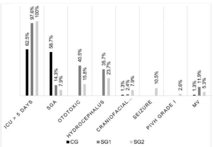
Figure 1 illustrates the occurrence of pre-, peri- and post-natal risk indicators of the 160 infants separated into groups.
Regarding the results of the Transient Evoked Otoacoustic Emissions (TEOAE), the Study Groups (SG1 and SG2) differed from the Control Group (CG), with higher occurrence of cochlear hearing loss in both ears (Tables 1 and 2).
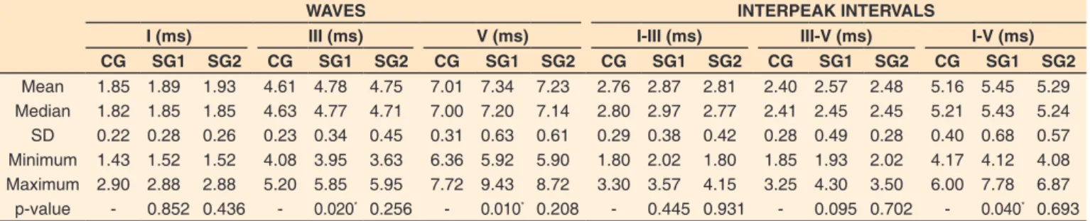
As for the search for Brainstem Auditory Evoked Potentials (BAEP), the mean values of absolute latencies and interpeak intervals did not differ between ears in the three groups. However, some differences between groups were observed: SG2 differed from CG by presenting prolonged absolute latencies of waves III and V and increased interpeak intervals III-V and I-V (Tables 3 and 4).
Altered results were classified in relation to the type of
impairment present in the auditory system. Higher occurrence of cochlear hearing loss, central auditory disorders, and auditory neuropathy spectrum was observed in SG1 and SG2 compared with CG. No differences were observed in the comparison between SG1 and SG2 (p>0.6) (Table 5).
Analysis of the type of central auditory disorder revealed higher occurrence of low brainstem impairment in both SG1 and SG2. High brainstem impairment and diffuse impairment of the auditory pathway also occurred in these groups, but to a lesser extent (Figure 2).
Figure 1. Distribution of the occurrence of risk indicators for hearing loss
Subtitle: CG = control group; SG1 = infants with Arnold-Chiari Type II Malformation;
SG2 = infants with Myelomeningocele; SGA = small for gestational age; PIVH = peri-intraventricular hemorrhage; MV = mechanical ventilation
Table 1. Results of Transient Evoked Otoacoustic Emissions for the right ear in the Study Groups
Absent Present
n % p-value n % p-value
CG 0 0% - 80 100%
-SG1 2 4.8% 0.049* 40 95.2%
-SG2 2 5.3% - 36 94.7% 0.038*
*Statistical significance (p≤0.05) in relation to the Control Group (CG); Test for the equality of two proportions
Figure 2. Incidence of auditory pathway impairment in the Study Groups
Subtitle: SG1 = infants with Arnold-Chiari Type II Malformation; SG2 = infants
with Myelomeningocele
DISCUSSION
Myelomeningocele (MMC) is the most common posterior
neural tube closure defect. According to the last DataSUS survey of December 2016, 650 children were born with MMC in Brazil in 2014(17). This malformation occurs between the
third and fifth weeks of intrauterine life because of a fusion
failure in the posterior elements of the spine. Of unknown etiology, MMC presents multifactorial characteristics (genetic and environmental), and it is known that women with a diet low in folic acid (vitamin B9) are more likely to give birth to children affected by this disease(18). MMC is associated with a
number of other disorders, such as motor deficit and fecal and
urinary incontinence, as well as with several malformations of the central nervous system (CNS) resulting from herniation of the posterior fossa components to the medullary canal. This set
Table 2. Results of Transient Evoked Otoacoustic Emissions for the left ear in the Study Groups
Absent Present
n % p-value n % p-value
CG 0 0% - 80 100%
-SG1 2 4.8% 0.049* 40 95.2%
-SG2 3 7.9% - 35 92.1% 0.011*
*Statistical significance (p≤0.05) in relation to the Control Group (CG); Test for the equality of two proportions
Subtitle: n = sample size; CG = control group; SG1 = infants with Arnold-Chiari Type II Malformation; SG2 = infants with Myelomeningocele
Table 3. Characteristics of the absolute latencies of waves I, III, and V and interpeak intervals I-III, III-V, and I-V for the right ear according to group
WAVES INTERPEAK INTERVALS
I (ms) III (ms) V (ms) I-III (ms) III-V (ms) I-V (ms)
CG SG1 SG2 CG SG1 SG2 CG SG1 SG2 CG SG1 SG2 CG SG1 SG2 CG SG1 SG2
Mean 1.85 1.89 1.93 4.61 4.78 4.75 7.01 7.34 7.23 2.76 2.87 2.81 2.40 2.57 2.48 5.16 5.45 5.29 Median 1.82 1.85 1.85 4.63 4.77 4.71 7.00 7.20 7.14 2.80 2.97 2.77 2.41 2.45 2.45 5.21 5.43 5.24 SD 0.22 0.28 0.26 0.23 0.34 0.45 0.31 0.63 0.61 0.29 0.38 0.42 0.28 0.49 0.28 0.40 0.68 0.57 Minimum 1.43 1.52 1.52 4.08 3.95 3.63 6.36 5.92 5.90 1.80 2.02 1.80 1.85 1.93 2.02 4.17 4.12 4.08 Maximum 2.90 2.88 2.88 5.20 5.85 5.95 7.72 9.43 8.72 3.30 3.57 4.15 3.25 4.30 3.50 6.00 7.78 6.87 p-value - 0.852 0.436 - 0.020* 0.256 - 0.010* 0.208 - 0.445 0.931 - 0.095 0.702 - 0.040* 0.693
*Statistical significance (p≤0.05) in relation to the Control Group (CG); Analysis of Variance (ANOVA)
Subtitle: ms = milliseconds; CG = control group; SG1 = infants with Arnold-Chiari Type II Malformation; SG2 = infants with Myelomeningocele; SD = standard
deviation
Table 4. Characteristics of the absolute latencies of waves I, III, and V and interpeak intervals I-III, III-V, and I-V for the left ear according to group
WAVES INTERPEAK INTERVALS
I (ms) III (ms) V (ms) I-III (ms) III-V (ms) I-V (ms)
CG SG1 SG2 CG SG1 SG2 CG SG1 SG2 CG SG1 SG2 CG SG1 SG2 CG SG1 SG2
Mean 1.85 1.88 1.88 4.64 4.84 4.70 7.03 7.45 7.24 2.79 2.96 2.82 2.39 2.62 2.54 5.19 5.58 5.27 Median 1.82 1.82 1.80 4.66 4.87 4.72 7.00 7.30 7.09 2.83 3.01 2.81 2.37 2.47 2.42 5.21 5.48 5.27 SD 0.22 0.25 0.24 0.25 0.41 0.42 0.30 0.69 0.65 0.28 0.44 0.44 0.26 0.57 0.48 0.35 0.78 0.75 Minimum 1.49 1.57 1.52 4.05 3.48 3.52 6.42 5.88 6.05 1.88 1.75 1.85 1.92 1.80 1.92 4.17 4.07 3.33 Maximum 2.73 2.48 2.67 5.17 5.80 5.97 7.60 9.60 8.88 3.30 3.90 4.20 3.17 4.32 3.92 5.88 7.97 7.13 p-value - 0.914 0.899 - 0.028* 0.842 - 0.001* 0.259 - 0.107 0.984 - 0.026* 0.378 - 0.014* 0.933
*Statistical significance (p≤0.05) in relation to the Control Group (CG); Analysis of Variance (ANOVA)
Subtitle: ms = milliseconds; CG = control group; SG1 = infants with Arnold-Chiari Type II Malformation; SG2 = infants with Myelomeningocele; SD = standard deviation
Table 5. Occurrence of peripheral and central disorders according to group
Diagnosis Typical CHL CAD ANS Total
n % p-value n % p-value n % p-value N % p-value n %
CG 80 100% - 0 0.0% - 0 0.0% - 0 0.0% - 80 100%
SG1 22 52.4% <0.001* 2 4.7% 0.166 13 31.0% <0.001* 5 11.9% 0.002* 42 100% SG2 21 55.3% <0.001* 2 5.2% 0.145 10 26.3% <0.001* 5 13.2% <0.001* 38 100% *Statistical significance (p≤0.05) in relation to the Control Group (CG); Test for the equality of two proportions
Subtitle: n = sample size; % = percentage; CHL = cochlear hearing loss; CAD = central auditory disorder; ANS = auditory neuropathy spectrum; CG = control group;
of malformations of the CNS, known as Arnold-Chiari Type II Malformation (ACMII), can lead to progressive dilation of the cerebral ventricles and consequent need for ventriculoperitoneal shunt (VPS) for the treatment of hydrocephalus(19).
In this study, associated risk indicators were collected in this population, observing that most infants with MMC and ACMII, in the Study Groups 1 and 2 (SG2 and SG1), respectively,
remained in neonatal intensive care unit (NICU) longer than five days. Indeed, this finding was expected, considering that
newborns who present this malformation need corrective surgeries and intensive care at birth. Moreover, of the 80 infants in the
Study Groups, 35.7% of SG1 and 23.7% of SG2, presented
hydrocephalus (Figure 1) - accumulation of cerebrospinal
fluid within the ventricles that leads to progressive dilation,
which may cause functional impairment of the brainstem and auditory pathway(20). In a survey conducted in Bahia state, the
authors verified that 85% of patients with MMC presented
hydrocephalus(21). Lower occurrence of hydrocephalus was
observed in the present study, probably due to preventive measures such as intra-uterine hydrocephalus correction and VPS
placement in the first days of life. Children with hydrocephalus
present higher occurrence of central auditory disorders, mainly
increased interpeak interval III-V, characterizing high brainstem
impairment(22).
In this research, 10.5% of the patients in SG2 presented
convulsions (Figure 1), a result lower than that described in the
literature, which reports that 20-25% of individuals with MMC have convulsive episodes, justified by cortical dysgenesis(23).
This difference can be attributed to early intervention of neurology and neurosurgery teams and administration of anticonvulsive medication.
Transient Evoked Otoacoustic Emissions (TEOAE) were collected from 160 newborns. The infants in the Study Groups presented grater absence of TEOAE: two bilateral fails were found in SG1 and two bilateral fails and one unilateral fail were detected in SG2 (Tables 1 and 2). This finding is in agreement with those of a recent study, in which the authors compared the
results of NHS in 90 newborns in NICU, 40 with MMC and
50 without the disease, and found greater absence of TEOAE in
the first(24). Cochlear hearing loss was observed in 4.8% of the
infants in SG1 and in 5.3% of those in SG2, with no difference
between the groups or ears (Table 5). These values are similar
to those obtained in newborns who remained in NICU. A recent
study conducted with 140 newborns at risk for hearing loss
who remained in NICU reported that 11.42% of this population failed the first stage of NHS and, after retest with TEOAE and Brainstem Auditory Evoked Potentials (BAEP), 6.25% of them
presented diagnosis compatible with hearing loss(25).
In the present study, search for BAEP was conducted in all participants, with analysis of the absolute latencies and interpeak intervals and comparison between the groups. Concerning the
type of hearing loss, 31% of the infants in SG1 and 26.3%
of those in SG2 presented central auditory disorders. In fact, several studies in the literature have indicated that this population presents higher incidence of this type of impairment(8,24,26).
Despite presenting 26.3% of central auditory disorders,
SG2 did not differ from CG with respect to the values of absolute latencies and interpeak intervals. A study addressing the values of BAEP in patients with isolated MMC showed that
74% of this population presents increased absolute latency of
wave V and interpeak interval I-V(8). This result differs from
those obtained in the present study, considering that the group
with such malformation (SG2) did not present statistically
significant difference for the values of absolute latency and interpeak interval compared with CG. However, 26.3% of the
infants with isolated MMC presented central auditory disorders,
with 18.4% of impairment in the low brainstem and 7.9% in
the high brainstem.
This study also aimed to classify, based on the values of absolute latency and interpeak interval, the type of impairment present in the auditory pathway. Both Study Groups presented higher occurrence of examinations with normal results (Figure 2). Low brainstem impairments were the most prevalent in both
groups (30.9% in SG1 and 18.4% in SG2). High brainstem impairments occurred more often in SG1 (11.9%), which
explains the increase in the absolute latencies of waves III and V.
Diffuse impairment of the auditory pathway occurred in 15.8%
of the individuals in SG2 (Figure 2). In a study conducted with
48 patients with ACMII, 36% of the individuals presented low brainstem impairment and 15% of them showed high brainstem
impairment(27), corroborating the findings of the present survey
(30.9% and 11.9%). No studies conducted with patients with
isolated MMC were found in the literature.
In SG1, increased absolute latencies of waves III and V and of the interpeak intervals III-V and I-V were observed,
which characterize high brainstem impairment. In fact, there is evidence in the literature that 41.7% of patients with ACMII
presented prolonged absolute latencies and interpeak intervals(27).
Auditory neuropathy spectrum (ANS) was observed in
11.9% of the patients in SG1 and in 13.2% of those in SG2.
ANS is described in the literature as presence of TEOAE and absent or altered BAEP, generating hearing impairment mainly for speech recognition and comprehension(28). Several studies
have shown the locations of hearing lesions that coincide with ANS, such as the tectorial membrane, inner and outer hair cells,
impaired synapses between inner hair cells and fibers of spiral ganglion neurons, impaired electrical transmission of fibers of
the cochlear nerve, and problems related to myelination of the cochlear nerve(29,30). Thus, ANS mainly affects the path of hair
cells external to the brainstem, which would justify the presence of this type of impairment in this population, considering that
MMC isolated or associated with ACMII is characterized by
anatomical alteration throughout the CNS(28).
Therefore, these findings suggest a need for otorhinolaryngologic
and audiological evaluation, including BAEP, in children with MMC and/or ACMII because of the risk of impaired speech and language development.
Main limitations to this study were as follows: need for further research addressing auditory disorders in this population and eventual consequences; regular monitoring of auditory development, with reassessment every three, six, and/or 12 months;
specific monitoring of speech and language development.
CONCLUSION
ACKNOWLEDGEMENTS
The first author is grateful to Professor Marisa Frasson de Azevedo, adviser, and Elaine Colombo Sousa Maruta, co-adviser,
for their guidance throughout this project.
REFERENCES
1. Bizzi JWJ, Machado A. Mielomeningocele: conceitos básicos e avanços
recentes. J Bras Neurocirurg. 2012;23(2):138-51.
2. Cameron AH. The Arnold-Chiari and other neuroanatomical malformations associated with spina bifida. J Pathol. 1957;73(1):195-211. http:// dx.doi.org/10.1002/path.1700730124.
3. Gilbert JN, Jones KL, Rorke LB, Chernoff GF, James HE. Central nervous system anomalies associated with meningomyelocele, hydrocephalus, and the Arnold-Chiari malformation: reappraisal of theories regarding the pathogenesis of posterior neural tube closure defects. Neurosurgery. 1986;18(5):559-64. http://dx.doi.org/10.1227/00006123-198605000-00008. PMid:3714003.
4. Fobe JL, Rizzo AM, Silva IM, Silva SP, Teixeira CE, Souza AM, Fernandes A. QI em pacientes com hidrocefalia e mielomeningocele: implicações do tratamento cirúrgico. Arq Neuropsiquiatr. 1999;57(1):44-50. http:// dx.doi.org/10.1590/S0004-282X1999000100009. PMid:10347723.
5. Tambaquim MLM, Lamônica DAC, Whitaker ME. Avaliação neuropsicológica e fonoaudiológica em crianças com mielomeningocele. In: Anais do Congresso Hispano-Português de Psicologia; 2005; Lisboa. Lisboa: Universidade de Lisboa; 2005.
6. Araújo AE, Galvão C. Desordens neuromotoras. In: Cavalcanti A, Galvão C. Terapia ocupacional: fundamentação teórico prática. Rio
de Janeiro: Guanabara Koogan; 2007. p. 328-37.
7. Lütschg J, Meyer E, Jeanneret-lseli C, Kaiser G. Brainstem auditory evoked potentials in meningomyelocele. Neuropediatrics. 1985;16(4):202-4. http://dx.doi.org/10.1055/s-2008-1059537. PMid:4080095.
8. Taylor MJ, Boor R, Keenan NK, Rutka JT, Drake JM. Brainstem
auditory and visual evoked potentials in infants with myelomeningocele.
Brain Dev. 1996;18(2):99-104. http://dx.doi.org/10.1016/0387-7604(95)00136-0. PMid:8733898.
9. American Academy of Pediatrics. Joint Committee on Infant Hearing. Year 2007 position statement: principles and guidelines for early hearing detection and intervention programs. Pediatrics. 2007;120(4):898-921. http://dx.doi.org/10.1542/peds.2007-2333. PMid:17908777.
10. Onoda RM, Azevedo MF, Santos AMN. Triagem auditiva neonatal: ocorrência de falhas, perdas auditivas e indicadores de riscos. Rev
Bras Otorrinolaringol. 2011;77(6):775-83.
11. Rosa LAC, Suzuki MR, Angrisani RG, Azevedo MF. Potencial Evocado Auditivo de Tronco Encefálico: valores de referência em relação à idade. CoDAS. 2014;26(2):117-21. http://dx.doi.org/10.1590/2317-1782/2014469IN. PMid:24918504.
12. Russo ICP, Valente CHV, Lopes LQ, Brunetto-Borginanni LMB.
Medidas de imitância acústica. In: Santos TMM, Russo ICP. Prática da audiologia clínica. 7. ed. São Paulo: Cortez; 2009. p. 183-216.
13. Keefe DH, Levi H. Maturation of the middle and external ears: acoustic
power-based responses and reflectance tympanometry. Ear Hear. 1996;17(5):361-73. http://dx.doi.org/10.1097/00003446-199610000-00002. PMid:8909884.
14. Margolis RH, Bass-Ringdahl S, Hanks WD, Holte L, Zapala DA. Tympanometry in newborn infants: 1KHz norms. J Am Acad Audiol.
2003;14(7):383-92. PMid:14620612.
15. Garcia MV, Azevedo MF, Testa JR. Medidas de imitância acústica em lactentes com 226hz e 1000hz: correlação com as emissões otoacústicas e o exame otoscópico. Rev Bras Otorrinolaringol. 2009;75(1):80-9.
http://dx.doi.org/10.1590/S0034-72992009000100013.
16. Castro NP Jr, Figueiredo MS, Mesquita OS No, Périco RAN. Avaliação eletrofisiológica da audição. In: Lopes O Fo, Campiotto AR, Levy
CCAC, Anelli W. Novo Tratado de Fonoaudiologia. 3. ed. Barueri: Manole; 2013. p. 201-19.
17. DATASUS: Departamento de Informática do Sistema Único de Saúde. Brasil: Sistema de Informações sobre Nascidos Vivos (SINASC) [Internet]. Brasília: DATASUS; 2016 [citado em 2016 Dez 11. Disponível em: http://tabnet.datasus.gov.br/cgi/tabcgi.exe?sinasc/
cnv/nvuf.def
18. Fernandes AC. Malformações do tubo neural. In: Hebert S, Xavier R,
Pardini AG Jr, Barros TEP Fo. Ortopedia e traumatologia: princípios e prática. 3. ed. Porto Alegre: Artmed; 2003. p. 839-57.
19. Bevilacqua NS, Pedreira DAL. Cirurgia fetal endoscópica para correção de mielomeningocele: passado, presente e futuro. Einstein.
2015;13(2):283-9. http://dx.doi.org/10.1590/S1679-45082015RW3032. PMid:26154549.
20. Cavalcanti DP, Salomão MA. Incidência de hidrocefalia congênita e o papel do diagnóstico pré-natal. J Pediatr. 2003;79(2):135-40. http://
dx.doi.org/10.2223/JPED.965.
21. Marques VB. Mielomeningocele: avaliação do acompanhamento
multidisciplinar [tese]. Bahia: Faculdade de Medicina da Bahia,
Universidade Federal da Bahia; 2014.
22. Rosa LAC. Avaliação eletrofisiológica da audição em crianças com hidrocefalia [dissertação]. São Paulo: Universidade Federal de São
Paulo; 2013.
23. Salomão JFM. Contribuição aos estudos dos disrafismos espinais císticos cervicais e torácicos altos com proposta de classificação [tese]. São Paulo: Escola Paulista de Medicina, Universidade Federal de São
Paulo; 2002.
24. Satzer D, Guillaume DJ. Prognostic value of newborn hearing
screening in patients with myelomeningocele. J Neurosurg Pediatr. 2014;14(5):495-500. http://dx.doi.org/10.3171/2014.7.PEDS14168. PMid:25216288.
25. Rechia IC, Liberalesso KP, Angst OVM, Mahl FD, Garcia MV, Biaggio EPV. Intensive Care Unit: results of the Newborn Hearing
Screening. Rev Bras Otorrinolaringol. 2016;82(1):76-81. http://dx.doi. org/10.1016/j.bjorl.2015.06.004. PMid:26712635.
26. Docherty TB, Herbaut AG, Sedgwick EM. Brainstem auditory evoked
27. Henriques OS Fo, Pratesi R. Abnormalities in auditory evoked potentials of 75 patients with Arnold-chiari malformations types I and II. Arq Neuropsiquiatr. 2006;64(3A):619-23. http://dx.doi.org/10.1590/ S0004-282X2006000400019. PMid:17119806.
28. Starr A, Picton TW, Sininger Y, Hood LJ, Berlin CI. Auditory neuropathy. Brain. 1996;119(Pt3):741-53. http://dx.doi.org/10.1093/ brain/119.3.741. PMid:8673487.
29. Moser T, Predoehl F, Starr A. Review of hair cell synapse defects in sensorineural hearing impairment. Otol Neurotol. 2013;34(6):995-1004. http://dx.doi.org/10.1097/MAO.0b013e3182814d4a. PMid:23628789.