A RANDOMIZED CLINICAL TRIAL OF ORAL
STEROIDS FOR ULNAR NEUROPATHY IN TYPE
1 AND TYPE 2 LEPROSY REACTIONS
José Antonio Garbino
1, Marcos da Cunha Lopes Virmond
2,
Somei Ura
1, Manoel Henrique Salgado
2, Bernard Naafs
3Abstract – Background: Steroids regimens in leprosy neuropathies are still controversial in botth types of reactions. Method: For this trial, 21 patients with ulnar neuropathy were selected from 163 leprosy patients, 12 with type 1 reaction (T1R) and nine with type 2 (T2R). One experimental group started with prednisone 2 mg/kg/day and the control group with 1 mg/kg/day. A clinical score based on tests for spontaneous pain, nerve palpation, sensory and muscle function was used. Neurophysiological evaluation consisted on the motor nerve conduction of the ulnar nerve in three segments. Student “t” test for statistical analysis was applied on the results: before treatment, first week, first month and sixth month, between each regimen and types of reaction. Conclusion: In both reactions during the first month higher doses of steroids produced better results but, earlier treatment with lower dose was as effective. Short periods of steroid, 1 mg/Kg/day at the beginning and,tapering to 0,5 mg/Kg/day or less in one month turned out to be efficient in T2R.
Key wORdS: leprosy, ulnar neuropathy, steroids, neurophysiology.
Ensaio clínico sobre o tratamento com esteróides via oral da neuropatia ulnar em reação tipo 1 e tipo 2 da hanseníase
Resumo – Introdução: O tratamento da neuropatia da hanseníase com esteróides é ainda controverso nos dois tipos de reações. Método: Neste ensaio, de 163 pacientes foram selecionados 21 com neuropatia ulnar, 12 com reação tipo 1 e 9 com tipo 2. Um grupo experimental iniciou com 2 mg/kg/dia e o grupo controle com 1 mg/ kg/dia. Foi composto um escore clínico pela avaliação da sensação dolorosa espontânea, palpação de nervos e funções sensitiva e motora. Realizou-se a condução nervosa motora do nervo ulnar em três segmentos. Aplicaram-se os estudos estatísticos com o teste t de Student nos resultados: antes do tratamento, primeira semana, primeiro mês e sexto mês. Conclusão: em ambas as reações dosagens mais elevadas iniciais produziram melhores resultados, mas a dose menor quando administrada precocemente foi igualmente efetiva. Períodos curtos com doses efetivas, 1 mg/Kg/dia no início e reduzindo-se para 0,5 mg/Kg/dia ou menos em um mês foram eficientes na reação tipo 2.
PAlAvRAS-chAve: hanseníase, neuropatia ulnar, esteróides, neurofisiologia.
1Instituto lauro de Souza lima, Bauru SP, Brasil; 2departamento de Produção da Faculdade de engenharia de Bauru, Universidade do estado de São
Paulo, Bauru SP, Brasil; 3departamento de dermatologia do centro Médico da Universidade de leiden, holanda.
Received 2 June 2008, received in inal form 17 September 2008. Accepted 3 October 2008.
Dr. José Antônio Garbino – Instituto Lauro de Lima - Caixa Postal: 3021 - 17034-971 Bauru SP - Brasil. E-mail: [email protected]
Immune response to Mycobacterium leprae may lead to disability due to nerve damage, which occurs mostly during the acute inlammatory episodes named reactions. According to the immunity, leprosy patients can devel-op distinct clinical groups1: polar tuberculoid tuberculoid (TT), borderline tuberculoid (BT), borderline borderline (BB), borderline lepromatous (Bl) and polar lepromatous (ll). Mainly two types of reactions are known, the type1 leprosy reactions (T1R) or reversal response, which hap-pens in groups that have cell-mediated immune reaction,
Arq Neuropsiquiatr 2008;66(4)
leprosy neuropathy: oral steroids Garbino et al.
a few studies the standard starting dose of prednisone/ prednisolone for ield use was established to be approx-imately 40 mg/day9,10. however, there are no references available relating body weight to steroid doses.
The follow-up of motor nerve function using voluntary Muscle Testing (vMT)gives insight into behavior of nerves during reactions and treatment11. Magora12 suggested using motor nerve conduction (MNc) for nerve monitoring in leprosy neuropathy. Naafs and dagne13 and later Naafs and van droogenbroeck14 compared MNc with vMT and grad-ed sensory testing (GST). The last two authors gathergrad-ed all the parameters to compose a nerve severity index. In this study, measurement of the MNc along the ul-nar nerve was selected to investigate differences in nerve responses in the two types of leprosy reactions, type 1 and type 2, under two different steroid regimens advised by the Brazilian guidelines for leprosy control15, initial dos-es of 1 mg/kg/day to 2 mg/kg/day.
METHOD
Patients presenting leprosy reaction and with active ulnar nerve involvement were selected at the outpatient department of the Instituto lauro de Souza lima in Bauru, Brazil. All pa-tients provided informed written consent and the study was ap-proved by the institutional ethical committee. Patients at risk of a neuropathy other than leprosy neuropathy were excluded, i.e. diabetes, alcoholism, hIv infection, also with family histo-ry of hereditahisto-ry neuropathy and over 60 years of age. Patients with inactive neuropathy and/or chronic neuropathic pain, with nerve abscess and with nerves that had been submitted to neu-rolysis, as well as patients with a contraindication for steroids were also excluded.
Reactions were deined as:
Type 1 reaction (T1R) in the TT, BT, BB and Bl patients: an in-creased inlammation of existing lesions with or without non-tender new lesions and/or acro-edema. Nerves may be enlarged, tender and show loss of function.
Type 2 reaction (T2R) in the ll patients: a sudden appear-ance of inlamed papules, nodules and plaques that are tender on palpation. The patient may be ill and run a mild fever. There may be signs of involvement of other organs, e.g., eyes, testes, joints, lymph glands and periosteum. Nerves may be enlarged, tender and show loss of function.
Out of 163 leprosy patients examined during the period of September 2003 to August 2005, 21 patients were eligible, and in-cluded in the study: 17 men and 4 women (ages 21–60, mean: 41.5). Twelve patients had T1R (3 BT and 9 BB) and nine T2R, all were ll. Patients of both reaction types were assigned to treatment groups: experimental or control. The patients were assessed im-mediately prior to the beginning of treatment (1st), after one week (2nd), after one month (3rd) and thereafter monthly, the last follow-up occurred after six-months (8th assessment). All patients were submitted to clinical and dermatological
exami-nation in order to classify the leprosy groups, a general clinical evaluation and laboratory tests were performed to assess blood cells, blood sugar levels, liver and kidney functions.
Clinical evaluation
1. Assessment of spontaneous pain – this utilizes a visual an-alog scale (vAS), in which zero represents no pain and 10 repre-sents unbearable, incapacitating pain16. 2. Nerve palpation (NP) – this is done at the elbow. Size and tenderness were evaluated and graded: 0 (normal palpation) –5 (maximum nerve enlarge-ment)17. 3. Graded sensory testing (GST) – nylon monoilaments (SORRI – Bauru/ Brazil Kit18, 0.5, 1, 2, 4, 10, 100 g), were used in two areas innervated by the ulnar nerve, one in the hypothe-nar region and one in the little inger. The results were comput-ed to a maximum of 12 points, when no ilament was felt in a completely damaged nerve, and zero when all ilaments were felt for a normal nerve. 4. voluntary muscle testing (vMT) – The tested muscles were the abductor digiti minimi muscle and the irst dorsal interosseus. A normal score would be 10 points (2 ×
5) and when paralyzed, 0 points. In this study it was employed a reverse scale in order to align the results and build the score. These results were comparable with the other tests, in which the greater values relate to diminishing function. A inal clini-cal score (cS) was clini-calculated by the somatory of the results of vAS, NP, GST and vMT. The vAS and NP were applied only by one examiner and the GST and vMT were applied by the insti-tution staff of therapists.
Neurophysiologic evaluation
Motor nerve conduction (MNc) studies were carried out over three segments of the ulnar nerve. Recording of the com-pound motor action potential (cMAP) was done with the active recording electrode on the abductor digiti minimi muscle belly and the reference electrode on a tendon or a bony surface19. The cMAP amplitude by supramaximal stimulation, measured from the base line to the negative spike, is a function of the number of functioning motor axons19.
1. The distal latency was measured over an 8 cm long segment from the active recording electrode to the wrist; the recording electrode was attached on the muscle belly. 2. The nerve was al-so stimulated just below the elbow and 11 cm proximal. The con-duction velocity over the forearm segment and across the elbow was computed. 3. The cMAP temporal dispersion (Td), i.e., the duration of cMAP19, was measured below and above the elbow. Its values, in percentage, were summated. 4. The minimum value of the F wave latency, related to demyelination in all segments of the nerve from stimulating electrode to spine and back to the recording electrode, was measured over a series of 20 stimuli. The period between the start of symptoms and the begin-ning of treatment was recorded as less than three months (<3 m) or more than three months but less than six (>3 <6 m).
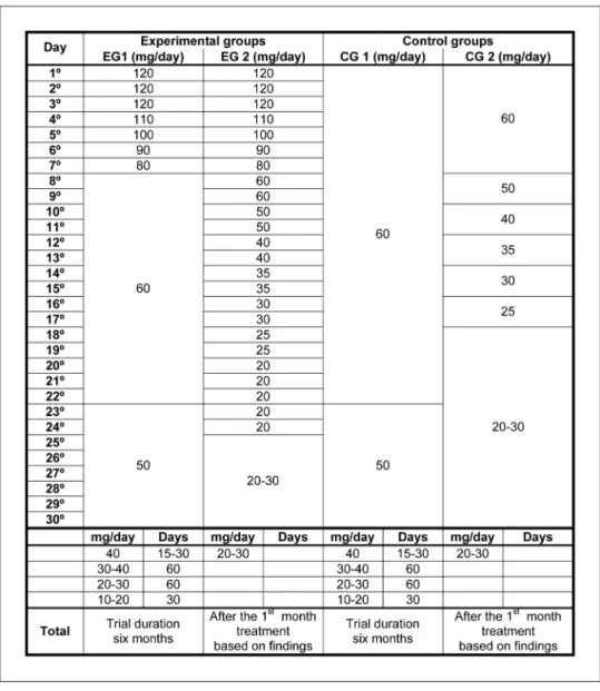
ex-perimental group (eG), one receiving prednisone 2 mg/kg (12 patients), or other control group (cG), which received 1 mg/kg (nine patients), as initial treatment. This resulted in four groups: eG T1R, cG T1R and eG T2R, cG T2R (Fig 1).
Results of the cS and the neurophysiologic parameters at the beginning of the study (1st assessment) were compared with the results obtained after the irst week (2nd assessment), with the results after the irst month (3rd assessment) and following the 6th month (8th assessment), of each reaction type in the
ex-perimental and control groups, using the Student “t” test for statistical analysis.
RESULTS
Out of 21 patients, 15 (71.4%) had inished multidrug-therapy around 17.7 months before the symptoms start-ed. eleven patients were taking inappropriate prednisone doses (mean 0.17 mg/kg/day) prior to inclusion in the protocol. The responses of their nerves were compared
Fig 1. Treatment regimes of experimental groups in T1R and T2R and control groups (1 and 2) in T1R and T2R, considering a patient with a 60 kg bodyweight.
Table 1. Distribution of nerves with active neural involvement, as type of reaction, grade of severity and duration of symptoms.
Grade T 1 R (time) T2 R (time) Total of nerves
<3 m >3 <6 m <3 m >3 <6 m
Partial lesion 12 5 8 3 28
complete paralysis 0 2 1 3* 6
Total of nerves 12 7 9 6 34
Arq Neuropsiquiatr 2008;66(4)
leprosy neuropathy: oral steroids Garbino et al.
with the results obtained in the patients without previous treatment. No signiicant differences were found. They were therefore included in the overall assessment.
Forty-two ulnar nerves from 21 patients were studied. eight nerves did not show any active neural involvement during the study, six nerves were completely damaged, and 28 nerves were followed by cS and neurophysiology. The distribution of nerves according to type of reaction, and duration of symptoms are demonstrated in Table 1.
during the study, six out of the nine T2R patients needed additional drug treatment 2–3 months after ini-tiation of treatment with prednisone. They received
tha-lidomide as an immunomodulator in doses of 100–200 mg/daily and a temporary slight increment of steroids. None of the T1R patients needed additional treatment. At the end of the time frame to develop this protocol all T1R patients were still on steroids, but tapering off, as well as six (out of 9) of the T2R group.
The values found for the cS within the four groups, when compared within each pair (before treatment, after one week, after 1 and 6 months), showed no signiicant dif-ferences, either for the eG compared with the cG (includ-ing both T1R and T2R) or for the T1R group compared with the T2R group (including both eG and cG). however, when the results before treatment (1st evaluation) and after 6 months (8th evaluation), were compared for each group, all groups showed signiicant improvement during the trial pe-riod (Student t test, p=0.000 for T1R and p=0.046 for T2R).
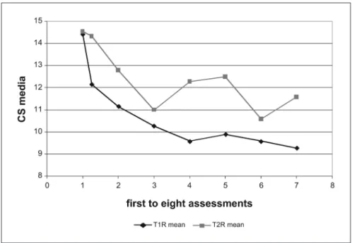
The graphic representation of the mean cS values (Fig 2) shows an obvious visual difference favouring T1R cas-es and a clear tendency to deterioration within the T2R group during dose reduction.
Neurophysiologic data
The most frequent abnormal indings observed were in the cv across the elbow (83.33%), F wave latency (69.44%) and in the Td at the elbow and above (across the elbow) (52.77%), followed by the cv along the forearm (38.89%) and the distal latency (30.56%).
The statistical differences between the parameters ob-tained in the eG and the cG are compared in Table 2, in-cluding all patients, independent on the type of reaction.
Fig 2. Mean of CS nerves results in patients with T1R and T2R (EG and CG together). The irst CS mean was prior to the treatment, the second was after at the end of the irst week, the third at the end of the irst month and thereafter monthly until the last occurence at the end of the sixth month.
Table 2. The neurophysiological results in patients of experimental group (EG) and control group (CG) (T1R and T2R) comparing the 1st evaluation with the 2nd, the 1st evaluation with the 3rd and
1st evaluation with the 8th (n=28). The highlighted data are the
results with statistical signiicance and the underlined data are the borderline results.
Steroid regimens eG × cG 1st× 2nd 1st× 3rd 1st× 8th
distal latency 0.057 0.082 0.095
cMAP wrist 0.968 0.380 0.663
cv in the forearm 0.023 0.057 0.787
cMAP at the elbow 0.981 0.279 0.310
cv across elbow 0.116 0.299 0.167
cMAP above elbow 0.322 0.680 0.267
Td (elbow + above) 0.095 0.032 0.703
The results showed a signiicant improvement of eG in the variables cv over the forearm and cMAP Td across the el-bow during the irst week and during the irst month. The F wave latency also showed greater improvement at the end of the irst week. In the irst week the distal latency showed a slight impairment in the eG group. Improvement was found at the end of irst month (p=0.082). The signii-cant differences disappeared after 6 months.
Statistical differences between the 2 types of reaction were seen only in the improvement of the cv (p=0.015)
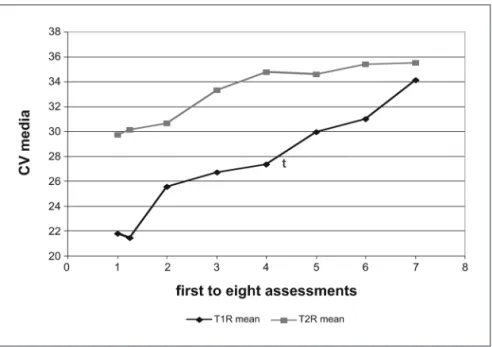
and the Td across the elbow (p=0.033) in nerves of pa-tients suffering from T1R compared with papa-tients with T2R after 6 months, i.e., at the end of the study. The graphic representation (Fig 3) of the cv in nerves in T1R compared with T2R, before treatment, shows cv markedly lower in T1R than in nerves in T2R.
Patients with symptoms lasting less than three months showed signiicantly greater improvement during the irst month in several of the parameters: cMAP at the elbow, cv across the elbow, cMAP above the elbow, Td across
Fig 3. Mean of CV across the elbow during a cohort in nerves of patients of groups T1 R and T2 R (n=28). The irst CV mean was prior to the treatment and the second was after at the end of the irst week, the third at the end of the irst month and thereafter monthly until the last occurence at the end of the sixth month
Table 3. The neurophysiological results in nerves with at two different treatment delay periods: less than three months (<3 m) or more than three months but less than six (>3 <6 m), in patients of EG and CG (T1R and T2R) comparing the 1st evaluation with the 2nd, the 1st
evaluation with the 3rd and 1st evaluation with the 8th (n=28). The
highlighted data are the results with statistical signiicance and the underlined data are the borderline results.
Treatment delay period (<3 m) × (>3 <6 m)
1st× 2nd 1st× 3rd 1st× 8th
distal latency 0.144 0.669 0.297
cAMP wrist 0.621 0.766 0.558
cv in the forearm 0.427 0.902 0.236
cAMP at the elbow 0.834 0.042 0.156
cv across elbow 0.114 0.014 0.043
cAMP above elbow 0.191 0.041 0.256
Td (elbow + above) 0.097 0.069 0.225
F wave 0.157 0.037 0.014
Table 4. The neurophysiological results in all nerves with symptoms for less than three months (<3 m) in patients of EG compared with CG (T1R and T2R) comparing the 1st evaluation with the 2nd, the 1st
evaluation with the 3rd and 1st evaluation with the 8th (n=20). The
highlighted data are the results with statistical signiicance. Treatment delay period
(<3 m) eG × (<3 m) cG
1st× 2nd 1st× 3rd 1st× 8th
distal latency 0.046 (T1R) 0.176 0.229
cAMP wrist 0.295 0.390 0.251
cv in the forearm 0.046 (T1R) 0.142 0.129
cAMP at the elbow 0.028 (T2R) 0.481 0.006 (T2R)
cv across elbow 0.196 0.081 (T1R) 0.319
cAMP above elbow 0.041 (T2R) 0.168 0.005 (T2R)
Td (elbow + above) 0.480 0.155 0.374
Arq Neuropsiquiatr 2008;66(4)
leprosy neuropathy: oral steroids Garbino et al.
the elbow and F wave latency, at the end of irst month (3rd assessment), when compared with patients whose treatment was delayed (Table 3).
Twenty out of the 28 nerves fell into the category of less than three months treatment delay (<3m). when only those 20 nerves were analysed, comparing the eG with the cG, minimal differences were observed. The same param-eters improved in eG (cv in the forearm at the irst week and F wave latency in the irst week and in the last evalu-ation) and in the cG (cMAP amplitude at the elbow and cMAP amplitude above) in the irst week and in the last evaluation. Other parameters, such as wrist cMAP am-plitude, vc and Td across elbow did not show statisti-cal differences.
The results show that there is a statistically a greater improvement in patients of the eG than cG, at the begin-ning of treatment, in the irst week, and at the 6th month of evaluation (Table 4).
Two of the patients developed adverse effects of ma-jor severity during the trial period, both of them in the eG: one patient developed osteoporosis with collapse of the 10th dorsal vertebra and another developed hyper-glycemia and cataracts. Patients of all groups had gained weight at the end of the study.
DISCUSSION
Several studies have discussed the duration of steroids treatment for reactions and there is evidence that the treatment period for T1R should be longer than the three months recommended by world health Organization, preferably six or, in some cases, even longer20. The dura-tion of treatment for a single episode of T2R is not clear, but there are indications that a reaction usually lasts one month or less21. The therapy with higher doses of steroids should be conined to this period in T2R.
Initial steroid dose has infrequently been discussed, although different standard regimens employ different doses. In fact, the initial dose for both reactions and the duration of treatment speciically in neural T2R, has not been fully studied22-24.
when the results for the cS were compared, no signii-cant differences were found, either for T1R and T2R (with-out considering the steroid regimen) or for eG and cG (with T1R and T2R grouped together). Similar results were seen in the literature25 when different steroid regimens were compared. Meanwhile, all groups showed signiicant improvement over time (p=0.000 for T1R and 0.046 for T2R), indicating the effectiveness of the chosen treat-ments (Fig 2). however, Figure 2 shows some differences between the T1R and T2R groups: nerves of T1R patients improve more and continuously while there is a tendency
to recur and to abate in T2R patients. In fact, when devel-oping a new reaction, either clinically or in the follow-up parameters, 6 out of the 9 patients in the T2R group need-ed adjustment of treatment as allowneed-ed by the protocol. This usually occurred after one to two months of treat-ment, when the steroids had reached the doses of 20–30 mg/day. when increasing the steroid doses and introduc-ing thalidomide improvement was again observed (Fig 2). In T1R group, relapses of reactions did not occur, con-trary to Manandhar’s et al.9 and Sundar Rao et al.26 reports, as the treatment period of T1R patients was adjusted to the true duration of 4 to 18 months20.
In the comparison of neurophysiologic parameters of the eG and cG, regardless of the type of reaction, statisti-cal differences were found at all three moments evaluat-ed: after the irst week (2nd), after the irst month (3rd) and after six months (8th assessment) (Table 4). After the irst week and at the end of the irst month the cv along the forearm and the Td across elbow were signiicantly better for the eG. These results relect remyelination. however, the improvement in the irst week is most likely a result of reduction of intraneural edema.
These results favor the eG during the first month; this is probably due to the more inlammatory, anti-edema effect of the higher steroid doses. After the irst month, when the same dose was given to both groups, statistical differences disappeared. however, the improve-ment of the parameters in the different groups continued. These results indicate a dose-response effect of steroid in the treatment of leprosy neuropathy during reactions, especially at the initial period, when inlammation with edema formation is a major component. The changes in cv at the elbow demonstrate graphically a remarkable reduction after the second month of the T1R compared to the T2R, showing more pronounced and continuous remy-elination in T1R than in T2R (Fig 3). The repetitive character of the T2R with neural involvement could be a major fac-tor inluencing the poor results of long-term treatment of a T2R neuropathy. when the two steroid regimens in T1R were compared, only the Td had signiicantly greater im-provement after 1 month of treatment in nerves of the eG (Table 2). This indicates that an early release of edema may lead to an earlier start of remyelination. In both reactions higher doses show better responses, but in T2R shorter treatment courses may be effective. The use of a higher dose for even an initial short period, as in pulse therapy, should be considered in severe nerve involvement27,28 .
slightly better overall eG results in this trial, it is clear that early treatment is more important than the higher dose of steroids.
The frequency of major adverse side-effects of ste-roids treatment29 in the patients of eG was relevant and it must always be taken into consideration.
In conclusion, the responses to steroid showed sig-niicance favoring the eG in both T1R and T2R nerves. The effect on nerve showed, at least initially, to be dose-de-pendent for both the T1R and T2R nerves. Short periods of high doses were effective in T2R, but additional doses and immunomodulating therapy are required between the reactional episodes. In nerves in which the treatment started early, i.e., less than three months after symptoms began, 1.0 mg/kg/day (cG) would be as effective as initial doses of 2 mg/kg/day (eG) for both reactions. Neurophys-iologic parameters showed to be more consistent than clinical tests for the outcome assessment in clinical trials.
REFERENCES
1. Riddley DS, Jopling WH. Classiication of leprosy according to immuni
-ty: a ive group system. Int J Lepr Other Mycobact Dis 1966;34:255-273.
2. Rose P, Waters MFR. Reversal reaction in leprosy and their manage
-ment. Lepr Rev 1991;62:113-121.
3. Lockwood DNJ, Vinayakumar S, Stanley JNA, McAdam KPWJ, Colston
MJ. Clinical features and outcome of reversal (type 1) reactions in Hy
-derabad, India. Int J Lepr 1993;61:8-15.
4. Wilder-Smith A, Wilder-Smith E. Effect of steroid therapy on parame
-ters of peripheral autonomic dysfunction in leprosy patients with acute neuritis. Int J Lepr 1997;65:20-27.
5. Srinivasan H, Rao KS, Shanmugam N. Steroid therapy in recent “qui
-et nerve paralysis” in leprosy. Lepr India 1982;54:412-419.
6. Thacker AK, Chandra S, Mukhija RD, Sarkari NB. Electro-physiological evaluation of nerves during reactions in leprosy. J Neurol 1996;243: 530-535. 7. Sugumaran ST. Steroid therapy for paralytic deformities in leprosy. Int.
J Lepr 1997;65:337-344.
8. Roche PW, Theuvenet J, LE Master J W, Butlin C R. Contribution of type 1 reaction to sensory and motor function loss in borderline leprosy patients and the eficacy of treatment with prednisone. Int J Lepr 1998;66: 340-347.
9. Manandhar R, Shrestha N, Butlin CR, Roche PW. High levels of inlam
-matory cytokines are associated with poor clinical response to steroid treatment and recurrent episodes of type 1 reactions in leprosy. Clin Exp Immunol 2002;128:333-338.
10. van Brakel WH, Anderson AM, Withington SG, et al. The prognostic im
-portance of detecting mild sensory impairment in leprosy: a random
-ized controlled trial (TRIPOD 2). Lepr Rev 2003;74:300-310.
11. Brandsma JW, Van Brakel WH, Anderson AM, Kortendijk AJ, Gurung
KG, Sunwar SK. Intertester reliability of manual muscle strength test
-ing in leprosy patients. Lepr Rev 1998:69:257-266.
12. Magora A, Sheshin J, Sagher F, Gonen B. The condition of the periph
-eral nerve in leprosy under various forms of treatment: conduction ve
-locity studies in long-term follow-up. Int J Lepr 1970;38:149-163. 13. Naafs B, Dagne T. Sensory testing: a sensitive method in the follow-up
of nerve involvement. Int J Lepr 1977;45:364-368.
14. Naafs B, van Droogenbroeck JBA. Décompression des névrites réac
-tionnelles dans la lèpre: justiicati on physio pathologi que et méthodes objectives pour en apprécier les résul tats. Med Trop 1977;37:763-770.
15. Brasil. Ministério da Saúde. Secretaria de Políticas de Saúde. Departa
-mento de Atenção Básica. Guia para o controle da hanseníase. Brasília.
(Cadernos de Atenção Básica n. 10, Série A: Normas e Manuais Técnic
-os, n .111). 2002.
16. Stump RNAG, Baccarelli R, Marciano LHSC, et al. Neuropathic pain in leprosy patients. Int J Lepr 2004;72:134-138.
17. Garbino JA, Opromolla DVA. Monitoração da neuropatia da han
-seníase. In Opromolla DVA, Baccarelli R (eds). Prevenção de incapaci
-dades e reabilitação em hanseníase. Bauru: ILSL; 2003:33-36. 18. Lehman LF, Orsini MBP, Nicholl ARJ. The development and adaptation
of the Semmes-Weinstein monoilaments in Brazil. J Hand Ther 1993; 6:290-299.
19. Stälberg E, Falck B. Motor nerve conduction study. Method Clin Neu
-rophysiol 1993;4:61-80.
20. Naafs B. Treatment duration of reversal reaction: a reappraisal: back to the past. Lepr Rev 2003;74:328-336.
21. Foss NT, Souza CS, Goulart IMB, Gonçalves HS, Virmond M. Han
-seníase: episódios reacionais. In Jatene FB, Cutait R, Cuce Nobre MR, Marques Bernardo W (eds). Projeto diretrizes. São Paulo: Associação
Médica Brasileira. Conselho Federal de Medicina; 2003:161-179. Dis
-ponível em: http:// www.amb.org.br.
22. Lockwood DNJ. Steroids in leprosy Type 1 (reversal) reactions: mech
-anisms of action and effectiveness. Workshop proceedings: leprosy re
-search at the new millennium. Lepr Rev 2000;71(Suppl):S111-S114.
23. Saunderson P. The epidemiology of reactions and nerve damage. Work
-shop proceedings. Leprosy research at the new millennium. Lepr Rev 2000;71(Suppl):S106-S110.
24. Van Brakel WH. Peripheral neuropathy in leprosy and its consequenc
-es. Workshop proceedings: leprosy research at the new millennium. Lepr Rev 2000;71(Suppl):S146-S153.
25. van Brakel WH. Peripheral neuropathy in leprosy: the continuing chal
-lenge. Utrecht: Universiteit Utrecht 1994.
26. Sundar Rao PSS, Sugumaran DST, Richard J, Smith WCS. Multi-centre,
double blind, randomized trial of three steroid regimens in the treat
-ment of type1 reactions in leprosy. Lepr Rev 2006;77:25-33.
27. Mahajan VK, Sharma NL, Sharma RC, Sharma A. Pulse dexamethason,
oral steroids and azathioprine in the management of erythema nodo
-sum. Lepr Rev 2003;74:171-174.
28. Schreuder P A M, Naafs B. Chronic recurrent ENL, steroid dependent: long-term treatment with high dose clofazimine. Lepr Rev 2003;74:386-388. 29. Richardus JH, Withington SG, Anderson AM, et al. Adverse events of
standardized regimens of corticosteroids of prophylaxis and treatment
of nerve function impairment in leprosy: results from the ‘TRIPOD’ tri