2 8 3
Arq Bras Oftalmol. 2012;75(4):283-5
Relato de Caso |
Case RepoRtunusual macular thickness in Alport syndrome: case report
Espessura macular atípica na síndrome de Alport: relato de caso
Thais Z. igami1, marcelo m. laveZZo2, Daniel a. FerraZ2, WalTer Y. Takahashi1, YoshiTaka nakashima2
INTRODUCTION
In 1927, Alport described the association of hereditary nephritis with sensorineural deafness observed in a family. In 1954, Sohar des-cribed the presence of crystalline lens malformation associated with renal and hearing impairment. Finally, in 1961, Julien Marie reported the binding of Alport syndrome (AS) in the presence of anterior len-ticonus(1).
As occurs in an approximate incidence of 1 for each 5,000 live births and their inheritance is linked to the X chromosome in 85% of cases, a reason why women still frequently develop disease although with less debilitating features since as lyonization offers females some protection(2,3). It consists of systemic changes due to mutation
in the COL4A genes responsible for synthesis of α-chains of type IV collagen present in basement membranes of many tissues. The COL4A5 gene is associated with X-linked disease as long as COL4A3 and COL4A4 genes occur mostly with autosomal recessive disease.
Among the tissues affected, some could be highlighted such as glomerular basement membrane, basement membrane of the organ of Corti of the cochlea, lens capsule and inner limi ting membrane as well as Bruch’s membrane in the eye(2,3). Gre gory et al., proposed that
diagnosis of AS was confirmed by the presence of 4 of the 10 crite-ria suggested by him(3). The main systemic presentations of Alport
syndrome are: renal failure, sensorineural hearing loss and early low visual acuity, manifested in the second decade of life. The decline of renal function begins with persistent hematuria and proteinuria and culminates in glomerulosclerosis, leading to the need of kidney
trans-plant. The most common ocular changes are anterior lenticonus and retinal dots and flecks. Posterior lenticonus, posterior polymorphous dystrophy, macular hole and macular atrophy are less commonly reported(3-6).
CASE REPORT
A 48 year-old white female was conducted to the Ophthalmology Day Clinic with the diagnosis of Alport syndrome (AS) for in -ves tigation of chronic visual changes in both eyes (OU). The patient complained of bilateral low visual acuity (LVA) since childhood, which improved after phacoemulsification in both eyes. As personal and ocular history, she reported kidney transplant 24 years ago, bilateral progressive hearing loss 20 years ago and extracapsular crystalline extraction in OU, 23 years ago in the right eye (OD) and 33 years ago in the left eye (OS). She also had history of consanguineous marria -ge between her paternal grandparents. Her paternal grandfather, aunt and uncle experienced the triad: renal disease, deafness and low vision.
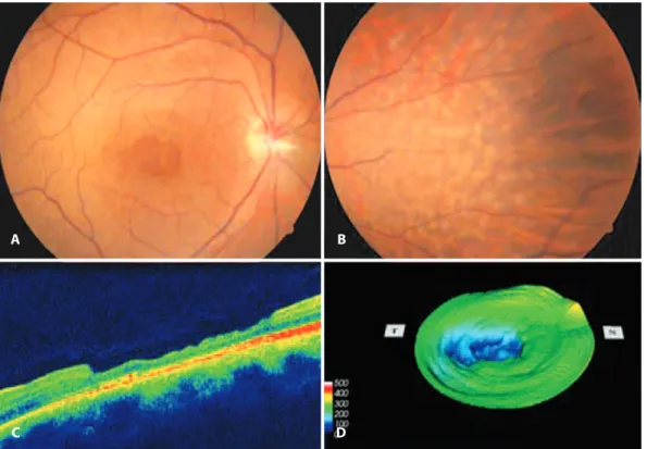
The ophthalmic exam demonstrated that best corrected visual acuity (BCVA) was 120/200 in OD and 140/200 in OS. Reflexes and external ocular motility were normal in OU. The slit lamp examination showed pseudophakia with posterior chamber intraocular lens (IOL) centered and without opacities in OU. At fundoscopy, there was symmetric, bilateral, discrete and circumscribed macular intense color of the retinal pigment epithelium (RPE) transmitted through
RESUMO
Este relato de caso descreve a presença de atrofia macular bilateral em uma paciente com síndrome de Alport e compara este achado com a literatura. Ao exame fundoscópico, havia discreto afinamento macular circunscrito demonstrando a coloração intensa do epitélio pigmentado da retina e a presença de lesões retinianas circulares esbranquiçadas (“dots” e “flecks”) na média periferia nasal em ambos os olhos. A tomografia de coerência óptica identificou atrofia parcial da retina neurossensorial bilateral na mácula, com maior extensão na área temporal. O caso descreve uma alteração oftalmológica rara da síndrome de Alport e de importante reconhecimento para precisar o diagnóstico e também para determinar o prognóstico visual.
Descritores: Nefrite hereditária/complicações; Mácula lútea/anormalidades; Distrofias retinianas; Doenças retinianas/diagnóstico; Tomografia de coerência óptica/ins -trumentação
ABSTRACT
This case report describes the presence of bilateral macular atrophy in a patient with Alport syndrome and compares this finding with literature. At fundoscopy, there was a discrete circumscribed macular thinning showing intense retinal pigment epithelium color and the presence of whitish circular retinal lesions (“dots” and “flecks”) at nasal mid periphery of both eyes. Optical coherence tomography showed bilateral partial atrophy of the neurosensory retina in the macula, with a greater extent in the temporal region. This case describes a rare ophthalmological finding in Alport syndrome and important to be recognized for a precise diagnosis as well as for determining visual prognosis.
Keywords: Nephritis, hereditary/complications; Macula lutea/abnormalities; Re tinal dystrophies; Retinal diseases/diagnosis; Optical coherence tomography/ins -trumentation
Submitted for publication: September 15, 2011 Accepted for publication: May 28, 2012
Study carried out at Departamento de Oftalmologia do Hospital das Clínicas da Faculdade de Me dicina da Universidade de São Paulo - HC/FMUSP - São Paulo (SP), Brazil.
1 Physician, Vitreoretinal Diseases Service, Department of Ophthalmology, Hospital das Clínicas, Universidade de São Paulo - USP - São Paulo (SP), Brazil.
2 Physician, Department of Ophthalmology. Hospital das Clínicas. Universidade de São Paulo - USP - São Paulo (SP), Brazil.
Funding: No specific financial support was available for this study.
Disclosure of potential conflicts of interest: T.Z.Igami, None; M.M.Lavezzo, None; D.A.Ferraz, None; W.Y.Takahashi, None; Y.Nakashima, None.
unusual macular thickness in Alport syndrome: case report
2 8 4 Arq Bras Oftalmol. 2012;75(4):283-5
the thinned retina, comparable to a macular pseudohole. In the na-sal mid-periphery, there were confluent ivory colored dots and flecks, apparently occupying intermediate levels of the retina in OU (Figure 1).
Fluorescein angiography revealed early hyperfluorescence in the well-defined macular region, which did not vary in intensity on late stages in OU. Optical coherence tomography (OCT) identified macular area with partial atrophy of neurosensory retina in OU, with a greater extent in the temporal region of the macula (Figure 1). The average foveal thickness was 112 µm in OD and 99 µm in the OS.
DISCUSSION
Ophthalmic signs in patients with AS include corneal endothe-lium, retinal and crystalline lens abnormalities. LVA is consequent to modifications in the crystalline lens as anterior lenticonus, posterior lenticonus, microspherophakia and cataract and may be reversible after phacoemulsification and IOL implantation in the capsular bag(4).
The most common retinal abnormality is a presence of peripheral and macular “dots” and “flecks”. These findings do not occur concomitantly
with the BCVA decline(4,5). Other fundoscopic sings reported more
rarely were: bilateral macular hole, vitelliform lesion and macular atrophy(5-9). The latter was first described by Usui et al.(7),and by other
authors (Table 1). Recently, Savige et al., evaluated a case series of patients with AS and macular changes and found similar alterations to the described case(8). The authors suggest that mutation of COLA4
gene causes a structural modification in the internal limiting mem-brane, inner nuclear and nerve fiber layer due to owing to a molecular alteration of type IV collagen. This change would cause a thinning and fragility of retinal layers resulting from the structural collagen changeover. The thinning was statistically significant at the foveal and temporal region of macula(8). Colville et al., described the same
macu-lar alteration named as “lozenge” or “dull macumacu-lar reflex” in a group of patients with AS, and highlighted the importance of acknowledging the findings with the aid of red-free image and its correlation with diagnosis and poor prognosis of the disease(10).
In the presented case, the patient was already pseudophakic and presented similar best-corrected visual acuity y (BCVA) in BE. Thus, there was no interference of the crystalline lens changes that could
Table 1. Characteristics of patients afected by Alport syndrome in the presence of bilateral macular atrophy
Author/year
Number of patients described
OD/OS BCVA
OD/OS
MT (µm) Age (years) Gender
Usui/2004(7) 01 20/20
20/20
177/186 38 M
Colville/2009(9) 06 NI NI 11-41 M
Fawzi/2009(5) 01 20/40
20/50
NI 19 M
Savige/2010(8) 010 NI 214 ± 46
(foveal)
14-74 7M
3F
BCVA= best corrected visual acuity; MT= macular thickness; OD= right eye; OS= left eye; NI= not informed by the author; M= male; f= female
Figure 1. Changes observed in the right eye. A) Presence of circumscribed pigmentary change in the macula. B) Presence of “dots” and “lecks” in the nasal retina. C, D) OCT identiied macular area with partial atrophy of neurosensory retina, with greater extent in the temporal region of macula. The average foveal thickness was 112 µm.
A
C
B
Igami TZ, et al.
2 8 5
Arq Bras Oftalmol. 2012;75(4):283-5 justify the reduction of BCVA. Partial atrophy in the macular region
while preserving the layer of photoreceptors could justify the relative interference on the visual potential. If her anterior lenticonus had been significant during early childhood, it is possible there could have been a slight degree of amblyopia contributing to poor vision. But probably, the lenticonus was not severe during childhood simply for the fact that renal failure requiring transplantation happened in her early adulthood (20 years old).
This patient could not have electrophysiology or visual field stu dies. There are evidences that electrophysiological analysis of patients with Alport syndrome shows no additional information that could con tri-bute on diagnosis or prognosis of this disease, since as renal disease alone can affect the results of electroretinogram (ERG)(10).
Histopathological studies of retina that provide detailed informa-tion are needed for possible increments in gene therapy as alternati-ve treatment of patients with AS
The case describes the presence of bilateral macular atrophy in AS, a rare ocular finding that is important to be recognized for the correct diagnosis of the disease as well as to transmit a visual progno-sis to the patient before or after cataract surgery. There are evidences that this kind of maculopathy can impair visual acuity (Table 1).
The finding of retinal changes related to Alport syndrome can also estimate the risk for renal bankruptcy(9).
REFERENCES
1. Perrin D. Le syndrome d’Alport (néphropathies héréditaires avec surdité et atteinte oculaire). Ann Ocul (Paris).1964;197:329-346.
2. Alves FRA, Ribeiro FAQ. Revisão sobre a perda auditiva na Síndrome de Alport, ana-lisando os aspectos clínicos, genéticos e biomoleculares. Rev. Bras. Otorrinolaringol. 2005;71(6):813-819.
3. Gregory MC, Terreros DA, Barker DF, Fain PN, Denison JC, Atkin CL. Alport syndrome-cli nical phenotypes, incidence, and pathology. Contrib Nephrol. 1996;117:1-28. 4. Seymenoglu G, Baser EF. Ocular manifestations and surgical results in patients with
Alport syndrome. J Cataract Refract Surg. 2009;35(7):1302-6.
5. Fawzi AA, Lee NG, Eliott D, Song J, Stewart JM. Retinal findings in patients with Alport Syndrome: expanding the clinical spectrum. Br J Ophthalmol. 2009;93(12): 1606-11.
6. Mete UO, Karaaslan C, Ozbilgin MK, Polat S, Tap O, Kaya M. Alport’s syndrome with bilateral macular hole. Acta Ophthalmol Scand. 1996;74(1):77-80.
7. Usui T, Ichibe M, Hasegawa S, Miki A, Baba E, Tanimoto N, Abe H. Symmetrical reduced retinal thickness in a patient with Alport syndrome. Retina. 2004;24(6):977-9. 8. Savige J, Liu J, DeBuc DC, Handa JT, Hageman GS, Wang YY, Parkin JD, Vote B, Fassett
R, Sarks S, Colville D. Retinal basement membrane abnormalities and the retinopathy of Alport syndrome. Invest Ophthalmol Vis Sci. 2010;51(3):1621-7.
9. Colville D, Wang YY, Tan R, Savige J. The retinal “lozenge” or “dull macular reflex” in Alport syndrome may be associated with a severe retinopathy and early-onset renal failure. Br J Ophthalmol. 2009;93(3):383-6.
10. Jeffrey BG, Jacobs M, Sa G, Barratt TM, Taylor D, Kriss A. An electrophysiological study on children and young adults with Alport’s syndrome. Br J Ophthalmol. 1994;78(1): 44-8.
