INTRODUCTION
Patients with celiac disease (CD) have an intolerance to the poli-peptide fragments of gluten, mediated by T lymphocytes. Gluten is a water-insoluble substance found in wheat lour, rye, barley and oats(30). CD depends on genetic, immunological and environmental factors and it is characterized by total or partial atrophy of the intestinal villi and consequent poor absorption of nutrients(6,9,27). Its prevalence in Brazil is shown to be 1/214(3).
CD diagnosis must be based on clinical, histopathological (gold standard) and serological examinations(3,27). There are few studies on the intestinal microbiota role in CD, even though gliadin (a gluten peptide) and microorganisms similarly activate pro-inlammatory routes(12). The information about the intestinal microbiota of celiac patients is mainly obtained from a stool sample examination(15).
Healthy subjects present a signiicantly higher concentration of biidobacteria when compared to celiac patients, while faecal pH seems to remain the same in both situations(8).
The only effective and possible treatment for CD is dietary, throughout the exclusion of gluten from the diet, which allows the remission of symptoms and the restoration of the regular mu-cosa(1). Without treatment, CD has a high morbi-mortality rate,
Effects of probiotic intake on intestinal
bifidobacteria of celiac patients
Flávia
MARTINELLO
, Camila Fontana
ROMAN
and Paula Alves de
SOUZA
Received 7/9/2016 Accepted 19/1/2017
ABSTRACT – Background – Healthy individuals exhibit a signiicantly higher concentration of faecal biidobacteria in comparison to celiac patients. Even though there are potential beneits in probiotic usage, they have been little explored as an adjunctive therapy in celiac disease. Objective – This study aimed at the comparison of faecal biidobacteria concentration and pH among celiac patients and healthy subjects before and after the daily intake of 100 g of yogurt containing probiotic for a thirty-day period. Methods – Feces from 17 healthy subjects and 14 celiac patients were analyzed, in which stool culture was performed for the isolation and quantiication of faecal biidobacteria. Furthermore, Gram’s method was employed for the microscopic analysis of the colonies, while the identiication of the Biidobacterium genus was made through determination of the fructose-6-phosphate
phosphoketolase enzyme. Faecal pH was measured using a calibrated pHmeter. Results – Faecal biidobacteria concentration before probiotic consump-tion was signiicantly higher in healthy individuals (2.3x10⁸±6.3x10⁷ CFU/g) when compared to celiac patients (1.0x10⁷±1.7x10⁷ CFU/g). Faecal pH
values did not show a signiicant difference. After the daily consumption of probiotic-containing yogurt both groups showed a signiicant increase in the concentration of faecal biidobacteria, but healthy subjects presented signiicantly higher biidobacteria concentrations (14.7x10⁸±0.2x10⁸ CFU/g)
than the celiac group (0.76x10⁸±0.1x10⁸ CFU/g). The obtained pH values from both groups were not signiicantly different, being 7.28±0.518 for the
celiac patients and 7.07±0.570 for healthy individuals after the probiotic intake. Conclusion – The probiotic supplementation signiicantly increased the number of biidobacteria in the feces of celiac patients, although it was not suficient to reach the concentration found in healthy individuals prior to its consumption.
HEADINGS – Probiotics. Celiac disease. Biidobacterium. Hydrogen-ion concentration. Feces, microbiology. Microbial colony count.
Declared conflict of interest of all authors: none Disclosure of funding: no funding received
Departamento de Análises Clínicas, Centro de Ciências da Saúde, Universidade Federal de Santa Catarina, Florianópolis, SC, Brasil.
Correspondence: Camila Fontana Roman. Departamento de Análises Clínicas. Centro de Ciências da Saúde. Bloco JK, sala 109. Universidade Federal de Santa Catarina. CEP: 88040-900 – Florianópolis, SC, Brasil. Email: [email protected]
with risks of developing complications such as anemia, infertility, osteoporosis and cancer, being the most prevalent the intestinal lymphoma(27). Alternative treatments are also available and can be used simultaneously as palliatives, for instance, the use of probio-tics, mainly Lactobacillus and Biidobacterium(7,15,24).
The presence of biidobacteria in the gastrointestinal tract seems to suffer variations throughout life and it is associated with beneicial effects to health, including the re-composition of the intestinal microbiota, the growth inhibition of pathogenic bacte-ria, regeneration of the epithelial barrier and anti-inlammatory effects(11,21,25,26,32). Some species have the capacity of inhibiting the increased permeability induced by gliadin, weakening its cytotoxic effect and the host autoimmune response(10). Smecuol et al. showed that celiac patients on a gluten diet had experienced beneicial ef-fects related to gastrointestinal tract symptoms (such as constipa-tion and gastroesophageal relux) when consuming biidobacteria in capsules before meals(28). Other beneicial effects of biidobacteria consumption with a gluten diet have been described, such as the reduction of human α-defensin 5 (HD-5) and paneth cells(20).
Biidobacterium and Lactobacillus are widely used in several
poorly explored as an adjunctive therapy in CD(17,32). In this context, the hypothesis is that the intestinal microbiota of patients with controlled CD can be restored by the daily intake of probiotic-containing yogurt. The results of this study will allow the analysis of the necessity and eficacy of supplementation with probiotics to restore the intestinal microbiota equilibrium, with consequent reduction of gastrointestinal complications and infections, improv-ing the quality of life of celiac patients.
METHODS
Study design
The Ethics Committee for Studies with Humans of the Federal University of Santa Catarina, Brazil, approved the experimental protocol for this study (number 772, 2010). The participants with CD were recruited in the local Association of Celiac People in Brazil (Associação dos Celíacos do Brasil – ACELBRA) during its monthly meetings. All celiac patients were on a controlled stage of the disease during the study, i.e., they were on a gluten free diet,
without signals and symptoms of CD. The non-celiac participants were randomly recruited from the population.
Volunteers were submitted to a clinical and sociodemographic questionnaire and the research started by collecting the irst stool sample to quantify biidobacteria and measure faecal pH. After-wards, each volunteer consumed one unit of probiotic-containing yogurt (100g) from Piá Essence, PIÁ®, Nova Petrópolis-RS) per day, having eaten in the fasting state at morning, during one month. The yogurt delivery was made weekly. After 30 days of consump-tion, feces were collected again in order to quantify biidobacteria and measure faecal pH.
Exclusion criteria
The following exclusion criteria for the participation in the study were adopted: individuals with suspicion or diagnosis of autoimmune diseases; suspicion or diagnosis of diabetes; lactose intolerance; allergy to any excipient present in the yogurt; individu-als who consumed products containing prebiotics and/or probiotics three months prior to the beginning of research, and individuals who presented fever, diarrhea and/or vomit three months prior to the beginning or during study.
Determination of faecal bifidobacteria content and pH
For the isolation and quantification of bifidobacteria and measurement of faecal pH, participants collected stool samples, which were sent to the laboratory and analyzed within 8 h after collection(13,29).
Feces aliquot (1 g) from each volunteer was diluted in 9 mL of distilled and deionized sterile water for the measurement of faecal pH in pHmeter PHTEK®. Another feces aliquot (1 g) from each stool sample was diluted in 9 mL of phosphate buffer. The mixture was homogenized ive times using the anaerobic technique. From this dilution (10-1), serial fold dilutions up to 10-7 were prepared. The stock phosphate buffer was previously prepared with 34 g of KH2PO4 in 500 mL of distilled and deionized water, having the pH adjusted to 7.2 with NaOH 1 N and the volume completed to 1 L with distilled water, being subsequently sterilized in an autoclave at 121°C during 18 minutes. For the dilution of the stool sample, the phosphate buffer was diluted a thousand times from the stock solution.
The culture media used for isolation of biidobacteria was the
RCA (Reinforced Clostridial Agar, DifcoTM BD) supplemented with antibiotics (nalidixic acid 2%, polymyxin B sulfate 0.85%, kanamycin sulfate 0.5%, iodoacetic acid 0.5%, 2,3,5-triphenyltetra-zolium chloride 0.5% and amphotericin B 0.001%)(14).
The spread-plating of 100 µL from each dilution was prepared and the plates were incubated at 37°C for 72 hours under anaerobic conditions(14) using a commercial anaerobic atmosphere genera-tion system (Anaerobac from Probac®), followed by counting of biidobacteria colonies in the plates containing between 30 and 300 colony-forming units (CFU). For the conirmation of the Biido-bacterium genus, Gram staining was made, as well as catalase proof
and fructose-6-phosphate phosphoketolase (F6PPK) reaction, as stated by Orban & Patterson (2000), for all isolated colony types(18).
The results from the biidobacteria quantiication were pre-sented as CFU per gram of feces (CFU/g). To obtain the results, the number of CFU counted in each plate was multiplied by its respective dilution factor and corrected for the sample volume spread. They are expressed as mean ± standard deviation (n=17 for the control group and n=14 for the celiac group).
Determination of yogurt bifidobacteria content and pH
All lots of the yogurt Piá Essence donated were analyzed for the isolation and quantiication of biidobacteria and measurement of pH. One pot containing 100 g of yogurt was randomly selected from each lot and 1 g was diluted in 9 mL of distilled and deion-ized sterile water for measurement of pH in pHmeter PHTEK® previously calibrated.
Another yogurt aliquot (1 g) was also diluted in 9 mL of phos-phate buffer. Serial dilutions were made from this solution as for the feces analysis. The spread-plating from each dilution, the counting of colonies, the conirmation of the genus and the expression of the results were made as previously described for the stool samples.
Statistical analysis
The statistical analysis was performed using the program GraphPad Prism® version 5.0 from 2007. For data distribution analysis, the D’Agostino normality test and Pearson Omnibus Normality Test were employed. Spearman’s correlation coeficient was employed to verify the correlation among the biidobacteria concentration, faecal pH and volunteers’ ages. Wilcoxon test was used for the comparison of the results between groups. A signii-cance level of 5% (P<0.05) was adopted for all tests.
RESULTS
The yogurt package informs that each 100 g of yogurt contains 108 CFU of Lactobacillus acidophilus and Biidobacterium lactis. Amongst the yogurt lots available to the volunteers, the average concentration of biidobacteria was 6.67x10⁸±10.3x10⁸ CFU/g of yogurt. The average yogurt pH was 4.28±0.15 and there was a signiicant correlation between the biidobacteria concentration and yogurt pH (P=0.0121). There was growth of Gram-positive
bacillus colonies in every lot of yogurt, being all catalase negative and showing F6PPK activity.
Amongst the 17 healthy control individuals, 10 were female and seven were male, aged between 18 and 58 years (average of 26 years old). This group of individuals did not have any relatives with CD.
higher in irst (father, mother and siblings) and second (grand-parents, aunts, uncles and cousins) degree relatives. Two (14.4%) volunteers had irst-degree celiac relatives, three (21.4%) had irst and second-degree celiac relatives, one (7.1%) could not answer and eight (57.1%) did not have celiac relatives. The average age in which the diagnosis was made was 36 years old, where 100% of the patients had the small intestine biopsy done for conirmation of CD. A relation between faecal biidobacteria concentration and age was not observed in any of the groups, either celiac or healthy subjects.
Seven (50%) of the 14 celiac patients received drug therapy after CD diagnosis, being calcium therapy the most prevalent in 57% of them, mainly related to women 30 years old or older.
During the stool sample examination from the volunteers, biidobacteria colonies were observed, presenting round shape, smooth surface, pink to wine color and small to medium size. The colony morphology was similar between both groups, celiac and control (Figure 1). Biidobacteria appeared in the form of short and long Gram-positive bacilli, with or without bifurcated ends V or Y-shaped, and as Gram-positive coccobacilli (Figure 2).
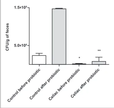
The results of biidobacteria quantiication in the stool samples are shown in Figure 3. Healthy individuals presented a signiicantly higher concentration of biidobacteria (2.3x10⁸ ± 6.3x10⁷ CFU/g) before the probiotic-containing yogurt intake when compared to the celiac group (1.0x10⁷±1.7x10⁷ CFU/g) (Figure 3). Celiac pa-tients presented, in average, 83% less biidobacteria than healthy individuals. Still, celiac faecal pH (7.19±0.521) was not signiicantly different from the faecal pH of the control group (7.18±0.522).
FIGURE 1. Biidobacteria colonies in Reinforced Clostridial agar media
supplemented with antibiotics. (A) Celiac patient plate. (B) Control subject plate.
A
C
FIGURE 2. Micromorphology of bifidobacteria colonies stained by
Gram method in optical microscopy in 1000 times increase. (A) Short Gram-positive bacilli, isolated, in pairs or grouped. (B) Gram-positive cocobacilli, isolated, in pairs or grouped. (C) Long Gram-positive bacilli with bifurcated ends V or Y-shaped.
A B
After the daily intake of 100 g of probiotic-containing yogurt for 30 days, healthy individuals presented a signiicantly higher biidobacteria concentration (14.7x10⁸±0.2x10⁸ CFU/g) than celiac patients (0.76x10⁸±0.1x10⁸ CFU/g) (Figure 3). However, faecal pH of celiac patients (7.28±0.518) did not show signiicant difference from the faecal pH of healthy individuals (7.07±0.570) after the yogurt intake.
DISCUSSION
Several probiotic supplements can be found on the market; meanwhile it is still hard to ind gluten free products for celiac patients. In this context, the product options for this research were limited. Amongst the companies for which support was requested, only PIÁ®, Nova Petrópolis-RS, provided the products. The average biidobacteria concentration provided for the research participants (6.67x10⁸±10.3x10⁸ CFU/g of yogurt) is enough to bring beneits to their health, according to Vinderola & Reinheimer(31).
A number of factors can affect probiotic bacteria viability in yogurts. High carbohydrate concentrations added to the product before its fermentation can inhibit the bacteria, leading to long periods of fermentation and an underdevelopment of acidity(16).
FIGURE 3. Number of colony forming units (CFU) of biidobacteria per
gram of feces from control and celiac groups, before and after probiotic intake. Results are expressed as mean ± standard deviation (control group n=17 and celiac group n=14). * P<0.05 nonparametric t test, when com-pared to the respective control group before probiotic intake or ** when compared to the respective control group after probiotic intake.
Oliveira & Damin(16) found that the number of probiotic bacteria remained stable for at least seven days of storage. However, in this study volunteers consumed the probiotics up to their expiration date, which simulates the acquisition of products commercialized for the general population. A yogurt of a lot provided for the vol-unteers was randomly tested six days after its expiration date, in which a biidobacteria concentration of 1.74x10⁶ CFU/g of yogurt was found. Coupled with the likely concentration of Lactobacillus,
this would still be a probiotic food and bring beneits to people’s health(19), including celiac patients.
The largest number of female celiac patients in this study is consistent with literature, which shows a higher prevalence of CD in women(3). About 30% of celiac patients evaluated in this study have a relative with CD, which is similar to a study made with patients from Association of Celiac People in Brazil, section from Santa Catarina, (ACELBRA-SC) in 2004, revealing that 27% of associates had relatives with CD(3). This data reinforces the idea that genetic determinants of CD are associated with environmental factors(15). It is important to note that 100% of the celiac patients who participated in the research had the intestinal biopsy done for their diagnosis, which is recommended by the literature(30).
The poor intestinal absorption of most nutrients resulting from the inlammatory response on CD can explain why most celiac patients reported having osteoporosis and osteopenia(3,30). It also explains why most participants of this research have replenished calcium and vitamin D after CD diagnosis. The supplementation with probiotic-containing yogurt could bring not only the beneits from the probiotics for celiac patients but also a greater amount of calcium absorbed from their diet.
The mechanisms of action of probiotics have not been com-pletely elucidated, even though many have been suggested and possibly operate individually or associated(32). There is evidence that probiotics have antimicrobial action, compete for limited nu-tritional resources from the intestinal microbiota, block adhesion of pathogens in the intestinal mucosa and have antitoxin effects of pathogens(22). Biidobacteria can also beneit people’s health by lowering intestinal pH through the production of short chain fatty acids (acetate and lactate), thus inhibiting pathogenic bacteria growth. This is a digestive system self-mechanism for popula-tion control and selectivity of bacterial colonizapopula-tion(13). Indeed, a signiicant correlation between faecal pH and biidobacteria concentration was not seen in this study.
Macro and micromorphology of biidobacteria colonies found in the stool samples were similar in both groups and were as de-scribed in the literature. However, the results show a signiicant lower quantity of biidobacteria CFU per gram of feces of celiac patients than in the control group. Some studies show that allergic children and patients with atopic diseases are frequently colonized by a reduced number of biidobacteria when compared to healthy children, showing a close relationship between biidobacteria con-centration and host immune disorders(5).
Nadal et al.(15) reported an imbalance in the intestinal biota of celiac children, especially the reduction of faecal Biidobacterium
spp. concentration. Similarly, Collado et al.(4) have reported that celiac children with active or inactive disease had inferior biido-bacteria counting than control groups for both analyzed samples, either feces or intestinal biopsy specimens. Therefore, this imbal-ance seems to be independent on the activity of the disease. This explains the lower biidobacteria concentration found in feces of adult celiac patients in this study, all in a controlled phase of CD.
The results found in this study for biidobacteria concentra-tion without probiotic consumpconcentra-tion show a signiicantly higher biidobacteria count in healthy subjects when compared to celiac patients, which is consistent with literature(8). Even after probiotic consumption, the faecal biidobacteria count in celiac patients from this study has not reached the counting in healthy individuals without probiotic consumption (Figure 3).
The values of faecal pH for both groups before probiotic intake had no signiicant difference, having them remained very similar even after probiotic intake. These results suggest that the higher faecal biidobacteria concentration after probiotic consumption did not increase intestinal fermentation, which would lower the pH and ease biidobacteria growth(13). However, it is worth noting that the pH from the control group was slightly more acidic than the pH from the celiac patients. The increase in biidobacteria count favors the lowering of faecal pH due to the fermentation done by these bacteria(13). The results of pH values from both groups, celiac and control, suggest that the smaller amount of biidobacteria in the intestine of celiac patients is probably not related to faecal pH, but to the pathogenesis of CD. Thus, the relationship between biidobacteria counting and CD has yet to be elucidated.
The maintenance of pH values before and after probiotic ingestion may be related to time or quantity/concentration of the daily-consumed probiotic, being suggested that probiotic effects are dose-dependent(19). However, the recommended dose by the literature was consumed in this study, which is between 106 e 1011 CFU/day, depending on the desired effect(22).
In order to have the metabolism and intestinal content relected in feces, variables must be taken into account, including intestinal motility, total iber ingestion, intestinal secretion, and duration of dietetic intervention. Because of that, faecal pH may not exactly relect colon pH. In fact, Bouhnik et. al.(2) have not considered the faecal pH as a good indicator of intestinal acidiication, since it has not changed after ingestion of nondigestible carbohydrates by 200 healthy volunteers, despite the increase in the number of faecal biidobacteria.
Although the healthy intestinal microbiota remains to be de-ined, there are many diseases related to its imbalance. In most cases, there is no information yet if microbiota imbalance has a triggering role or if it is a disease consequence. Anyway, both relationships lead to the hypothesis that an intervention to restore the microbiota to the healthiest state could mitigate the disease. The consumption of properly selected probiotics could be used with such role(23).
There is indication, amongst research to elucidate activity of biidobacteria, that intestinal microbiota change can inluence the typical inlammatory reactions in CD in a specie-speciic way(4). Therefore, it is thought that biidobacteria has a great therapeutic potential, and manipulation of intestinal biota, as with probiotic supplementation, might improve quality of life of celiac patients. However, it should be noted that the inclusion of a small num-ber of participants, the evaluation of pH and biidobacteria con-tents during a short period of time and the availability of molecular methods, more accurate to evaluate the intestinal microbiota, may be considered limitations of the present study. Therefore, we suggest that additional studies should be performed in order to evaluate all the aspects regarding intestinal microbiota and probiotic sup-plementation in CD.
CONCLUSION
The results obtained in the present study allow the conclusion that there is a lower biidobacteria count in the intestinal micro-biota of celiac patients, even when they are on a gluten free diet and consuming probiotic-containing food, when compared to the control group. This disturbance is independent on the faecal pH. Supplementation with probiotics increased the number of faecal biidobacteria, which relects its intestinal concentration. Further research must be performed in order to evaluate the equilibrium of other bacteria (for instance, the pathogenics); to verify how long
biidobacteria count remains elevated after probiotic consumption; to correlate small intestine biopsy results with biidobacteria con-centration, since celiacs were on a gluten free diet; and evaluate if the microbiota imbalance was due to gluten contamination in food. In summary, this information will help develop speciic dietetic recommendations to celiac patients based on their microbiota composition.
Authors’ contributions
Martinello F: survey execution. Roman CF: writing and trans-lation of text. Souza PA: statistical analysis and writing of text.
REFERENCES
1. Accomando S, Cataldo F. The global village of celiac disease. Dig Liver Dis. 2004; 36:492–8.
2. Bouhnik Y, Raskine L, Simoneau G, Paineau D, Bornet F. The capacity of short-chain fructo-oligosaccharides to stimulate faecal biidobacteria: a dose-response relationship study in healthy humans. Nutr J. 2006;5:8.
3. Cassol CA, Pellegrin CP, Wahys MLC, Pires MMS, Nassar SM. [Clinical proile of Santa Catarina members of Brazilian Celiac Association]. Arq Gastroenterol. 2007;44:257-65.
4. Collado MC, Calabuig M, Sanz Y. Differences between the fecal microbiota of coeliac infants and healthy controls. Curr Issues Intest Microbiol. 2007;8:9–14. 5. Collado MC, Donat E, Ribes-Koninckx C, Calabuig M, Sanz Y. Imbalances in
faecal and duodenal Biidobacterium species composition in active and non-active coeliac disease. BMC Microbiol. 2008; 8:232.
6. Di Cagno R, Rizzello CG, Gagliardi F, Ricciuti P, Ndagijimana M, Francavilla R, et al. Different fecal microbiotas and volatile organic compounds in treated and untreated children with celiac disease. Appl Environ Microbiol. 2009;75:3963-71. 7. Fontana L, Bermudez-Brito M, Plaza-Diaz J, Munoz-Quezada S, Gil A. Sources, isolation, characterisation and evaluation of probiotics. Br J Nutr. 2013; 109 Suppl 2:S35-50.
8. Golfetto L, Senna FD, Hermes J, Beserra BTS, França FS, Martinello F. Lower biidobacteria counts in adult patients with celiac disease on a gluten-free diet. Arq Gastroenterol. 2014;51:139-43.
9. Ivarsson A, Hernell O, Nyström L, Persson LA. Children born in the summer have increased risk for coeliac disease. J Epidemiol Community Health. 2003;57:36–9. 10. Laparra JM, Sanz Y. Biidobacteria inhibit the inlammatory response induced by gliadins in intestinal epithelial cells via modiications of toxic peptide generation during digestion. J Cell Biochem. 2010; 109:801-7.
Martinello F, Roman CF, Souza PA. Efeitos do consumo de probióticos sobre as biidobactérias intestinais de pacientes celíacos. Arq Gastroenterol. 2017;54(2):85-90.
RESUMO – Contexto – Indivíduos saudáveis apresentam uma concentração de biidobactérias fecais signiicativamente maior em comparação a pacientes celíacos. Apesar de haver benefícios potenciais no uso de probióticos na doença celíaca, estes têm sido pouco explorados como uma terapia adjuvante. Objetivo – Este estudo objetivou a comparação do pH e concentração fecal de biidobactérias entre pacientes celíacos e indivíduos saudáveis antes e após o consumo diário de 100 g de iogurte contendo probiótico por um período de 30 dias. Métodos – Foram analisadas fezes de 17 pessoas saudáveis e 14 pacientes celíacos, tendo sido realizada a coprocultura para o isolamento e quantiicação de biidobactérias fecais. Além disso, o método de Gram foi empregado na análise microscópica das colônias, enquanto a identiicação do gênero Biidobacterium foi feita através da determinação da enzima frutose-6-fosfato fosfocetolase. O pH fecal foi medido usando um pHmetro calibrado. Resultados – A concentração de biidobactérias fecais antes do consumo do iogurte probiótico foi signiicativamente maior em indivíduos saudáveis (2.3x10⁸±6.3x10⁷ UFC/g) quando comparada aos celíacos
(1.0x10⁷±1.7x10⁷ CFU/g). Por outro lado, o pH fecal de ambos os grupos não apresentou diferença signiicativa. Após o consumo diário de iogurte
contendo probiótico, ambos os grupos tiveram um aumento signiicativo na concentração de biidobactérias fecais, entretanto indivíduos saudáveis apresentaram concentrações de biidobactérias signiicativamente maiores (14.7x10⁸±0.2x10⁸ UFC/g) do que o grupo celíaco (0.76x10⁸±0.1x10⁸
UFC/g). Os valores de pH obtidos de ambos os grupos não foram signiicativamente diferentes, sendo de 7.28±0.518 para os pacientes celíacos e de 7.07±0.570 para os indivíduos saudáveis após o consumo do probiótico. Conclusão – A suplementação com probiótico aumentou signiicativamente o número de biidobactérias nas fezes dos pacientes celíacos apesar de não ter sido suiciente para alcançar a concentração encontrada em indivíduos saudáveis antes do consumo de probióticos.
DESCRITORES – Probióticos. Doença celíaca. Biidobacterium. Concentração de íons de Hidrogênio. Fezes, microbiologia. Contagem de colônia microbiana.
11. Leahy SC, Higgins DG, Fitzgerald GF, Sinderen DV. Getting better with biido-bacteria. J Appl Microbiol. 2005;98:1303-15.
12. Medina M, Palma GD, Ribes-Koninckx C, Calabuig M, Sanz Y. Biidobacteri-um strains suppress in vitro the pro-inlammatory milieu triggered by the large intestinal microbiota of coeliac patients. J inlamm. 2008;5:1-13.
13. Mohan R, Koebnick C, Schildt J, Mueller M, Radke M, Blaut M. Effects of Biidobacterium lactis Bb12 Supplementation on Body Weight, Fecal pH, Acetate, Lactate, Calprotectin, and IgA in Preterm Infants. Pediatr Res. 2008;64:418-22. 14. Muñoa FJ, Pares R. Selective medium for isolation and enumeration of
Biido-bacterium spp. Appl Environ Microbiol. 1988;54:1715-8.
15. Nadal I, Donant E, Ribes-Koninckx C, Calabuig M, Sanz Y. Imbalance in the composition of the duodenal microbiota of children with celiac disease. J Med Microbiol. 2007;56:1669–74.
16. Oliveira MN, Damin MR. [Effect of total solids and sucrose contents on acidity, irmness and viability of yogurt and probiotic bacteria in fermented milk]. Ciênc Tecnol Aliment. 2003;S23:172-6. Available from http://www.scielo.br/scielo. php?script=sci_arttext&pid=S0101-20612003000400032.
17. OMG. Organização Mundial de Gastroenterologia. Guias Práticas da OMGE: Probióticos e Prebióticos. 2008. Available from: http://www.serdigital.com.br/ gerenciador/clientes/biologicus/arquivos/25.pdf.
18. Orban JI, Patterson JA. Modiication of the phosphoketolase assay for rapid identiication of biidobactérias. J Microbiol Methods. 2000;40:221-4. 19. Pérez N, Iannicelli JC, Girard-Bosch C, González S, Varea A, Disalvo L.
20. Pinto-Sánchez MI, Smecuol EC, Temprano MP, Sugai E, González A, Moreno ML et al. Biidobacterium infantis NLS super strain reduces the expression of [alpha]-defensin-5, a marker of innate immunity, in the mucosa of active celiac disease patients. J Clin Gastroenterol. 2016. [Epub ahead of print].
21. Pokusaeva K, Fitzgerald GF, Sinderen DV. Carbohydrate metabolism in Biido-bacteria. Genes Nutr. 2011;6:285-306.
22. Saad SMI. Probióticos e prebióticos: o estado da arte. Rev Bras Cienc Saude. 2006;42:1-16. Available from: http://www.scielo.br/scielo.php?script=sci_arttex-t&pid=S1516-93322006000100002&lng=en.
23. Sanders M E. Impact of probiotics on colonizing microbiota of the gut. J Clin Gastroenterol. 2011;45:S115-9.
24. Sanz, Y. Novel perspectives in celiac disease therapy. Mini Rev Med Chem. 2009;9:359–67.
25. Satokari RM, Vaughan EE, Smidt H, Saarela M, Matto J, de Vos WM. Molecular approaches for the detection and identiication of biidobacteria and lactobacilli in the human gastrointestinal tract. Syst Appl Microbiol. 2003;26:572–84. 26. Shanahan F. Probiotics in perspective. Gastroenterology. 2010;139:1808-12.
27. Silva TSG, Furlanetto TW. Diagnóstico de doença celíaca em adultos. Rev Assoc Med Bras. 2010;56:122-6. Available from: http://www.scielo.br/scielo. php?script=sci_arttext&pid=S0104-42302010000100027&lng=en.
28. Smecuol E, Hwang HJ, Sugai E, Corso L, Chernavsky AC, Bellavite FP et al. Exploratory, randomized, double-blind, placebo-controlled study on the effects of Biidobacterium infantis natren life start strain super strain in active celiac disease. J Clin Gastroenterol. 2013;47:139-47.
29. Thitaram SN, Siragusa GR, Hinton AJr. Biidobacterium-selective isolation and enumeration from chicken caeca by a modiied oligosaccharide antibiotic-selective agar medium. Lett Appl Microbiol. 2005;41:355-60.
30. Van Heel DA, West J. Recent advances in coeliac disease. Gut. 2006;55:1037-46. 31. Vinderola CG, Reinheimer JA. Enumeration of Lactobacillus casei in the presence of Lactobacillus acidophilus and lactic starter in fermented dairy products. Int Dairy J. 2000;10:271-5.