ABCD Arq Bras Cir Dig
Original Article
2017;30(2):98-102
DOI: /10.1590/0102-6720201700020005
TISSUE EXPRESION OF THE GENES MUTYH AND OGG1 IN
PATIENTS WITH SPORADIC COLORECTAL CANCER
Expressão tecidual dos genes MUTYH e OGG1 em doentes com câncer colorretal esporádico
Enzo Fabrício Ribeiro NASCIMENTO1, Marcelo Lima RIBEIRO2, Daniela Oliveira MAGRO3, Juliana CARVALHO4, Danilo Toshio KANNO2,Carlos Augusto Real MARTINEZ2, 3, Cláudio Saddy Rodrigues COY3.
From the 1Programa de Pós-Graduação
em Ciências da Cirurgia, Faculdade de Ciências Médicas, Universidade Estadual de Campinas, Campinas, SP; 2Programa
de Pós-Graduação em Ciências da Saúde, Universidade São Francisco, Bragança Paulista, SP; 3Departamento de Cirurgia,
Faculdade de Ciências Médicas, Universidade Estadual de Campinas, Campinas, SP; 4Centro
Integrado de Assistência a Saúde da Mulher, Universidade Estadual de Campinas, (1
Faculty of Medical Sciences of the State University of Campinas (FCM-UNICAMP), Postgraduate Program in Surgery Sciences, Campinas, SP; 2São Francisco University,
Post-Graduation Program in Health Sciences, Bragança Paulista, SP; 3Faculty of Medical
Sciences of the State University of Campinas, Department of Surgery, Campinas, SP; 4State
University of Campinas, Integrated Center for Women’s Health Care), Campinas, SP, Brazil.
HEADINGS - Colorectal cancer. Genes. DNA repair. Oxidative stress. DNA Glycosolasys.
ABSTRACT - Background: MTUYH and OGG1 genes have importance in the base excision repair systems of oxidized DNA bases. Modification of the tissue expression of these genes is related to the increased risk of developing colorectal cancer. Aim: To evaluate the tissue expression of MUTYH and OGG1 comparing normal and neoplastic tissues of patients with sporadic colorectal cancer and to correlate it with clinical and histopathological variables. Method: MUTYH and OGG1 tissue expression was quantified by RT-PCR in patients with colorectal cancer and the values were compared in normal and neoplastic tissues. MUTYH and OGG1 expression was measured and normalized to the constitutive 18S gene. The level of expression of both genes was correlated with the variables: age, gender, tumor location, size of the tumor, histological type, degree of cell differentiation, invasion depth in the intestinal wall, angiolymphatic infiltration, lymph node involvement and TNM staging. Results: Was found downregulation of both genes in neoplastic when compared to normal tissue. There was downregulation of the MUTYH in larger tumors and in patients with angiolymphatic invasion. Tumors with more advanced TNM stages (III and IV) presented downregulation of both genes when compared to those with earlier stages (I and II). Conclusion: The MUTYH and OGG1 genes present downregulation in the more advanced stages of colorectal cancer.
RESUMO - Racional: Os genes MUTYH e OGG1 possuem importância nos sistemas de reparo por excisão de bases oxidadas do DNA. Modificação na expressão tecidual desses genes encontra-se relacionada ao maior risco do deencontra-senvolvimento do câncer colorretal. Objetivo: Avaliar a expressão tecidual dos genes MUTYH e OGG1 comparando tecidos normais e neoplásicos de portadores de câncer colorretal esporádico e correlacioná-la com variáveis clínicas e histopatológicas. Método: Avaliou-se por PCR, em tempo real, a expressão tecidual dos genes MUTYH e OGG1 em 49 portadores de câncer colorretal comparando tecidos normais e neoplásicos. A expressão dos genes MUTYH e OGG1 foi quantificada e normalizada com o gene constitutivo 18S. A intensidade de expressão de ambos os genes foi correlacionada as variáveis: idade, gênero, localização do tumor, tamanho do tumor, tipo histológico, grau de diferenciação celular, profundidade de invasão na parede intestinal, invasão angiolinfática, linfonodos comprometidos e estadiamento TNM. Resultados: Encontrou-se menor expressão de ambos os genes no tecido neoplásico quando comparado ao tecido normal. Houve menor expressão do gene MUTYH nos tumores com maiores dimensões e nos doentes que apresentavam invasão angiolinfática. Tumores com estadios mais avançados (III e IV) apresentavam expressão menor de ambos os genes quando comparados àqueles com estadios mais precoces (I e II). Conclusão: Os genes MUTYH e OGG1 apresentam menor expressão tecidual nos estadios mais avançados do câncer colorretal.
Correspondence:
Enzo Fabrício do Nascimento E-mail: enzonascimento@uol.com.br
Financial source: none Conflicts of interest: none
Received for publication: 12/12/2016 Accepted for publication: 14/03/2017
DESCRITORES - Câncer colorretal. Genes.
Reparo do DNA. Estresse Oxidativo. DNA Glicosilases.
INTRODUCTION
A
ccording to recent epidemiological inquiry, colorectal cancer (CRC) occurredin 2015, 1.4 million people worldwide being the third type of most common
malignant neoplasm among men and the second among women9. The National
Institute of Cancer (INCA) estimates that for the biennium 2016-2017 In Brazil, depending
on the geographical region considered, the CRC can affect of 3.35 to 28,15/100,000
inhabitants among men and 2.09 to 29,13/100,000 inhabitants among women13. With
the increase in life expectancy, the phenomena of globalisation at greater exposure to carcinogenic agents and, especially, the change of dietary habits it is expected that the
CRC has increasing importance in the profile of mortality from cancer throughout the
world considerably increasing the economic and social costs19,23.
Genetic studies have shown development of sporadic CRC is an evolutionary process
related to cell cycle21. It is already well established that the
development of the CRC from the normal mucosa, is mediated by a sequence of mutations of genes controllers of cell cycle
(the proliferation, differentiation, apoptosis and DNA repair)8,30.
Mutations in these genes can confer additional advantages for growth of tumor tissue in relation to normal tissue19.
Although the initial phenomena of colorectal carcinogenesis are still a reason for constant research, it has been demonstrated that DNA damage caused by reactive oxygen species (ROS) is related to the development of CRC34,27,31,20,4. The ROS are
produced in large quantities in chronic inflammatory processes
affecting the intestinal epithelium. These radicals induce persistent damage to DNA and, if they are not readily inhibited by antioxidant systems can cause mutations related to the development of the CRC16. One of the mechanisms of DNA
damage to more well-studied relates to the oxidation of the base fertilization guanine, forming the 8-oxoguanina (8-oxoG). The 8-oxoG is a highly oxidized mutagenic capable of
causing transversions of CC→TA tapes of DNA allowing, if not
repaired, the appearance of mutations. When these mutations compromise tumor suppressor genes or oncogenes may arise a clone of cells with proliferative autonomy and uncontrolled growth, changes inherent to the neoplastic cells31.
It is already well established that the oxidation of bases of DNA allows for the development of the CRC and that removal of these databases oxidized is vital to avoid the appearance of mutations26. A chronic imbalance between the mechanisms of
damage and DNA repair increases the risk of genomic instability and, therefore, it is important to understand the mechanisms by which the repair systems that remove the oxidized bases of DNA present in the genome. For both, living organisms have
specific genes to correct the errors of matching caused by
oxidative stress. The system of excision of bases (BER) is the primary way to repair DNA responsible for correction of oxidized bases, having a fundamental role in preserving the integrity of the DNA submitted to the deleterious action of ROS28,24. Among all the genes involved in repairing and removing the 8-oxoG, genes MUTHY and OGG1, components of the system BER, have a prominent role. These genes encode DNA-glicosilases which has the function of removing the oxidized bases paired erroneously, ensuring the maintenance of the integrity of the tapes of DNA. Recent studies suggest that patients with CRC have lower expression of genes OGG1 and mutyh in tissue cancer when compared to normal tissues suggesting that these systems operate without a disability33. However, the
correlation between the level of tissue expression and clinical variables and histopathological were still little studied33. The
objective of this study was to evaluate the tissue expression of genes MUTYH and OGG1 in normal tissue and neoplastic patients with CRC sporadic and correlate it to the main clinical and histopathological.
METHOD
This study was submitted to and approved by the Research Ethics Committee at the Universidade São Francisco in
Bragança Paulista (Project No. 0235.0.142.000-07). All patients
who have provided biological material for the present study signed an informed consent form agreeing to participate in all stages, and were informed of the purpose of the research.
Casuistry
Were studied prospectively 49 patients being 26 (53%) women with an average age of 65.8±11.3 years, suffering
from adenocarcinoma of the colon and rectum, surgery with curative intent. Were excluded by means of criteria of clinical and endoscopic patients suspected of hereditary CRC (adenomatous polyposis familial syndrome and Lynch), sick undergoing treatment neoadjuvant chemoradiation
therapy, patients with CRC associated with inflammatory
bowel disease, less than 18 years, sick submitted to surgical treatment in urgent circumstances, and those who refused to participate in the study. The patients selected for the study were submitted to clinical staging, laboratory and imaging examinations in accordance with the guidelines recommended by the American Society of Colorectal Surgeons5.
Sample collection
Immediately after the removal of the surgical specimen were collected fragments of tissue obtained from the mucosa normal colic (at least 10 cm from the proximal margin of the lesion), and the periphery of the neoplastic lesion. After
collection, the fragments were identified with the record of
the patient name, date and place of where they had been collected. The fragments were placed in tubes suitable for storage in ultra-cooling and immediately stored at -80° C until the time of processing. The data, epidemiological,
clinical and pathological findings were obtained from medical
records. Histopathological data such as location of the tumor, macroscopic aspect of the tumor, lesion size, histological
type, degree of cellular differentiation, depth of invasion
in the intestinal wall, presence of lymphatic invasion or perineural, number of lymph nodes resected, number of lymph nodes involved, reason of lymph nodes involved and staging TNM were extracted from the histopathological report prepared by a single pathologist with expertise in colorectal neoplasia. The staging of tumors followed the
TNM Classification according to the 6th edition proposed by
the UICC (International Union of Cancer Control). All the sick were treated at the outpatient clinic of a colorectal neoplasia of Hospital Universitário São Francisco, Bragança Paulista, SP, Brazil, by the same professional.
Extraction of RNA and RT-PCR (Real-time polymerase chain reaction)
Total RNA was isolated using the kit of tissue RNeasy (Qiagen). The purity was assessed using the NanoDrop 2000
spectrophotometer (Thermo Scientific, Wilmington, DE, USA).
The single strand of cDNA was synthesized from RNA using the kit for high capacity storage of cDNA (Applied Biosystems, Foster City, CA, USA) following the manufacturer’s protocol. Quantitative PCR was performed using a system of 7500 real time PCR (Applied Biosystems) using the numbers of cycles limit determined by the software RQ Study (Applied Biosystems). The reactions were racing in triplicate, and the numbers of cycle threshold were considered by the average. The mixture of the reaction containing a total of e 50µl It was prepared as follows: 25 µl SYBR Green™, PCR SuperMix UDG (Invitrogen Life Technologies, Alemeda, CA, EUA), 10 mM for each primer (Table 1), e 1 µl de cDNA (100 ng). The
first cycle was performed with a preliminary treatment with
UDG for 2 min at 50° C and denaturation by more 2 min to
95° C, followed by 45 cycles of denaturation at 95° C for 15s, annealing at 60º C for 15 s and extension of the primer at 72º C during 15 s. This step was followed by an analysis of
melting points of amplification products of dual tape consisting
of 40 cycles of 1° C decreased (15 s each) starting at 95° C.
The first derivative of this map, dF/dT, represents the rate of change of fluorescence in the reaction. A significant change in the fluorescence occurs at the point of fusion of free dual
chain found in the PCR. A map of dF/dT as a function of temperature shows these changes as distinct peaks.
The expression of genes MUTYH and OGG1 expression was measured and normalized for the gene incorporation 18S, which showed a constant expression in all samples tested. The expression on was calculated according to the formula 2 (-∆∆Ct). And the results were expressed as mean of
TABLE 1- Primers used for RT-PCR (attachment)
Gene
Sequence (5’-3’)
18S CGCGGTTCTATTTTGTTGGT
CGGTCCAAGAATTTCACCTC
MUTYH CCTGTGGGGACCTTATGCT
CCTTTGGAACCCTTTCTGC
OGG1 CCTGTGGGGACCTTATGCT
CCTTTGGAACCCTTTCTGC
Statistical analysis
Were performed using statistical software version 13.0 SPSS (SPSS, Chicago, IL). The values found for the expression of the genes in the normal tissues and in the considered variables were
identified by descriptive statistics and expressed as the average
expression of the gene with its respective standard error. The comparison of the gene expression between the normal and neoscopic tissues, as well as between the proposed variables was performed using the parametric Mann-Whitney test. Values
of p≤0.05 were significant values obtained in the comparison between normal and neoplastic tissues were identified with a cross (†), while the values obtained comparing the stratifications between
the clinical and histopathological variables with an asterisk (*).
RESULTS
Population of patients
Table 2 shows the distribution of the sample in relation to the variables considered in the study.
TABLE 2 - Relationship of the variables studied
Characteristics Number of Patients (%) Genre
Male Female
23/49 (47%) 26/49 (53%)
Age
41-51 years
52-62 years >63 years
5/49 (10%) 9/49 (19%) 35/49 (71%)
Location Colon Rectum
17/49 (35%) 32/49 (65%)
Tumor size
≤ 3 cm
> 3 cm
14/30 (47%)
16/30 (53%) Histological type
Usual Mucoprodutor
47/49 (96%) 2/49 (4%)
Degree of cell differentiation
Good Moderately Little
5/49 (10%) 38/40 (78%)
6/49 (12%)
Degree of invasion of the intestinal wall T1 - T2
T3 -T4 38/49 (78%)11/49 (22%)
Resected lymph nodes ≤12
>12
27/49 (55%) 32/49(65%)
compromised lymph nodes N0
N1-N2
29/49 (59%) 20/49 (41%)
Reason for compromised lymph nodes 0 - ≤ 10%
> 11% - ≤ 20%
> 21%
24/49 (49%) 7/49 (14%) 18/49 (37%)
Angiolymphatic invasion Yes
No
17/49 (35%) 32/49 (65%)
Perineural Invasion Yes No
27/49 (55%) 22/49 (45%)
Staging TNM I and II III and IV
19/49 (39%) 30/49 (61%)
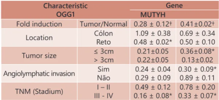
Table 3 shows the expression of genes MUTYH and OGG1 with respect to the characteristics of the studied samples, as well as between the normal tissue and neoplastic, considering
only the variables where there was a statistically significant difference in one of the genes.
TABLE 3 - Gene expression OGG1 and MUTH according to the characteristics of the sample
Characteristic OGG1
Gene MUTYH
Fold induction Tumor/Normal 0.28 ± 0.12† 0.41±0.02†
Location Cólon
Reto
1.09 ± 0.38
0.48 ± 0.02* 0.69 ± 0.340.50 ± 0.10
Tumor size ≤ 3cm
> 3cm
0.21±0.05 0.22±0.05
0.36±0.08* 0.13±0.02
Angiolymphatic invasion Sim Não
0.24 ± 0.04 0.29 ± 0.09
0.30 ± 0.09* 0.89 ± 0.11
TNM (Stadium) I – II
III - IV 0.16 ± 0.08*0.49 ± 0.12
0.78 ± 0.20 0.33 ± 0.07*
† = p < 0.05 When compared to normal tissue; ** p < 0.05 When comparing the
characteristics
DISCUSSION
The human genome is particularly vulnerable to oxidative stress. The ROS in the cells of the mucosa colic are formed as products of the normal aerobic metabolism or as a result of exposure of the cells to ionizing radiation, chemicals,
ischemia, inflammation acute and chronic or, even, for the simple modification in the supply of energy substrate to normal
colonocites represented by short-chain fatty acids20, 12. As they
are electrophilic molecules with the ability highly reactive, ROS attack substances with high electron density such as the nitrogenous bases that make up DNA3. These radicals can cause
breakage single or double quote in the tapes of the DNA or oxidize their nitrogenous bases allowing for the development of errors of matching and, consequently, the development of mutations7,2. When we consider the environmental factors
related to the development of the CRC, studies have shown that oxidative stress is one of the main mechanisms responsible for the appearance of mutations related to the development of the disease6,2. It has been demonstrated that the chronic
inflammation and continuously in the intestinal mucosa, as occurs in the intestinal inflammatory diseases, increasing the
production of ROS, increases the possibility of developing of CRC31. Further strengthens this possibility the results of studies
showing that the levels of oxidative stress in the neoplastic tissue
of patients with CRC is significantly higher when compared to
the normal tissue26,27.
The oxidative damage most often caused in the DNA by exposure to the RLO is the formation of 8-oxoG. The neoplastic tissue of patients with CRC has on average two times more 8-oxoG when compared to normal mucous17.
In order to correct the errors of matching caused by 8-oxoG and prevent the emergence of mutations, the body has
different systems of DNA repair. The system BER is the main
responsible for excision of oxidized bases22. The genes MUTYH,
OGG1, MTH1, NEIL1, APEX1, PARP1, LIG3, MGP e NTHL1 are the main components of the system BER.
The genes MUTYH It is located on the short arm of
chromosome 1 (1p32-34) and has 16 exons. Encodes a
instead of cytosine. MUTY-glycosylase acts by removing from the DNA strand adenine bases poorly coupled with 8-oxoG. The enzyme corrects this error so that the development of mutations by type G:C>T:A which can lead to tumor formation if they compromise cell cycle controlling genes. The importance of mutations in the MUTYH gene in colorectal carcinogenesis was highlighted by the Al-Tassan et al. In 2002, where the authors
demonstrated for the first time, individuals from the same family
who presented biallelic germline mutations in the MUTYH gene and who had recessive forms of familial adenomatous polyposis (FAP), associated with the development of colorectal adenomas presenting progression to CRC in age when compared to the classic forms of FAP1. When analyzing the neoplastic tissues of
these patients they found a high index of transversions of the type G:C>T:A in commonly mutated genes in the CRC (APC e
K-RAS). Later, other authors confirmed these findings showing that mutations in the MUTYH gene, reducing the effectiveness
of the BER system, predisposes to the appearance of CRC1,14,15. The OGG1 gene is located on the short arm of the chromosome 3 (3p25.3). It consists of 12 exons and encodes the enzyme 8-oxoguanine glycosylase. It is estimated that the
enzyme contains 424 amino acids in its molecule and molecular
weight of 47kD25. It was demonstrated that the gene OGG1
has an essential role in repairing BER correcting transversions G:C>T:A. The enzyme transcribed, promotes the hydroxylation of connections glicofílicas formed between the 8-oxoG and the fraction of sugar from the basis of fertilization, removing the molecule of 8-oxoG and forming a site apurinic on tape
of DNA that after the excision is subsequently filled with the
correct fertilization. By its fundamental role in the system BER, mutation and polymorphisms in the gene OGG1 are considered events that increase the susceptibility to various forms of cancer. Studies have shown that reducing the activity of the gene could contribute to the development of CRC32.
A considerable number of hereditary CRC shows genetic or epigenetic genes defects on the system repair. The possibility that also occur changes in the system BER in the CRC sporadically
is a plausible hypothesis. So, the research profile of expression
of genes of the system BER related to correction of oxidized bases comparing normal tissue and neoplastic, can improve the understanding of the role of these genes in patients with sporadic CRC32. Previous studies have demonstrated that there
is less efficiency of the system BER in patients with CRC when
compared to healthy volunteers15,33. However, the mutations
of the MUTYH gene and OGG1 in patients with sporadic
CRC are not frequent and are influenced by the ethnic group
studied14,15,33. Thus, in order to evaluate the mutations in these genes it is necessary to study a large contingent of patients belonging, if possible, the same ethnicity. A study evaluating the importance of mutations in the MUTYH to examine their
prevalence in polish ethnicity found that among 1042 patients only 0.4% of the patients had a mutation biallelic associated
with the development of adenomas and CRC15. Other authors
by analyzing the contribution of mutations in germ MUTYH in the development of the CRC have evaluated 358 patients were not selected10. They found two patients (0.6%) with germ
mutations biallelics and eight (2.2%) monoallelics mutations in the gene MUTYH. Patients with biallelic mutation had multiple adenomas, but not adenomatous polyposis profuse and, in both cases, the tumors were located in the distal colon. These results suggest that mutations biallelics of gene despite increasing the formation of adenomas and, consequently, the chance of developing cancer is likely to represent less than 3% of cases of sporadic CRC10.
This study evaluated the expression of tissue several genes of the system BER and the ability to repair DNA of these genes by comparing normal tissue and neoplastic and correlating them with clinical variables and pathological32. The
authors found that the expression of tissue gene OGG1 was
significantly lower in tissues, neoplastic while the gene MUTYH
showed no significant differences. They found that, to relate
the expression of genes OGG1 and MUTYH when gender, age, location of the tumor histological grade stadiums of the TNM
classification (I+II+x+II+IV) found no significant differences32.In
Brazil, only one study evaluated the behavior of the gene OGG1 in patients with sporadic CRC29. Using samples from the same
population of patients the authors found a lower expression of OGG1 in tumoral tissue29. With regard to location of the tumor
identified lower gene expression only in patients with cancer
of the rectum and in tumors with more advanced stages29.
In the present study we found similar results. When evaluating the expression of OGG1 we found reduced expression in tumor tissue, similar to the studies previously cited32,29.
We also found a reduction in the tissue expression of the gene in patients with tumors located in the rectum and in
patients classified in more advanced stages of the disease.
With respect to expression of the gene MUTYH, despite being
lower in neoplastic tissue, we found no statistically significant differences (p=0.06). It is possible that these values could present significant differences if they were included a larger
number of patients. When you relate gene expression MUTYH with variables selected for the present study found that there was less expression of the gene in tumors that had diameter greater than 3 cm, in presenting angiolymphatic invasion and
those classified in the more advanced stages (T3-T4) of the TNM classification. These findings suggest that the lower expression
in these patients may contribute to the worse prognosis of the disease, since it is related to variables that confer a worse prognosis of the disease.
When we consider the results of the present study, and having science that there is a higher degree of oxidative stress in tissues neoplastic, it is possible that the lower expression of genes OGG1 and MUTYH in tumor tissue, as well as in the variables related to a worse prognosis of the disease, may occur due to the accumulation epigenetic mutations or hypermethylation in these genes by reducing their capacity to repair and favoring the development of a phenotype more aggressive as shown above11. The hypermethylation of the
promoter region of genes that make up the system BER has been found in a variety of tumors (thyroid, bladder, ovaries, the brain as well as in the CRC)18.
Considering that genes OGG1, MUTYH, PARP-1 and XRCC1 are part of the same repair system (BER) it is possible that the presence of polymorphisms in these genes may interfere with
their tissue expression. Recent study evaluating the influence of the APE1 T2197G polymorphism (asp148Glu) showed that
this alteration interferes in the expression of the genes of the BER system when comparing normal and neoplastic tissues. The
results found in the present study not only confirm previous
results but also reinforce the importance of repair integrity by excision of bases in the etiopathogenesis and progression of sporadic CRC.
CONCLUSION
The MUTYH and OGG1 genes present downregulation in the more advanced stages of colorectal cancer.
REFERENCES
1. Al-Tassan N, Chmiel NH, Maynard J, Fleming N, Livingston AL, Williams GT, et al. Inherited variants of MYH associated with somatic G:C-->T:A mutations in colorectal tumors. Nat Genet. 2002;30(2):227-32. 2. Ames BN, Shinegawa MK, Hagen TM. Oxidants, antioxidants, and the
degenerative diseases of aging. Proc Natl Acad Sci USA. 1993;90(17):7915-22. 3. Battacharya PK, Barton JK. Influence of intervening mismatches on long range
5. Chang GJ, Kaiser AM, Mills S, Rafferty JF, Bui WD, on behalf of Standards Practice Task Force of American Society of Colon and Rectal Surgeons. Dis Colon Rectum. 2012; 55(8):831-43.
6. Demple B, Harrison L. Repair of oxidative damage to DNA: enzimology and biology. Annu Rev Biochem. 1994;63:915-48.
7. Dizdaroglu M. Chemical determination of free radical-induced damage to DNA. Free Radical Biol Med. 1991;10(3-4):225-42.
8. Fearon ER, Vogelstein B. A genetic model for colorectal tumorigenesis. Cell. 1990;61(5):759-67.
9. Ferlay J, Soerjomataram I, Dikshit R, Eser S, Mathers C, Rebelo M, et al. Cancer incidence and mortality worldwide: Sources, methods and major patterns in GLOBOCAN 2012. Int J Cancer. 2015; 136(5): 359-86. 10. Fleischmann C, Peto J, Cheadle J, Shah B, Sampson J, Houlston RS.
Comprehensive analysis of the contribution of germline MYH variation to early-onset colorectal cancer. Int J Cancer. 2004;109(4): 554-8. 11. Gao D, Herman JG, Guo M. The clinical value of aberrant epigenetic
changes of DNA damage repair genes in human cancer. Oncotarget. 2016;7(24):37331-46.
12. Hwang BJ, Shi G, Lu AL. Mammalian MutY homolog (MYH or MUTYH) protects cells from oxidative DNA damage. DNA Repair (Amst). 2014; 13:10-21.
13. Instituto Nacional de Câncer (Brasil). Estimativa 2016. Incidência de Câncer no Brasil. Disponível em: http://www.inca.gov.br/estimativa/2016/index. asp?ID2. Acesso em: 18 out. 2016.
14. Jones S, Emmerson P, Maynard J, Best JM, Jordan S, Williams GT, et al. Biallelic germline mutations in MYH predispose to multiple colorectal adenoma and somatic G:C-->T:A mutations. Hum Mol Genet. 2002;11(23):2961-7. 15. Kabzinski J, Mucha B, Cuchra M, Markiewicz L, Przybylowska K, Dziki A,
et al. Efficiency of base excision repair of oxidative DNA damage and its impact on the risk of colorectal cancer in the Polish population. Oxid Med Cell Longev. 2016; 2016: 3125989.
16. Kidane D, Chae WJ, Czochor J, Eckert KA, Glazer PM, Bothwell AL, et al. Interplay between DNA repair and inflammation, and the link to cancer. Crit Rev Biochem Mol Biol. 2014; 49(2):116-39.
17. Krokan HE, Nilsen H, Skorpen F, Otterlei M, Slupphaug G. Base excision repair of DNA in mammalian cells. FEBS Lett. 2000; 476(1-2):73–77. 18. Lahtz C, Pfeifer GP. Epigenetic changes of DNA repair genes in cancer.
J Mol Cell Biol 2011; 3(1): 51-8.
19. Malheiros APR, Teixeira MG, Habr-Gama A, Alcântara PSM. Resultados do tratamento cirúrgico do câncer colorretal em doentes de idade até 64 anos e de 65 anos ou mais. Rev. bras Coloproct. 2005; 25:128-36. 20. Martinez CA, Ribeiro ML, Gambero A, Miranda DD, Pereira JA, Nadal
SR. The importance of oxygen free radicals in the etiopathogenesis of diversion colitis in rats. Acta Cir Bras. 2010;25(5):387-95.
21. Martinez CAR, Cordeiro AT, Priolli DG, Miranda DDC, Bartchewsky Junior W, Margarido NF, et al. Avaliação da expressão tecidual do gene de reparo MLH1 e dos níveis de dno oxidativo ao DNA em doentes com câncer colorretal. Rev. bras. colo-proct. 2009;29(3):303-13.
22. Mol CD, Parikh SS, Putnam CD, Lo TP, Tainer JA. DNA repair mechanisms for the recognition and removal of damaged DNA bases. Annu REv Biophys Biomol Struct. 1999; 28:101-28.
23. Nahas, SC, et al. Prognostic factors of surgically-treated patients with cancer of the right colon: a ten years’ experience of a single universitary institution. ABCD, arq. bras. cir. dig., 2015; 28(1):3-7. ISSN 0102-6720. 24. Peterson CL, Cote J. Cellular machineries for chromosomal DNA repair.
Genes Dev. 2004, 18(6): 602-16.
25. Radicella JP, Dherin C, Desmaze C, Fox MS, Boiteux S. Cloning and characterization of hOGG1, a human homolog of the OGG1 gene of Saccharomyces cerevisiae. Proc. Nat. Acad. Sci. 1997; 94(15): 8010-15. 26. Ribeiro ML, Priolli DG, Miranda DCC, Arçari DP, Pedrazzoli Júnior J,
Martinez CAR. Analysis of oxidative DNA damage in patients with colorectal cancer. Clin Colorectal Cancer. 2008;7(4):267-72.
27. Ribeiro ML, Priolli DG, Miranda DDC, Paiva DA, Pedrazzoli Júnior J, Martinez CAR. Avaliação do dano oxidativo ao DNA de células normais e neoplásicas da mucosa cólica de doentes com câncer colorretal. Rev bras. colo-proctol. 2007; 27(4):391-402.
28. Robertson AB, Klungland A, Rognes T, Leiros I. DNA repair in mammalian cells: base excision repair: the long and short of it. Cell Mol Life Sci. 2009; 66(6):981-3.
29. Santos JC, Funck A, Silva-Fernandes IJL, Rabenhorst AHB, Martinez CAR, Ribeiro ML. Effect of APE1 T2197G (Asp148Glu) polymorphism on APE1, XRCC1, PARP1 and OGG1 expression in patients with colorectal cancer. Int J Mol Sci. 2014; 15:17333-17343
30. Schumutte C, Yang AS, Nguyen TT, Beart RW, Jones PA. Mechanisms for the involvement of DNA methilation in colon carcinogenesis. Cancer Res. 1996;56(10):2375-81.
31. Seril DN, Liao J, Yang GY, Yang CS. Oxidative stress and ulcerative colitis-associated carcinogenesis: studies in human and animals models. Carcinogenesis. 2003;24(3):353-62.
32. Slyskova J, Korenkova V, Collins AR, Prochazka P, Vodickova L, Svec J, et al. Functional genetic and epigenetic aspects of base nucleotide excision repair in colorectal carcinomas. Clin Cancer Res. 2012;18(21):5878-87. 33. Slyskova J, Naccarati A, Pardini B, Polakova V, Vodickova L, Smerhovsky
Z, et al. Differences in nucleotide excision repair capacity between newly diagnosed colorectal cancer patients and healthy controls. Mutagenesis 2012; 27(4):225-32.
34. Wheeler JM. Epigenetics, mismatch repair and colorectal cancer. Ann R Col Surg Eng. 2005;87(1):15-20.