Rev Odontol UNESP. 2014 Jan-Feb; 43(1): 41-45 © 2014 - ISSN 1807-2577
ORIGINAL ARTICLE
Doi: http://dx.doi.org/10.1590/S1807-25772014000100007
Digital evaluation of the inluence of interruption of the ixation
process on radiographic contrast and base-plus-fog density
in three commercial brands of radiographic ilms
Avaliação digital da inluência da interrupção da ixação no contraste radiográico e na densidade base e
velamento em três marcas comerciais de ilmes radiográicos
Paula Verona Ragusa da SILVA
a, Renan Roberto da COSTA
b, Mariliani Chicarelli da SILVA
c,
Lilian Cristina Vessoni IWAKI
c, Wilton Mitsunari TAKESHITA
daACCCC – AC Camargo Cancer Center, São Paulo, SP, Brasil
bFaculdade de Odontologia, UNESP – Univ Estadual Paulista, Araraquara, SP, Brasil cUEM – Universidade Estadual de Maringá, Maringá, PR, Brasil
dUFS – Universidade Federal de Sergipe, Aracaju, SE, Brasil
Resumo
Introdução: Com a preocupação em antecipar o acesso ao resultado de radiografias intrabucais, o processamento radiográfico é muitas vezes negligenciado, comprometendo a qualidade da imagem. Objetivo: O objetivo deste trabalho foi avaliar a influência da interrupção do processo de fixação no contraste radiográfico e na densidade base e velamento (DBV) em 3 marcas comerciais de filmes periapicais. Material e método: Foram realizadas 90 radiografias de um stepwedge de alumínio e uma placa de chumbo para cada marca, e as mesmas foram divididas de acordo com o tempo de imersão inicial no fixador em: grupo controle (sem interrupção na fixação), 5, 10, 20, 30 e 40 segundos. Durante o processamento, os filmes tiveram a fixação interrompida e foram expostos à luz de um negatoscópio por 1 minuto, e então completaram o tempo de fixação de 10 minutos. As radiografias foram digitalizadas e analisadas no software Image Tool 3.0. Resultado: O filme Kodak
não apresentou diferenças estatísticas significantes entre os grupos, enquanto que o filme Agfa
apresentou diferença na DBV em relação ao grupo de 5 segundos, e o filme Dentix
apresentou diferença estatística em todos os grupos comparando-os ao grupo controle. Conclusão: Sob as condições estudadas, o filme Kodak
não sofreu influência da interrupção da fixação na DBV e no contraste da imagem, possibilitando o acesso antecipado ao resultado do exame radiográfico, enquanto que o filme Agfa
necessita de pelo menos 10 segundos de fixação inicial e o filme Dentix
obtém melhores resultados quando não interrompido o processo de fixação.
Descritores: Intensificação de imagem radiográfica; radiografia dentária; filme para raios X.
Abstract
Introduction: With the interest in anticipating access to the result of intraoral radiography, the radiographic processing is frequently neglected, compromising image quality. Objective: The aim of this study was to evaluate the influence of interrupting the fixation process on the radiographic contrast and base-plus-fog density (BPFD) in three brands of periapical films. Material and method: Ninety radiographs were taken of an aluminum stepwedge and a lead plate for each brand, and they were divided according to the time of initial immersion in the fixative in: control group (without interrupting the fixing), 5, 10, 20, 30 and 40 seconds. During processing, films had the fixing stage stopped and were exposed to a negatoscope for 1 minute, then the fixation time of 10 minutes was completed. The radiographs were digitized and exported to Image Tool 3.0.software. Result: Kodak
film showed no statistically significant differences between groups, while Agfa
film presented difference in BPFD compared with Group 5 seconds, and Dentix
film showed statistical difference in all groups in comparison with the control group. Conclusion: Under the conditions studied, Kodak
film is not influenced by disruption of fixation as regards BPFD and image contrast, enabling early access to the results of radiographs, whereas Agfa
film requires at least 10 seconds of initial fixation, and Dentix
film obtains better results when the process of fixation is not interrupted.
INTRODUCTION
A good quality radiographic exam is fundamental for correct diagnosis in various situations in clinical dentistry1. It
allows detection of the presence of pathologies, anomalies, in addition to conirmation of the integrity of dental structures2,3.
For adequate radiographic interpretation, strict quality control of image capture is fundamental4. his consists of continued
evaluation of the energy factors related to exposure to X-rays, image processing, patient protection and gain of working time. Command of all the stages of the radiographic exam and a careful technique may diminish the time of attending patients, as these factors prevent unnecessary repetitions5.
herefore, adequate contrast and base-plus-fog density (BPFD) are fundamental criteria for correct interpretation of the images obtained, since radiographic contrast is translated by the graduation of the diferent densities of ilms in diferent areas of a radiograph1*
; and BPFD represents the optical density inherent
to the radiographic ilm6, which may result from the quality of
the emulsion and its interaction with the processing solutions, secondary radiation, and safety light5**.
Continuing studies must be conducted to evaluate diferent brands of ilms with regard to their respective BPFDs, with a view to quality control in dental radiography6,7, bearing in mind
that with technological advancement, new commercial brands of ilms are introduced into the market.
Moreover, it has been noted that many professionals neglect the radiographic processing procedure in daily practice, particularly the stage of ixation, although they are aware that in order to obtain a quality image, it is important for all the stages of the radiographic exam to be performed correctly, starting with ilm storage and through to its inal drying. his is mainly due to the need for reducing the working time and the apparent ease of performing the technique.
As a result of this, and concern about anticipating access to intraoral radiographic results, without compromising their quality, the aim of this study was to make a digital evaluation of the inluence of interrupting the ixation process on radiographic contrast and base-plus-fog density (BPFD) of 3 commercial brands of periapical ilms.
MATERIAL AND METHOD
For this research a stepwedge (aluminum equivalence scale)8,9, made of a speciic and internationally standardized
alloy (aluminum alloy 2026, ABNT), consisting of eight steps measuring 1 mm in height each, in addition to a lead plate with an area of 1cm2, 30 Kodak
Insight periapical ilms, sensitivity F (Eastman Kodak Company, Rochester, USA), 30 ilms Agfa
Dentus M2 Comfort periapical ilms, sensitivity E (Agfa-Gevaert,
* Goaz PW, White SC. Processing X-ray ilm. In: Goaz PW, White SC. Oral radiology, principles and interpretation. St. Louis: Mosby-Year Book; 2009. ** Dezotti MSG. Avaliação de ilmes radiográicos periapicais em diferentes condições de processamento pelos métodos sensitométrico, digital e morfométrico [tese doutorado]. Bauru: Faculdade de Odontologia da USP; 2003.
Mortsei, Belgium) and 30 Dentix
periapical ilms, sensitivity E (Foma Bohemiaspol. s R.O, HradecKrálové, Czech Republic) (Figure 1), totaling 90 radiographic ilms for the study.
he ilms were exposed to radiation under standardized technical conditions, on the same day and by the same operator, using the DabiAtlante
(Model Spectro 70x electronic) intraoral radiographic appliance, with mechanical calibration previously veriied, and with the technical factors of exposure ixed at 70 kVp and 8 mA, for the time of 0.4 seconds determined by a previous pilot test, containing a lead plate and a stepwedge. he main X-ray beam fell perpendicularly on the ilm/stepwedge/lead plate, at a distance of 40 cm (Figure 2).
In the act of processing, a random subdivision was performed for each commercial brand of ilms, to group them according to the times of initial immersion in the ixer, with the following groups being obtained: Control (without interrupting ixation), 05, 10, 20, 30 and 40 seconds.
Each ilm was individually processed by one and the same examiner, by the temperature-time method, in a portable VH
dark chamber (Essence Dental, Araraquara, Brazil), using the ready-to-use Kodak
(Eastman Kodak Company, Rochester, USA) solutions that were at a temperature of 22 °C. According to the tables provided by the ilm manufacturers, the immersion time in the developer was 4 minutes, intermediate washing for 20 seconds, complete ixation time of 10 minutes, in addition to inal washing for another 20 minutes. To control the method, a thermometer for liquids and a stopwatch were used. So that the deterioration of the solutions due to the quantity of ilms
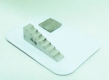
Figure 1. Radiographic Films of three diferent brands.
processed and oxidation would not interfere in the processing quality of the following radiographs, the processing liquids were changed for each commercial brand.
In accordance with the previously described standards, the radiographs were processed individually. he ilms of the experimental groups were immersed in the ixer, and when the initial immersion time was up, the stop-watch was stopped, the ilms were removed from the ixer solution, and then exposed to the light of a negatoscope without coming into direct contact with it, for a period of 60 seconds. Without returning to the receptacle with water, ixation of the ilms was resumed until the time of 10 minutes was up, then they were immersed in water for 20 minutes. Ater complete drying, the radiographs were digitized in a scanner with a transparency reader, of the HP
Scanjet G4050 brand, with an optical digitization resolution of up to 4800 d.p.i. (dots per inch). All the images were captured with a ixed resolution of 300 d.p.i., saved in TIFF (Tagged Image File Format) format and sent to the Image Tool 3.0 sotware program for analysis of the image of the irst and last step of the stepwedge, for the purpose of calculating the contrast and measuring the DPFD.
It is important to point out that for the measurement of the contrast and base-plus-fog density, a scale that ranges from 0 to 255 is used, with 0 being equivalent to black and 255 to white. he intermediate numbers represent the various gray tones present in the image, with the high-density values corresponding to lighter images, and the lower values to darker images7.
he data were inserted in the Excel 2010 and SPSS 11.0 sotware programs, and submitted to the analysis of variance (ANOVA) and Tukey statistical tests, at a 5% level of signiicance.
RESULT
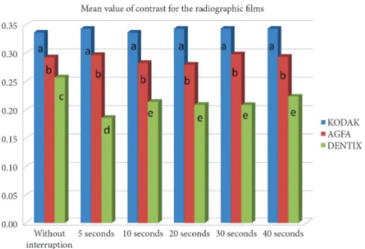
For presentation of the data, two graphs were constructed of mean BPFD and contrast values, in which equal letters correspond to no statistically signiicant diference at a level of 5% between the studied means, with the purpose of evaluating the homogeneity between the groups.
When the contrast in the diferent radiographic ilms was analyzed, the Kodak
and Agfa
brands presented no statistically signiicant diferences between the diferent groups with interruption of ixation. However, the ilms in the experimental groups of the Dentix
brand presented considerable diference in comparison with the control group, with this diference being more signiicant in the group of interruption for 5 seconds (Figure 3).
When analyzing the BPFD, constant values were observed for Kodak
ilm. he Agfa
brand presented values close to those of the Kodak
brand, however, in the group in which there was an interruption of 5 seconds, a diference was observed in comparison with the control group and other groups. When the results with reference to the Dentix
ilm were analyzed, it was observed that all the groups presented diferences in comparison with the control, with these being more signiicant in the groups with interruptions of 5 and 10 seconds (Figure 4).
DISCUSSION
Daily dental practice demands the use of radiographs with speed, with a view to reducing the time of work. Both in specialized procedures and those in the general clinical, the professional encounters situations in which there is a need for obtaining good quality radiographs quickly. However, interrupting the ixation of the radiograph, frequently done by clinicians for this purpose, results in the loss of details in the radiographic image. Over time, the poorly ixed radiographs present a yellowed color and the image formed tends to disappear, by virtue of the absence of an efective protective barrier8.
Moreover, the use of good quality radiographs is imperative for making a correct diagnosis and treatment in the various situations of the dental clinic, and particularly for documentation3,9. herefore, the dentist must use means that
optimize processing, but continue to guarantee a satisfactory result. Knowing the importance of the use of radiographs as an auxiliary diagnostic method, there is the challenge of reducing the risk ofered by radiation doses, avoiding unnecessary repetitions, and diminishing the clinical working time5, and nevertheless
Figure 3. Mean value of contrast for the diferent radiographic ilms. Equal letters correspond to no statistically signiicant diference at the level of 5%.
continue to obtain radiographs rich in details by means of a well performed technique.
In seeking better results and greater speed, some authors have suggested the use of careful automatic processors with respect to all the stages and conditions of processing2. However, in Brazil,
the low low of patients or high cost of acquiring an automatic processor make it unfeasible for all the private dental oices or those of the national health system to purchase such a unit. herefore, all that remains is to carefully comply with all the steps involved in obtaining a high quality radiograph, and this is classiied as such when it presents the maximum amount of details, medium contrast and density and minimal distortion3.
hese criteria may be inluenced by various factors, among them the choice of radiographic ilm used and the radiographic processing technique. Although radiographic processing is considered an easy-to-perform procedure, it is responsible for a large portion of imaging errors10,11. here continues to be
controversy about the ideal time of immersion of the radiographic ilm in the ixation liquid3, however, it is recommended that the
time demanded by the radiographic ilm manufacturer should be respected, which suggests a mean of 2 to 4 minutes, while the literature recommends the time corresponding to double the amount of time necessary for the milky image to become transparent, which occurs in around 8 to 10 minutes3.
he quality of radiographs largely depends on their correct manipulation in accordance with the ilm manufacturer`s instructions. Casanova et al.9 and Lourenço et al.3 have made
reference to various factors, such as the type of ilm, types of solutions, forms of processing, development temperature, inal washing, drying, sensitivity, contrast and BPFD, as factors potentially responsible for the poor quality of radiographs.
Among the means of processing most commonly used by dentists, the manual inspection method has been observed to be the most frequently used. However, some researches have shown that under-processing occurs in a large portion of dental oices10-14,
which may undoubtedly induce diagnostic errors. Lemke et al.15,
in their research, veriied that the majority of professionals evaluated were performing radiographic overexposure, in order to under-process the radiographs aterwards, with the aim of reducing working time.
In seeking image quality we may use the BPFD as a means of measurement. he BPFD is the speciic density of the ilm itself, and may be inluenced by the processing solutions, secondary radiation and safety light, which interact with this ilm. Costa et al.6 mentioned that studies analyzing BPFD of the
diferent radiographic ilms should be continually conducted in order to obtain quality control of dental radiographs.
According to Lourenço et al.3, literature on the subject is
scarce, however, as has been mentioned, it is fundamental to maintain radiographic quality within the dental oice. In view of the foregoing, the present study sought to evaluate the BPFD of three commercial brands (Kodak
, Agfa
and Dentix
),as well as the contrast of ilms by means of using a digital sotware program that enabled richer details to be obtained. he Kodak
and Agfa
brands were chosen because they have been on the market for many years, and Dentix
because it is a relatively new brand in Brazilian industry.
In the present research, no statistically signiicant diferences were observed as regards BPFD and contrast in the group of Kodak
ilms, however, there was evidence of some variations when analyzing ilms of the Agfa
and Dentix
brands, with these results being in agreement with the above-mentioned literature. However, it is pointed out that ilms with diferent sensitivities were used (Kodak
sensitivity F, Agfa
sensitivity E and Dentix
sensitivity E), due to the restricted availability on the market, and we do not discard the hypothesis that this fact may have contributed to the diferent performance of the radiographic brands with reference to contrast and BPFD.
Some studies have investigated the interruption of ixation of the radiograph by the automated16,17 and manual method of
processing, and when they used the automated method with Kodak
ilms, they observed acceptable BPFD values for all the types of ilms. Araujo et al.2 (2009), when interrupting manual
processing, did not observe interference in BPFD and contrast of the radiographic image, provided that the ilms were returned to the solution for complete ixation. However, the study of Kaugars et al.18, analyzing diferent types of ilms, showed
evidence of a trend towards increase in BPFD as a result of type of ilm and processing condition.
Lourenço et al.3, in their study, observed that the interruption
of ixation followed by reading in the negatoscope did not interfere in the base-plus-fog density and contrast of the radiographic image, even ater a period of six months of iling the ilms, a fact that difers from the inding of the research here presented. According to this same author, the results found by them suggest the involvement of two factors: the ixing solution remaining on the ilm during the period of reading in the negatoscope, which allows continuation of the action of sodium hypochlorite in removal of the silver grains, and the return to the ixer for conclusion of ixation ater the time the ilm stayed in the negatoscope. Even if this hypothesis is sound, in the present study these suppositions could not be conirmed, bearing in mind the discrepancy in the result found for the Dentix
ilm.
Although there are recommendations from the manufacturers that ixation should be performed in an uninterrupted manner, in this search it was found that suspension of ixation for a few seconds could be indicated for the ilms from the manufacturers Kodak
and Agfa
, provided that for the latter, a minimum time of 10 seconds of initial ixation is respected, and that both return to the ixer to complete the pre-determined time of 10 minutes. On the other hand, the results of this study point out that the processing of the Dentix
ilms must be carried out faithfully in accordance with the manufacturer`s instructions.
CONCLUSION
According to the conditions studied, it could be concluded that:
• For Kodak
ilm there was no inluence of the interruption of ixation on BPFD and contrast of the image, making it possible to anticipate access to the result of the radiographic exam;
• Agfa
ilm needs a minimum time of 10 seconds of initial ixation in order to have no interference in the BPFD;
• Dentix
REFERENCES
1. Changizi V, Jazayeri E, Talaeepour A. Study of densitometry comparison among three radiographic processing solutions. Iran J Radiat Res. 2006;4(2):81-6.
2. Araujo AMM, Pontual AA, Silveira MMF, Brasileiro IV, Pontual MLA. Inluência da interrupção da ixação no contraste radiográico e na densidade base e velamento. Rev Assoc Paul Cir Dent. 2009;63(5):409-14.
3. Lourenço ADA, Pontual AA, Silveira MMF, Pontua, MLA. Radiographic image quality ater interruption of the ixing stage to view the image with a viewbox. Rev Odonto Ciênc. 2010;25(1):78-82. http://dx.doi.org/10.1590/S1980-65232010000100016
4. hornley PH, Stewardson DA, Rout PGJ, Burke FJT. Assessing the quality of radiographic processing in general dental practice. Br Dent J. 2006;200:515-9. http://dx.doi.org/10.1038/sj.bdj.4813527
5. Brücker MR, Tavano O, Costa NP. Análise do comportamento das soluções RPX-Omat da Kodak através do método sensitométrico. Rev Odonto Ciênc. 1992;7(13):37-52.
6. Costa C, David AF, David SMN, Matsui RH, Castilho JCM, Varoli FP. Estudo das densidades base e velamento obtidas de ilmes radiográicos em diferentes condições de processamento. Ciênc Odontol Bras. 2005;8(1):90-6.
7. Travessas JAC, Mahl CRW, Fontanella, VRC. Avaliação da densidade radiográica digital de quatro ilmes periapicais. Rev Fac Odontol Porto Alegre. 2004;45(2):17-20.
8. White SC, Pharoah MJ. Radiologia oral: princípios e interpretação. St Louis: Mosby; 2007.
9. Casanova MA, Haiter-Neto F, Bóscolo FN, Almeida SM. Sensitometric comparisons of insight and Ektaspeed Plus Films: efects of chemical developer depletion. Braz Dent J. 2006;17(2):149-54. http://dx.doi.org/10.1590/S0103-64402006000200013
10. Pontual ML, Veloso HHP, Pontual AA, Silveira MMF. Errores en radiograias intrabucales realizadas en la Facultad de Odontología de Pernambuco-Brasil. Acta Odontol Venez. 2005;43:19-24.
11. Gasparini D, Vaz EMS, Haiter Neto F, Boscolo FN. Análise de erros radiográicos cometidos por alunos da Faculdade de Odontologia de Piracicaba, no período de 1975 a 1988. Rev Odontol Univ São Paulo. 1992;6:107-14.
12. Sur J, Endo A, Matsuda Y, Itoh K, Katoh T, Araki K, et al. A measure for quantify the radiopacity of restorative resins. Oral Radiol. 2011;27:22-27. http://dx.doi.org/10.1007/s11282-010-0055-4
13. De Paula MV, Fenyo-Pereira M. Controle de qualidade em radiograias periapicais – padrões de exposição e revelação. Rev Assoc Paul Cir Dent. 2001;55:355-60.
14. Beltrame M, Oliveira AE, Spyrides KS, Cordeiro PV. Análise do processamento radiográico nos consultórios de Feira de Santana - BA. Rev Fac Odontol Univ Passo Fundo. 2003;8:50-4.
15. Lemke F, Tavano O, Mezadri AC. Veriicação das condições de exposição e processamento de ilmes radiográicos em consultórios odontológicos. RPG: Rev Pós-Grad. 2006 abr-jun;13(2):175-80.
16. Costa C, Yamamoto CM, Barbosa J, Saraceni CHC, Armonia PL. Avaliação das densidades óticas de ilmes radiográicos digitalizados quando processados pelo método roller. Rev Inst Ciênc Saúde. 2007;25(4):399-402.
17. Geist JR, Brand JW, Pink FE. he efect of automated nonroller processing on the sensitometric characteristics of 3 intraoral ilm types. Oral Surg Oral Med Oral Pathol Oral Radiol Endod. 2003;96(1):102-11. http://dx.doi.org/10.1016/S1079-2104(03)00221-X
18. Kaugars GE, Broga DW, Collett WK. Dental radiologic survey of Virginia and Florida. Oral Surg Oral Med Oral Pathol. 1995;60(2):225-229. http://dx.doi.org/10.1016/0030-4220(85)90300-7
CONFLICTS OF INTERESTS
he authors declare no conlicts of interest.
CORRESPONDING AUTHOR
Paula Verona Ragusa da Silva
Rua Professor Antônio Prudente, 211, Liberdade, 01509-900 São Paulo - SP, Brasil e-mail: [email protected]