728
Revista da Sociedade Brasileira de Medicina Tropical 43(6):728-730, nov-dez, 2010
Communication/Comunicação
1. Program in Experimental Pathology, Department of Pathological Sciences, Londrina State University, Londrina, PR, Brazil. 2. Department of Pathological Sciences, Londrina State University, Londrina, PR, Brazil. 3. Department of Cell Biology, Brasilia University, Brasília, DF, Brazil.
Address to: Dr. Emerson José Venancio. Deptº Ciências Patológicas/UEL. Rodovia
Celso Garcia Cid (PR-445), KM 380 Campus Universitário, 86051-990 Londrina, PR, Brazil.
Phone: 55 43 3371-5732 e-mail: [email protected] Received in 03/05/2010 Accepted in 13/07/2010
A semi-nested PCR assay for molecular detection of
Paracoccidioides
brasiliensis
in tissue samples
Semi-nested PCR para a detecção molecular de
Paracoccidioides brasiliensis
em amostras de
tecido
Andrea Cristine Koishi
1, Débora Fonseca Vituri
1, Pedro Sebastião Raimundo Dionízio Filho
2,
Alexandre Augusto Sasaki
1, Maria Sueli Soares Felipe
3and Emerson José Venancio
2ABSTACT
Introduction: Paracoccidioidomycosis is a systemic infection caused by
Paracoccidioides brasiliensis. Methods: In this study, a semi-nested PCR for paracoccidioidomycosis diagnosis was developed. he primers ITS1 and ITS4 were used in the irst reaction, while the primers MJ03 and ITS1 primer were used in the second reaction. he semi-nested PCR was used to investigate biopsies of ive patients with oral lesions that resembled paracoccidioidomycosis. Results: he semi-nested PCR was positive for four samples and negative for a sample from a patient later diagnosed with leishmaniasis. Conclusions: he new semi-nested PCR describe is useful for paracoccidioidomycosis diagnosis.
Key-words:Paracoccidioides brasiliensis. Semi-nested PCR. Molecular detection.
RESUMO
Introdução: A paracoccidioidomicose é uma infecção sistêmica causada pelo Paracoccidioides brasiliensis. Métodos: Neste estudo, uma semi-nested PCR foi desenvolvida para o diagnóstico da paracoccidioidomicose. Os oligonucleotídeos iniciadores ITS1 e ITS4 foram usados na primeira reação, enquanto os oligonucleotídeos iniciadores MJ03 e ITS1 foram usados na segunda reação. A semi-nested PCR foi usada para investigar biopsias de cinco pacientes com lesões orais que se assemelhavam a paracoccidioidomicose. Resultados: A semi-nested PCR foi positiva para quatro amostras e negativa para a amostra de um paciente, posteriormente diagnosticado com leishmaniose. Conclusões: A semi-nested PCR descrita aqui é útil para o diagnóstico da paracoccidioidomicose.
Palavras-chaves: Paracoccidioides brasiliensis. Semi-nested PCR. Detecção molecular.
he thermal dimorphic fungus Paracoccidioides brasiliensis is the etiologic agent of paracoccidioidomycosis (PCM), a systemic mycosis endemic in Latin America1.
PCM diagnosis can be determined by direct observation or the culture of clinical samples, histological analyses and serological methods. However, these techniques each have concerns: using direct observation, P. brasiliensis yeast cells may not be observed or could be mistaken for other dimorphic fungi1; sample culture
is slow and frequently negative; in histological analysis, the pseudoepitheliomatous hyperplasia typical of PCM resembles squamous cell carcinoma so closely that it is possible to mistake one for the other; inally, serological assays are highly sensitivity, but not totally speciic.
Alternative diagnostic methods for PCM have been developed, including polymerase chain reactions (PCR). he most frequently used target sequences for molecular detection of P. brasiliensis by PCR are the gp432-4 and ribosomal DNA genes5-8. PCR assays have been
used experimentally to detect P. brasiliensis in the serum and tissues of infected mice2,4, in artiicially contaminated soil and in environmental
samples8. In PCM patients, PCR has been used on sputa, cerebrospinal
luid and parain-embedded tissues3,9,10. However, molecular diagnosis
of PCM is not used in clinical routine.
Here, we report a semi-nested PCR (snPCR) assay for the molecular detection of P. brasiliensis using two universal primers for fungi and a speciic primer designed to detect a speciic DNA ribosomal sequence of P. brasiliensis.
Two strains of P. brasiliensis (LDR1 and Pb18) and one isolate of each of the following fungi, Candida albicans (CR15), Histoplasma capsulatum, Sporothrix schenckii,Cryptococcus sp and Tricophyton rubrum were used. All fungi isolates were maintained on Sabouraud dextrose agar at room temperature, with the exception of P. brasiliensis
and Cryptococcus sp, which were maintained at 35ºC.
DNA from fungal cells and tissues from mice or patients were extracted by maceration in liquid nitrogen followed by phenol-chloroform-isoamyl alcohol treatment and sodium acetate-ethanol precipitation. DNA concentration and purity were determined by spectrophotometry.
Swiss male mice (n= 5) were injected with 1.5 x 10⁶ yeast cells
of P. brasiliensis Pb18 via the tail vein. he mice were killed 16h ater fungal inoculation and the lungs were removed under aseptic
conditions, weighed and manually homogenized in PBS (100μL
to 20mg of tissue). Lung samples were cultured on BHI agar plates
supplemented with 4% horse serum, 5% growth factor11 and 1%
penicillin and streptomycin solution and the number of CFU/g of tissue was calculated. For histological analysis, the lungs were routinely processed for the preparation of parain-embedded tissue sections and stained with hematoxylin-eosin (HE).
he following primers ITS1 (5’-TCCGTAGGTGAACCTGCGG-3’) and ITS4 (5’-TCCTCCGCTTATTGATATGC-3’), described
elsewhere12, were used in the snPCR . The primer MJ03
729
Koishi AC et al - An improved method for Paracoccidioidomycosis diagnosis
TABLE 1 - Detection of P. brasiliensis in lung tissue of mice 16 hours ater
inoculation of the fungus.
Results for
P. brasiliensis histopathology P. brasiliensis mice Mouse no. CFU/g of lung (HE stain) semi-nested PCR GAPDH PCR
C1 0 - - +
C2 0 - - +
3 1×10⁴ + + +
4 0 + + +
5 0 + + +
6 4×10³ + + +
7 2.3×10³ + + +
C: control, +: positive, -: negative, GAPDH: glyceraldehyde 3-phosphate dehydrogenase gene.
TABLE 2 - Patient clinical data.
Patients
sex occupation tobacco alcohol biopsy histopathology molecular
(age in years) smokers consumption PCM diagnosis
T1 M (35) machine operator no chronic alveolar ridge oral PCM positive
T2 M (33) driver no not chronic palate leishmaniosis negative
T4 M (38) farmer yes chronic lip oral PCM positive
T5 M (44) bricklayer yes not chronic vestibular fornix oral PCM positive
T7 M (61) farmer yes nonalcoholic alveolar ridge oral PCM positive
comparing sequences from P. brasiliensis and genetically close species,
B. dermatitidis, C. immitis, H. capsulatum and S. schenckii. he MJ03 primer was used with ITS1 primer in the second round PCR to generate a fragment of 212bp. he irst round PCR consisted of 5µl of DNA sample in a total volume of 25µl, with 20mM Tris-HCl, (pH 8.4; 50mM KCl), 1.5mM of MgCl2, 0.2μM of primers ITS1 and ITS4,
1U of Taq polymerase (Invitrogen, Brazil), and 0.25mM of dNTP
(Amresco). he reaction mixture of the second round PCR was identical, except that 1µl of the irst reaction product and the inner primer pair ITS1 and MJ03 were used. he PCR was performed in a thermal cycler (MWG Biotech) programmed as follows: 95°C for 2min; 30 cycles of 95°C for 30sec, 55°C for 30sec, 72°C for 1min; and 72°C for 5min. he quality of DNA extracted from mice tissues
was evaluated by PCR using GAPDH primers2. PCR products were
analyzed by electrophoresis on an 8% polyacrylamide gel, stained with silver nitrate.In order to evaluate the speciicity of the primers,
genomic DNA templates (25ng) from all fungi cited abovewere
tested. he lower detection limit was determined using genomic DNA of P. brasiliensis LDR1, and using mice lungs spiked with serial dilutions of P. brasiliensis.
To evaluate snPCR with clinical biopsy, samples of lesions from 5 patients with an initial clinical diagnose of PCM from University Dentistry Center at the State University of Londrina, Brazil were used. he samples were stored at -20ºC until DNA extraction. his study was approved by the Ethics Commitee of the Londrina State University, Brazil.
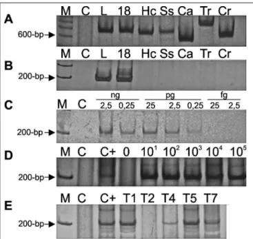
As expected, in the irst reaction, amplicons of diferent sizes were generated with DNA of all the fungi tested (Figure 1A). he second round PCR only ampliied a fragment of 212bp with DNA of
P. brasiliensis, thus showing the speciicity of this reaction (Figure 1B). In relation to sensitivity, the test was able to detect 0.25pg of P. brasiliensis
DNA (Figure 1C) and in mice lungs samples spiked with yeast cells from
P. brasiliensis, it detected as few as 10 yeast cells (Figure 1D).
he histopathological analysis and snPCR were able to detect
P. brasiliensis in all the samples tested, while culture was positive in three out of ive mice samples (Table 1).
In clinical samples from patients with an initial clinical diagnose of PCM, the snPCR for P. brasiliensis was positive in four out of ive patients. he negative sample was from a patient later diagnosed with leishmaniasis (Figure 1E and Table 2).
Molecular biological methods for detection and characterization of microorganisms have revolutionized diagnostic microbiology and the PCR technique is of great importance in this respect. he snPCR described here uses the universal to fungi ITS1 and ITS4 primers and the MJ03 primer that is speciic for P. brasiliensis. his speciic primer was design comparing sequences from GenBank database and anneals to the internal transcribed spacer 1 (ITS1) variable region.
FIgURE 1 - Speciicity and sensitivity of the semi-nested PCR assay. A) First round of nested with primers ITS1 and ITS4. B) Second round of semi-nested with primers ITS1 and MJ03. C) he detection limit of semi-semi-nested PCR (ITS1 and MJ03) with DNA from P. brasiliensis Ldr1 isolate. D) Detection limit
of lung tissue spiked with 0 to 105 yeast cells of P. brasiliensis. E) Detection in
clinical samples. M: 100bp DNA Ladder (Invitrogen, Brazil), C: Negative control, C+: Positive control, L: P. brasiliensis Ldr1 isolate, 18: P. brasiliensis Pb18 isolate; Hc: H. capsulatum, Ss: S. schenckii, Ca: C. albicans (CR15), Tr: T. rubrum, Cr: Cryptococcus sp, T1- T7: tissue samples from patients, 8% polyacrylamide gel stained with silver nitrate.
In a preview study using the ITS1-5.8S-ITS2 region as target, an unexpected cross-reaction with H. capsulatum was observed6.
730
Rev Soc Bras Med Trop 43(6):728-730, nov-dez, 2010
reports10 and similar to a nested PCR assay for S. schenckii13. he
high sensitivity in the present test is probably the result of choosing the high copy number ITS1-5.8S-ITS2 region14 in association
with snPCR, recognized as having a 1,000-fold greater sensitivity than conventional PCR. Furthermore, detection of amplicons by polyacrylamide gels/silver nitrate methodology is recognized as more sensitive than agarose and ethidium bromide detection.
To show the potential application of this snPCR for the molecular detection of P. brasiliensis in clinical samples, mouse lung samples spiked with yeast cells from P. brasiliensis and positive results were obtained with as few as 10 yeast cells. Similar results were observed with a PCR assay for the detection of P. brasiliensis in spiked sputum samples3.
Furthermore, the comparative test among culture, histopathological analysis and snPCR performed using experimentally infected mice, showed that histopathological analysis and snPCR were slightly more sensitive than culture.
In clinical samples from patients with an initial clinical diagnose of PCM, the snPCR for P. brasiliensis was positive for four patients, and negative for a sample, later diagnosed as leishmaniasis, a disease with lesions that can resemble oral PCM lesions. he diagnoses were later conirmed by histopathological analysis.
The semi-nested PCR described in this study is a rapid (approximately 12 hours), speciic and sensitive method and was useful for detecting the presence of P. brasiliensis DNA in culture and tissue.
ACKNOWLEDGMENTS
he authors declare that there is no conlict of interest.
CONFLICT OF INTEREST
FINANCIAL SUPPORT
REFERENCES
he authors would like to thank MA Ono, HO Saridakis, I Felipe and RMB Quesada for providing the fungi strains and isolates used in this study.
This study was partial supported by the Coordenação de Aperfeiçoamento de Pessoal de Nível Superior and the Conselho Nacional de Desenvolvimento Cientíico e Tecnológico, Brazil.
1. Lacaz CS. Mycological diagnosis. In: Franco F, Lacaz CS, Restrepo A, Negro GD, editors. Paracoccidioidomycosis. Boca Raton: CRC Press; 1994. p.339-344. 2. Bialek R, Ibricevic A, Aepinus C, Najvar LK, Fothergill AW, Knobloch J,
et al. Detection of Paracoccidioides brasiliensis in tissue samples by a nested PCR assay. J Clin Microbiol 2000; 38:2940-2942.
3. Gomes GM, Cisalpino PS, Taborda CP, Camargo ZP. PCR for diagnosis of paracoccidioidomycosis. J Clin Microbiol 2000; 38:3478-3480.
4. Nakagawa EI, Uno J, Sano A, Yarita K, Kamei K, Miyaji MK, et al. Detection of the gp43 gene and (1-3)-beta-D-glucan of Paracoccidioides brasiliensis in the blood of experimentally infected mice. Nippon Ishinkin Gakkai Zasshi 2002; 43:29-35.
5. Imai T, Sano A, Mikami Y, Watanabe K, Aoki FH, Branchini ML, et al. A new PCR primer for the identiication of Paracoccidioides brasiliensis based on rRNA sequences coding the internal transcribed spacers (ITS) and 5 x 8S regions. Med Mycol 2000; 38:323-326.
6. Motoyama AB, Venâncio EJ, Brandao GO, Petrofeza-Silva S, Pereira IS, Soares CM, et al. Molecular identiication of Paracoccidioides brasiliensis by PCR ampliication of ribosomal DNA. J Clin Microbiol 2000; 38:3106-3109.
7. Sano A, Yokoyama K, Tamura M, Mikami Y, Takahashi I, Fukushima K, et al. Detection of gp43 and ITS1-5.8S-ITS2 ribosomal RNA genes of Paracoccidioides brasiliensis in paraffin-embedded tissue. Nippon Ishinkin Gakkai Zasshi 2001; 42:23-27.
8. heodoro RC, Candeias JM, Araujo JP, Bosco SM, Macoris SA, Padula LO, et al. Molecular detection of Paracoccidioides brasiliensis in soil. Med Mycol 2005; 43:725-729.
9. Ricci G, Silva ID, Sano A, Borra RC, Franco M. Detection of Paracoccidioides brasiliensis by PCR in biopsies from patients with paracoccidioidomycosis: correlation with the histopathological patern. Pathologica 2007; 99:41-45. 10. San-Blas G, Nino-Veja G, Barreto L, Hebeler-Barbosa F, Bagagli E, Briceno RO,
et al. Primers for clinical detection of Paracoccidioides brasiliensis. J Clin Microbiol 2005; 43:4255-4257.
11. Castañeda E, Brummer E, Perlman AM, McEwen JG, Stevens DA. A culture-medium for Paracoccidioides brasiliensis with high plating eiciency, and the efect of siderophores. J Med Vet Mycol 1988; 26:351-358.
12 White TJ, Bruns T, Lee S, Taylor J. Ampliication and direct sequencing of fungal ribosomal RNA genes for phylogenetics. In. Innis MA, Gelfand DH, Sninsky JJ, White TJ, editors. PCR Protocols: A Guide to Methods and Applications. San Diego: Academic Press; 1990. p. 315-322.
13. Hu S, Chung WH, Hung SI, Ho HC, Wang ZW, Chen CH, et al. Detection of Sporothrix schenckii in clinical samples by a nested PCR assay. J Clin Microbiol 2003; 41:1414-1418.