ABSTRACT: Huanglongbing (HLB), a devastating citrus disease caused by the bacterium “ Candi-datus Liberibacter spp.”, is now responsible for significant economic losses worldwide. Yet, no effective disease control has been found, and the non-cultivability of the bacterium has severely hampered studies on the pathogen. The 16S rDNA gene is a well-characterized sequence, essen-tial for cell survival, and is used for bacterial identification or assignment of close relationships at the genus and species levels. Quantitative Real-Time PCR (qPCR) assays based on 16S rDNA genes are widely used in the detection of “Ca. Liberibacter spp.” in multiplex reactions. We have developed for the first time a set of qPCR primers based on the conserved 16S rDNA gene, which specifically and simultaneously detects in a singleplex reaction, all three bacterial species associated with HLB, and can differentiate Ca. Liberibacterasiaticus or africanus from ameri-canus by their characteristic melting curves. The assay is very sensitive, and it was possible to amplify expected DNA fragments with an efficiency of 98 % using the Syber Green system and a Ct value lower than tested methods for HLB diagnosis. The application of this fast, simple and efficient detection methodology could also be important in the detection of all species of HLB-associated Liberibacters and could contribute to early pathogen detection, a crucial step in the development of preventive strategies aimed at avoiding the dissemination of this devastating disease in HLB-free areas.
Keywords: Candidatus Liberibacter spp., Syber Green qPCR, Argentina, greening, pathogen detection
Introduction
Huanglongbing (HLB) is the most serious cit-rus disease that gives rise to global economic losses (Gottwald et al., 2007). Its causal agent is a Gram nega-tive and phloem-inhabiting bacterium, “Candidatus Li-beribacter spp.” (Jagoueix et al., 1994). For many years, two species have been associated with HLB: “Ca. Li-beribacter asiaticus” (Las), first known in Asia and later found in South, North and Central America, and “Ca. Liberibacter africanus” (Laf), only reported in Africa (Gottwald, 2010). However, in 2004, a third bacterial species was detected in Brazil, “Ca. Liberibacter ameri-canus” (Lam), (Teixeira et al., 2005). Liberibacter can be transmitted by grafting, dodder or two insect vectors:
Diaphorina citri for Las and Lam, and Trioza erytreae for Laf (Bové, 2006).
HLB is a significant threat to all regions not yet infested. In Argentina, D. citri was first reported in the province of Entre Ríos (32°02'52" S and 60°16'52" O)
(Vaccaro, 1994) and later found in Salta (23º21'28" S and 64º09'63" W) and Jujuy (23°45' S and 65°30' O) (Augier et al., 2006). In June 2012, positive detection of HLB was confirmed in backyard trees in Misiones (26°55' S and 54°31' O), located in the northeast of Argentina, but no HLB has been detected yet in com-mercial citrus groves in those regions (Dr. Outi, per-sonal communication, 2013). No direct disease control is available, and the little knowledge we have of the
disease, combined with the aggressive infection be-havior and difficulties to control the bacteria, makes tree protection the most important management mea-sure.
HLB diagnosis based on disease symptoms is dif-ficult because of similarities to nutritional deficiencies (Bové, 2006). Microscopy, serology and DNA–DNA hy-bridization used to detect the pathogens (Bové, 2006; Villechanoux et al., 1990) are either time consuming or lacking in sensitivity. Methods based on PCR have been widely used to detect HLB-associated Liberibacter, mainly based on the 16S rDNA and the beta operon sequences (Hocquellet et al., 1999; Hung et al., 1999;
Jagoueix et al., 1996; Teixeira et al., 2005), and several real-time quantitative PCR assays (qPCR) have been designed to detect and quantify low Liberibacter titers, both in plants (Li et al., 2006; Teixeira et al., 2008) and insects (Manjunath et al., 2008).
With the aim of detecting simultaneously the three Liberibacter species associated with HLB, we have de-signed a qPCR primer set that amplifies a region of the 16S rDNA gene, considering the high degree of conser-vation of this gene for its importance in the functioning of cells (Clarridge, 2004). This primer set is as sensitive or even more sensitive than other reported primers, which makes this methodology a simple and efficient tool for the early detection of the pathogen and the de-velopment of management strategies to avoid the rapid dissemination of HLB.
1Obispo Colombres Agro-Industrial Experimental Station/ National Council for Scientific and Technical Research – Institute of Agro-Industrial Technology of Northwestern Argentina, Av. William Cross 3150, C.P. T4101XAC – Las Talitas, Tucumán – Argentina.
2National University of Rosario/Faculty of Biochemistry and Pharmacy – Institute of Molecular and Cellular Biology of Rosario, Suipacha 590, S2002LRK – Rosario – Argentina. 3Institute of Science and Technology "Dr. Cesar Milstein"/ CONICET, Saladillo 2468, C.P. C1440FFX – Ciudad Autónoma de Buenos Aires – Argentina.
*Corresponding author <[email protected]>
Edited by: Luís Eduardo Aranha Camargo
Novel set of real-time PCR primers for simultaneous detection of Liberibacter
Ingrid Georgina Orce1, Lorena Noelia Sendín1, María Rosa Marano2, Adrián Alberto Vojnov3, Atilio Pedro Castagnaro1, María Paula
Filippone1*
Received December 19, 2013 Accepted September 07, 2014
Materials and Methods
DNA sample and plasmid preparation
The pGEM-T Easy vector containing the 16S rDNA sequence from Las, Lam and Laf was used to construct a standard curve for the different sets of qPCR-primers. Quantification of the plasmid DNA based on the copy number (CN) of the target gene was determined as previ-ously described by Wang et al. (2006). The plasmid solu-tion was serially diluted from 108 CN to 1 CN.
DNA samples of symptomatic leaves from 65 citrus trees and 40 insects were collected as microbial-free, non-infectious DNA samples. The Las/Lam-infected leaves were from Sao Paulo, Brazil (23°30' S and 46°37' O), and the Laf-infected leaves were from Pretoria, South Africa (25°43'32" S and 28°14'38" E).
As a negative control for the PCR and qPCR assays, total DNA was extracted from the midribs (about 250 mg) of healthy greenhouse-grown sweet orange (Citrus sinensis
L.) trees (about five years old) by using the CTAB method (Murray and Thompson, 1980) and suspended in 100 µL of water. As a non-template control (NTC), water was in-cluded.
Primer design and validation of primer-specificity A new set of qPCR primer pair, named p3G, was de-signed based on 16S rDNA sequences of “Ca. Liberibacter spp.” obtained from the DNA database GenBank (acces-sion numbers: NC_020549 of Las, EU921621 of Laf and AY742824 of Lam). Multiple sequence alignments were performed using DNAMAN software and from these analyses primers were designed for a specific sequence region in order to amplify the DNA from all three spe-cies of different sizes. The p3G primers were empirically designed in this region using the software Primer3 (http:// frodo.wi.mit.edu/). The sequences of forward primer f-p3G and reverse primer r-f-p3G were
5’-CTTATCACCGGCAGTCCCTATAAAG-3’ and 5’-CAGCTCGTGTCGTGAGATGTTG-3’,respectively, and led to an amplicon of 122 bp for Las, 121 bp for Laf and 105 bp for Lam.
Analyses for the detection of Ca. L. solanacearum (Lso, accession numbers EU834130.1) were carried out by
in silico PCR using the program http://insilico.ehu.es/PCR/. To make sure that the designed primers were unique to “Ca. Liberibacter spp.”, a search was performed against available microbial sequences in the GenBank and in silico PCR (http://insilico.ehu.es/PCR/). The speci-ficity of primer pairs were also PCR-evaluated with DNA prepared from a number of citrus-related pathogens, in-cluding Xanthomonas citri subsp. citri, the causal agent of citrus canker; Xylella fastidiosa strain 9a5c, the causal agent of citrus variegated chlorosis, and Guirnardia citri-carpa, the causal agent of black spot. Genomic DNA from pure cultures of other bacterial species like Enterococcus
and citrus endophytes, Burkholderia sp. and Methylobac-terium sp., were also included. Twenty samples of citrus leaves (4 of C. sinensis and 16 of C. limon) collected in
non-infected areas in Tucumán (27º0'0" S, 65º30'0" W), Argentina, were tested with the p3G primers.
Conventional PCR and Real-time PCR for the new p3G primers
To determine optimal annealing temperatures, a conventional PCR experiment was conducted by using a MyCycler thermal-cycler with a temperature gradient heating block. Primer annealing temperatures were set from 58 ºC to 72 °C. The PCR reaction was performed in a total volume of 25 uL containing 1 × master mix, 0.4 uM of each primer (f-p3G and r-p3G) and 500 ng of DNA template.
For real-time PCR, SYBR Green (SG) systems were used (SG qPCR). Real-time PCR assays with the new sets of primers were performed by using a Miniopticom Real-time PCR. All reactions were generated in triplicate and each run contained one negative (-DNA, HLB-free citrus DNA) and one positive DNA control (plasmid DNA). PCR amplification conditions were 95 °C for 10 min followed by 40 cycles of 95 °C for 30 s and, 70 °C for 45 s. The reactions were generated using a total volume of 25 uL containing 1 × master mix (iQ SYBR Green, BioRad), 0.4 uM of each primer (f-p3G and r-p3G) and the appropriate amount of DNA template. Data were analyzed using CFX Manager Software Version 2.1 from BioRad.
Sensitivity assay of the qPCR method
To compare the assay sensitivity of the qPCR, dif-ferent HLB-detection PCR reactions were chosen. For a SYBR Green real-time PCR comparison, the primer pair f-rplLAs/r-rplLAs for “Ca. Liberibacter asiaticus”, previ-ously published by Teixeira et al. (2008) was used. The standard curve for rplLas was constructed using ten-fold serial dilutions of plasmid DNA containing the sequence of the rplKAJL-rpoBC gene cluster (β-operon) from Las. A Taqman based real-time PCR assay was performed us-ing an HLBasp 16S rDNA primer-probe set as previously described (Li et al., 2006). To compare the different qPCR systems, optimization and standardization were carried out with ten-fold dilutions of total DNA samples obtained from field-grown plants infected with Las.
Results
Novel qPCR primers designed to detect HLB Liberibacter species
da-tabases at NCBI and an in silico PCR amplification experi-ment did not identify any additional homologous DNA sequences, suggesting high specificity of the designed primers (Figure 1). Even though these primers were spe-cifically designed for HLB “Ca. Liberibacters spp.”, p3G was able to amplify “Ca. Liberibacter solanacearum”. In silico PCR performed with this bacterium gave the expect-ed fragment (121 bp) with one mismatch at the beginning of the sequence, thus indicating the potential broad ap-plication of the p3G primers.
Optimizations of SG qPCR conditions and standard curve
To attain an efficient and solid SG qPCR, the optimization of the primer’s annealing temperature is important for determining high endpoint fluorescence and low Ct values. To determine optimal annealing temperatures, conventional PCR experiments were conducted by using plasmid DNA containing the 16S rDNA gene sequence from Las. Annealing test tem-peratures were set from 62 ºC to 72 °C. Distinct and single DNA bands were amplified at annealing temper-atures between 65 ºC and 70 °C (Figure 2). The higher temperature of 70 °C was considered to be the optimal annealing temperature for the qPCR primers, since it allowed for higher stringency of the PCR reaction.
The SG qPCR yielded positive amplification results only for plasmid DNA corresponding to Las, Lam or Laf with Ct values between 12 and 14. In the absence of DNA (NTC) or with DNA from healthy sweet orange, no Ct values were obtained. Melt-curve analysis showed that samples detected by SG qPCR were Liberibacter-specific amplicons with a melting peak at 82 °C for Lam and 83 °C for Las and Laf (Figure 3).
Ten-fold serial dilution series of the plasmid pGEM-T Easy vector containing the 16S rDNA gene sequence from either Las, Lam or Laf ranging from 108 to 1 CN
uL–1, were used to make a standard curve with the p3G
primers. DNA amplification was carried out using three replicates for each dilution tested. The standard curve obtained for Las had an average slope value of 3.370, in-dicating an efficiency of the PCR of 98 %. Similar results were obtained when standard curves were made for Lam and Laf. The detection limit for all three Liberibacters was equivalent to ten copies of 16S rDNA. A linear re-lationship was observed between Ct values and the log concentrations of recombinant plasmid DNA (Figure 4).
Quantification of Liberibacter species in plant and insect samples by using p3G primers
Positive DNA microbial-free, non-infectious DNA samples of leaves from 65 citrus trees and 40 insects were checked and species were identified by conven-tional PCR (data not shown) by using established pro-tocols (Jagoueix et al., 1996; Teixeira et al., 2005). Next, the population of each Liberibacter was quantified by SG qPCR using the p3G primer set. Quantification of Liberibacter in target samples was performed as an indirect measurement of CN of the 16S rDNA inserts deduced from the standard curve.
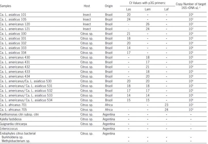
Out of 65 vegetal samples tested positive by qPCR for Liberibacter, 30 were positive for Las, 20 were positive for Lam, 13 samples tested positive for both species and only two were positive for Laf, which corresponded to the Laf positive control used in the assay. All Ct values of positive DNA leaf samples were between 14 and 24 (Table 1). Where a mix of Las and Lam was present in DNA samples, Ct val-ues between 14 and 20 were observed (Table 1). When DNA samples extracted from insects were evaluated, 27 samples were positive for Las and 13 were posi-tive for Lam. The Ct value was between 20 and 26 for both, Las and Lam pathogens. No discrepancy was observed between the results from real-time PCR with those obtained with conventional PCR (data not shown). The copy number of 16S rDNA obtained with these Ct values in DNA samples varied from 102 to 106
CN (Table 1).
Specificity of SG qPCR assay for the detection of “Ca. Liberibacter spp.”
The p3G set of primers amplified the specific target from DNA extracts of HLB infected plants with an optimal temperature of 70 ºC, without any ampli-fication of negative bacterial controls and NTC. The primer pair p3G was specific to Las, Lam and Laf, and did not cross-react with endophytes commonly resident in citrus or other citrus pathogens including Xylella fastidiosa, Xanthomonas citri subsp. citri or the fungus
Guirnardia citricarpa. Additionally, as expected these primers did not cross-react with other bacterial species like Enterococcus sp., isolated from the soil of sugarcane fields (Table 1), nor amplified samples of citrus leaves collected in non-infected growth areas in Tucumán, Ar-gentina.
Las as described by Teixeira et al. (2008). The standard curve for rplLas was made by using ten-fold serial dilu-tions of plasmid DNA containing the sequence of the
rplKAJL-rpoBC gene cluster (β-operon) from Las. The ef-ficiency of the PCR reaction was 110 % for Las with a detection limit of 10 CN (Figure 5). When comparing both primer sets in SG qPCR assays, rplLas and p3G had the same detection limit. Both primer sets succeeded in detecting 10 CN with an efficiency of 98 % and 110 % for p3Gand rplLas, respectively. However, the lowest Ct was observed for the p3G primers when the same copy number of targeted DNA was compared (Figure 5). For 108 CN the Ct value for p3G was 7, whereas the Ct value
for the rplLas primers was 13.
In vegetal samples, the detection limit was 10 CN for both, p3G and rplLas primers with a Ct value of 33 and 35, respectively. The PCR efficiency was 103.3 % for rplLas primers and 98 %, for p3G primers (Figure 6). The Ct values obtained with vegetal samples correlates with the Ct value observed for plasmid DNA. To further evaluate the p3G assay, another comparison was made against a detection method based on 16S rDNA ampli-fication; this time the TaqMan system was applied by using a primer-probe set previously reported by Li et al. (2006). Vegetal samples containing DNA templates rang-ing from 105 to 1 CN had Ct values between 23 and 38.
The efficiency of the PCR reaction was 98.2 % for Las and the detection limit of TaqMan PCR for HLB patho-gens was therefore equal to 10 copies. Figure 6 shows curves obtained with one of the plant samples, named AS13.
Discussion
To prevent the outbreak and spreading of HLB-associated Liberibacter species in unaffected citrus production areas, it is necessary to have sensitive and reliable diagnostic methods for the early detection of the pathogen. This is particularly critical in areas where Comparison of sensitivity between PCR diagnostic
methods
The sensitivity of the SG qPCR assay developed in this study was compared to a certain number of previously established protocols. For this purpose, the β-operon primer set f-rplLAs/r-rplLAs was selected for
Figure 3 – SYBR Green quantitative PCR amplification curves and melting curves with primers p3G for detection of “Ca. Liberibacter spp.” in DNA samples of Liberibacters (Las: “Ca. L. asiaticus”, Lam: “Ca. L. americanus” or Laf: “Ca. L. africanus”). Melting curve peak observed was 82 °C for Lam and 83 °C for Las and Laf.
Figure 4 – Standard curve obtained with p3G primers using tenfold serial dilution ranging from 108 to 1 copy number (CN) for the
pGEM-T Easy vector containing 16S rDNA sequence from “Ca. L. asiaticus” (Las), “Ca. L. americanus” (Lam) and “Ca. L. africanus” (Laf).
psyllids are present and HLB has not yet been reported, as in the northwest of Argentina.
The rapid expansion of HLB in Brazil and Flor-ida are examples where the confirmation of the dis-ease occurred much later than the infection by the pathogen (Coletta-Filho et al., 2004; Manjunath et al., 2008), which clearly attests that the plant had been infected for a long period of time when disease
symp-toms appeared. It is rather difficult to diagnose HLB on the basis of disease symptoms, and when it hap-pens, it is too late to prevent it from spreading. In areas where the HLB pathogen is unknown or unde-tected, a strategy that allows for both early detection of any Liberibacter species and low inoculum loads is necessary in order to detect the disease in time. Thus, it is imperative to be able to confirm the presence of
Table 1 – Specificity of SYBR Green quantitative PCR assays for “Candidatus Liberibacter spp.” in 21 of 105 analyzed DNA samples.
Samples Host Origin Ct Values with p3G primers: Copy Number of target 16S rDNA uL–1
Las Lam Laf
Ca. L. asiaticus 101 Insect Brazil 20 - - 104
Ca. L. asiaticus 105 Insect Brazil 24 - - 103
Ca. L. americanus 120 Insect Brazil - 26 - 102
Ca. L. americanus 121 Insect Brazil - 24 - 103
Ca. L. asiaticus 330 Citrus sp. Brazil 21 - - 104
Ca. L. asiaticus 331 Citrus sp. Brazil 18 - - 105
Ca. L. asiaticus 332 Citrus sp. Brazil 20 - - 104
Ca. L. asiaticus 333 Citrus sp. Brazil 14 - - 106
Ca. L. asiaticus 334 Citrus sp. Brazil 17 - - 105
Ca. L. americanus 430 Citrus sp. Brazil - 18 - 105
Ca. L. americanus 431 Citrus sp. Brazil - 17 - 105
Ca. L. americanus 432 Citrus sp. Brazil - 21 - 104
Ca. L. americanus 433 Citrus sp. Brazil - 18 - 105
Ca. L. americanus 434 Citrus sp. Brazil - 20 - 104
Ca. L. americanus/Ca. L. asiaticus 530 Citrus sp. Brazil 20 20 - 104
Ca. L. americanus/ Ca. L. asiaticus 531 Citrus sp. Brazil 18 18 - 105
Ca. L. americanus/ Ca. L. asiaticus 532 Citrus sp. Brazil 17 17 - 105
Ca. L. americanus/ Ca. L. asiaticus 533 Citrus sp. Brazil 14 14 - 106
Ca. L. americanus/ Ca. L. asiaticus 534 Citrus sp. Brazil 15 15 - 106
Ca. L. africanus 701 Citrus sp. Africa - 23 103
Ca. L. africanus 705 Citrus sp. Africa - - 24 103
Xanthomonas citri subsp. citri Citrus sp. Argentina - - -
-Xylella fastidiosa Citrus sp. Argentina - - -
-Guignardia citricarpa Citrus sp. Argentina - - -
-Enteroccocus Argentina - - -
-Endophytes citrus bacterial Burkholderia sp. Methylobacterium sp.
Citrus sp. Argentina
-Las: “Ca. L. asiaticus”; Lam: “Ca. L. americanus”; Laf: “Ca. L. africanus”.
the pathogen in order to act as quickly as possible, and then additional measures can be taken to identify the species present.
Recently, new diagnosis systems such as biosen-sors and optical sensing techniques have been devel-oped. However, the most sensitive and rapid method for detecting the pathogen is to use various PCR-based molecular techniques. This method is currently being used in most laboratories for HLB diagnosis in dif-ferent parts of the world. Continuous improvements have been introduced in these diagnostic strategies, particularly to make possible detection of more than one Liberibacter species. For this purpose, Li et al. (2006) have developed a multiplex real-time PCR (Taq-Man) assay using a probe-primer set for detection of the three known species of Liberibacter causing HLB. This test uses a 16S rDNA-based TaqMan probe and a set of three different forward primers for the detection of the three different species of Liberibacter and one
common reverse primer. This set has been successfully applied in the detection, identification and quantifica-tion of Las in host plants of citrus (Li et al., 2009; Tati-neni et al., 2008) and in the psyllid vector (Manjunath et al., 2008).
In order to simplify methods currently used in the detection of HLB-associated pathogens, a region of the 16S rDNA gene has been used to design a new set of primers; they hybridize perfectly to the genome of all three species of Liberibacter causing HLB in a single-plex real-time PCR reaction. The 16S rDNA sequences are highly conserved among the species of Liberibacter but variation between them is sufficient to prompt de-signing primers capable of detecting and identifying the bacterium in PCR assays (Coletta-Filho et al., 2005; Ja-goueix et al., 1996; Teixeira et al., 2005). The gene is large enough to provide distinctly and statistically valid measurements as it has sufficient interspecific polymor-phisms of the 16S rDNA gene.
The sensitivity of the qPCR assay development in this study was tested against a standard qPCR method-ology and the Ct values of the p3G primers were com-pared with PCR-primers targeting the 16S rDNA gene (Li et al., 2006) or the β-operon region (Teixeira et al., 2005) of “Ca. Liberibacter spp.”. qPCR protocols tested showed the same detection limit, ten copies, but the Ct value obtained with the p3G primers was lower than that obtained by the other methods tested. Kim and Wang (2009) indicate that Ca. Liberibacter asiaticus contains three copies of 16S rDNA and, compared to qPCR as-says targeting β-operon, a 16S rDNA based qPCR assay which is likely to be more sensitive, due to its higher copy number per genome. This could help to define diag-nostic situations where a very high Ct is obtained, close to the safety limit.
Ananthakrishnan et al. (2013) have reported the design of a single pair of degenerate primer-probes tar-geting the rpoB gene of Liberibacter enabling the de-tection of “Ca. Liberibacter spp.” at the genus level in one PCR reaction. In a similar way, like the method developed for the rpoB gene, the p3G primers described in this analysis detect all three Liberibacter species as-sociated with HLB with high sensibility and specific-ity. Both methods can confirm whether Libericabter is present in a plant or insect sample; however, with the p3G primers Las/Laf from Lam samples can be differ-entiated by their characteristic melting curves. This is especially interesting in the continents of North and South America where two forms of HLB have been re-ported, Las and Lam (Teixeira et al., 2005; Teixeira et al., 2008).
If necessary, the Liberibacter species present in a sample could be determined by an electrophoresis by us-ing a DNA analyzer system like a sequencer; the ampli-fication produces three different sizes of DNA for each Liberibacter species. In contrast, this is not possible for the rpo assay because the size of the amplified fragment is the same for all three species of Liberibacter.
Further-Figure 6 – Comparison of qPCR systems for HLB diagnostic. The standard curves for SYBR Green (p3G and rplLas, upper and middle, respectively) and TaqMan qPCR assays (HLBasp, down) were obtained by using as a template a ten-fold serial dilution from a quantified DNA extracted from a vegetal sample infected with “Ca. L. asiaticus”, (named AS13), ranging from 105 to 1 copy
more, the comparison of the 16S rDNA gene sequences allows for differentiation between organisms at the ge-nus level across all major phyla of bacteria, in addition to classifying strains at multiple levels, including at spe-cie and subspespe-cie level (Clarridge, 2004). The degree of conservation is assumed to result from the importance of the 16S rDNA as a critical component of cell func-tion, a fact which contrasts with other genes which can usually tolerate mutations with more frequency since they may affect structures not as unique and essential as rDNA (Clarridge, 2004).
Although it is possible to design three fluores-cent probes to detect the three species of Liberibacter in the same reaction tube using the p3G assay, our main goal was to design a method that is sensitive, simple and low-cost for diagnosis. The addition of more probes to the reaction tube could not only inter-fere and hamper the efficiency of the test but would also increase the cost of reaction per sample. To avoid this, a representative region of the 16S rDNA gene was chosen; this area allowed to design of highly specific primers, without decreasing the sensitivity and speci-ficity, as well as producing a new cost-effective tool for HLB diagnosis.
Reaction in silico was performed using the 16S rDNA sequence from “Ca. Liberibacter solanacearum”, which is the causal agent of diseases of high economic impact on solanaceous crops (mainly potatoes and toma-toes) and carrots (Munyaneza et al., 2012). Thus, the p3G primers designed for HLB Liberibacter detection were expected to amplify the expected fragment (121 bp) with one mismatch at the 5’ end, showing the possible broad application of these primers.
The singleplex assay was designed using the p3G primers hybridizing to a very conserved sequence of the Liberibacter genomes associated with HLB. This strate-gy could be of importance for the detection of new spe-cies which cause the diseases, especially after two HLB causing species were identified in Brazil, one of which had never been reported previously. The present analy-sis offers an efficient technique for the diagnoanaly-sis of HLB which, combined with a thoroughly designed sample testing program, would help to prevent the outbreak and spread of this devastating disease in major citrus produc-tion areas not yet affected.
Acknowledgments
This project was partially supported by the EE-AOC Citrus Program. MRM, AAV, APC and MPF are members of the CONICET. LNS and IGO are CONICET fellows. We especially thank Dr H. Coletta-Filho (Cen-tro Apta de Citricultura Sylvio Moreira, SP, Brazil), Dr. D. Teixeira (FUNDECITRUS, SP, Brazil) for providing us the 16S rDNA and rplLas plasmid DNA and HLB posi-tive samples and Dr Welin (EEAOC, Argentina) for the critical reading of this manuscript and for reviewing the English version of the manuscript.
References
Ananthakrishnan, G.; Hu, H.; Brlansky, R. 2013. Development and applications of primers and probe for genus specific detection of 'Candidatus Liberibacter species' by real-time PCR. Plant Disease 97: 1235-1243.
Augier, L.; Gastaminza, G.; Lizondo, M.; Argañaraz, M.; Willink, E. 2006. Presence of Diaphorinacitri (Hemiptera: Psyllidae) in Northwestern Argentina (NOA). Revista de la Sociedad Entomológica Argentina 65: 67-68 (in Spanish).
Bové, J.M. 2006. Huanglongbing: a destructive, newly-emerging, century-old disease of citrus. Journal of Plant Pathology 88: 7-37. Clarridge, J.E. 2004. Impact of 16S rRNA gene sequence analysis
for identification of bacteria on clinical microbiology and infectious diseases. Clinical Microbiology Reviews 17: 840-862. Coletta-Filho, H.; Takita, M.; Targon, M.; Machado, M. 2005.
Analysis of 16S rDNA sequences from citrus huanglongbing bacteria reveal a different “Ca. Liberibacter” strain associated with citrus disease in Sao Paulo. Plant Disease 89: 848-852. Coletta-Filho, H.; Targon, M.; Takita, M. 2004. First report of
the causal agent of Huanglongbing (“Candidatus Liberibacter asiaticus”) in Brazil. Plant Disease 88: 1382.
Gottwald, T.R.; Da Graça, J.V.; Bassanezi, R.B. 2007. Citrus huanglongbing: the pathogen and its impact. Plant Health Progress 6.
Gottwald, T.R. 2010. Current epidemiological understanding of citrus Huanglongbing. Annual Review of Phytopathology 48: 119-139.
Hocquellet, A.; Toorawa, P.; Bové, J.M.; Garnier, M. 1999. Detection and identification of the two “Candidatus Liberobacter sp” associated with citrus huanglongbing by PCR amplification of ribosomal protein genes of the beta operon. Molecular and Cellular Probes 13: 373-379.
Hung, T.; Wu, N.M.; Su, H. 1999. Development of a rapid method for the diagnosis of citrus greening disease using the polymerase chain reaction. Journal of Phytopathology 147: 599-604. Jagoueix, S.; Bove, J.M.; Garnier, M. 1994. The phloem-limited
bacterium of greening disease of citrus is a member of the a subdivision of the Proteobacteria. International Journal of Systematic Bacteriology 44: 379-386.
Jagoueix, S.; Bové, J.M.; Garnier, M. 1996. PCR detection of the two “Candidatus liberobacter species” associated with greening disease of citrus. Molecular and Cellular Probes 10: 43-50. Kim, J.; Wang, N. 2009. Characterization of copy numbers of 16S
rDNA and 16S rRNA of Candidatus Liberibacter asiaticus and the implication in detection in planta using quantitative PCR. BioMed Central Research Notes 2: 37.
Li, W.; Hartung, J.S.; Levy, L. 2006. Quantitative real-time PCR for detection and identification of Candidatus Liberibacter species associated with citrus huanglongbing. Journal of Microbiological Methods 66: 104-115.
Li, W.; Levy, L.; Hartung, J.S. 2009. Quantitative distribution of 'Candidatus Liberibacter asiaticus' in citrus plants with citrus Huanglongbing. Phytopathology 99: 139-144.
Manjunath, K.; Halbert, S.; Ramadugu, C.; Webb, S.; Lee, R. 2008. Detection of 'Candidatus Liberibacter asiaticus' in
Munyaneza, J.E.; Sengoda, V.G.; Stegmark, R.; Arvidsson, A.K.; Anderbrant, O.; Yuvaraj, J.K. 2012. First Report of “Candidatus Liberibacter solanacearum” associated with Psyllid-affected carrots in Sweden. Plant Disease 96: 453. Murray, M.; Thompson, W.F. 1980. Rapid isolation of high
molecular weight plant DNA. Nucleic Acids Research 8: 4321-4326.
Tatineni, S.; Sagaram, U.S.; Gowda, S. 2008. In planta distribution of 'Candidatus Liberibacter asiaticus' as revealed by polymerase chain reaction (PCR) and real-time PCR. Phytopathology 98: 592-599.
Teixeira, D.C.; Ayres, A.J.; Kitajima, E.W.; Tanaka, F.A.O.; Danet, J.L.; Jagoueix-Eveillard, S.; Saillard, C.; Bove, J.M. 2005. First report of a huanglongbing-like disease of citrus in São Paulo state, Brazil, and association of a new liberibacter species,
Candidatus Liberibacter americanus, with the disease. Plant Disease 89: 107.
Teixeira, D.C.; Saillard, C.; Couture, C.; Martins, E.C.; Wulff, N.A.; Jagoueix-Eveillard, S.; Yamamoto, P.T.; Ayres, A.J.; Bove, J.M. 2008. Distribution and quantification of Candidatus
Liberibacter americanus, agent of huanglongbing disease of citrus in São Paulo state, Brasil, in leaves of an affected sweet orange tree as determined by PCR. Molecular and Cellular Probes 22: 139-150.
Vaccaro, N.C. 1994. Behavior and dissemination of “sharpshooter of Citrus” Diaphorina citri (Homoptera-Diaphoridae) in the citrus area of Entre Rios. In: Carpeta de información citrícola. v. 2. Sección G (27). EEA/INTA, Concordia, Argentina (in Spanish).
Villechanoux, S.; Garnier, M.; Bové, J.M. 1990. Purification of the bacterium-like organism associated with greening disease of citrus by immunoaffinity chromatography and monoclonal antibodies. Current Microbiology 21: 175-180.