online | memorias.ioc.fiocruz.br
Improved reverse transcription-polymerase chain reaction assay
for the detection of flaviviruses with semi-nested primers for
discrimination between dengue virus serotypes and Zika virus
Allan RD Nunes1,3, Brenda Elen B Alves2,4,5, Hannaly WB Pereira2,4,5, Yasmin M Nascimento2,5,
Ingryd C Morais2,3,5, José Veríssimo Fernandes2,4, Josélio MG Araújo2,4,5, Daniel CF Lanza1,3/+
1Universidade Federal do Rio Grande do Norte, Departamento de Bioquímica, Laboratório de Biologia Molecular Aplicada, Natal, RN, Brasil 2Universidade Federal do Rio Grande do Norte, Departamento de Microbiologia e Parasitologia, Laboratório de Biologia Molecular de
Doenças Infecciosas e Câncer, Natal, RN, Brasil
3Universidade Federal do Rio Grande do Norte, Programa de Pós-Graduação em Bioquímica, Natal, RN, Brasil 4Universidade Federal do Rio Grande do Norte, Programa de Pós-Graduação em Biologia Parasitária, Natal, RN, Brasil 5Universidade Federal do Rio Grande do Norte, Instituto de Medicina Tropical, Laboratório de Virologia, Natal, RN, Brasil
BACKGROUND The genus Flavivirus includes a variety of medically important viruses, including dengue virus (DENV) and Zika virus (ZIKV), which are most prevalent in Brazil. Because the clinical profile of patients affected by different DENV serotypes or ZIKV may be similar, the development of new methods that facilitate a rapid and accurate diagnosis is crucial.
OBJECTIVES The current study aimed to develop an improved reverse transcription-polymerase chain reaction (RT-PCR) protocol for universal detection of flaviviruses by using semi-nested primers that discriminate between DENV serotypes and ZIKV.
METHODS The bioinformatics workflow adopted for primer design included: (1) alignment of 1,442 flavivirus genome sequences, (2) characterisation of 27 conserved regions, (3) generation of a primer set comprising 77 universal primers, and (4) selection of primer pairs with greatest coverage and specificity. Following primer design, the reaction was validated in vitro. The same approach was applied to the design of primers specific for DENV and ZIKV, using a species-specific sequence database.
FINDINGS The new assay amplified an 800-806 nt variable region of the NS5 gene and allowed discrimination of virtually all flavivirus species using reference-sequence comparison. The 800-806 nt fragment was validated as a template for a semi-nested multiplex PCR using five additional primers for the detection of DENV and ZIKV. These primers were designed to generate amplicons of different sizes, allowing differentiation of the four serotypes of DENV, and ZIKV using agarose gel electrophoresis.
MAIN CONCLUSIONS The bioinformatics pipeline allowed efficient primer design, making it possible to identify the best targets within the coding region of the NS5 protein. The multiplex system proved effective in differentiation of DENV1-4 and ZIKV on a 2% agarose gel. The possibility of discriminating DENV serotypes and ZIKV in the same reaction provided a faster result consuming less sample. In addition, this simplified approach ensured the reduction of the cost per analysis.
Key words: arbovirus - Flaviviridae - West Nile virus - yellow fever virus
doi: 10.1590/0074-02760170393 Financial support: CAPES, CNPq.
+ Corresponding author: [email protected] Received 25 September 2017
Accepted 23 January 2018
The genus Flavivirus (family Flaviviridae) is com-prised of 53 virus species (Simmonds et al. 2017), in-cluding human pathogens such as West Nile virus, Japa-nese encephalitis virus, tick-borne encephalitis virus, yellow fever virus, dengue virus (DENV) and Zika vi-rus (ZIKV) that infect populations of several countries (Mackenzie et al. 2004, Guzman et al. 2010).
Dengue and Zika infections are major problems faced by the Brazilian public health today. DENV has been con-sidered a significant problem since 1986, when serotype 1 (DENV-1) was identified in the state of Rio de Janeiro (Schatzmayr et al. 1986). The serotypes DENV-2 and DENV-3 were introduced in Brazil via Rio de Janeiro in 1990 and 2000, respectively. In subsequent years, these DENV serotypes spread throughout the country reaching
25 of the 26 Brazilian states by the end of 2006 (Nogueira et al. 2007). Serotype DENV-4 re-emerged in Brazil in the Roraima state of the Amazon region in 2010, 28 years after the first outbreak was reported. Following reports of DENV-4 in Roraima in 1982 and 2010, the virus was identified in 2011 in other states of the northern region of Brazil including Amazonas, Amapá, and Pará. DENV-4 was actually detected serologically in populations from several Brazilian states (Nogueira et al. 2011).
Currently, the laboratory diagnosis of diseases caused by flaviviruses is carried out using specific serological as-says, commonly based on enzyme-linked immunosorbent assays (ELISAs). These tests detect virus-specific IgM and IgG antibodies 5-7 days after onset of infection, which makes it unfeasible for a rapid diagnosis in most cases. In contrast, polymerase chain reaction (PCR) and it deriva-tions can be used during the acute phases of infection and are known to be rapid, specific, and capable of pathogen detection with a great sensitivity. Several reported PCR assays for flaviviruses are only of use as confirmatory tests after the clinical testing is completed and the differ-ential diagnosis made because the PCR primers may only amplify one, or a limited range of closely related species (Fulop et al. 1993, Tanaka 1993, Pierre et al. 1994).
Since the publication of the first tests using universal primer pairs for flaviviruses in 1993 (Fulop et al. 1993, Tanaka 1993), several universal primer sets have been de-veloped for the same purpose (Eldadah et al. 1991, Chow et al. 1993, Pierre et al. 1994, Puri et al. 1994, Figueiredo et al. 1998, Kuno 1998, Scaramozzino et al. 2001, Sán-chez-Seco et al. 2005, Chao et al. 2007, Dyer et al. 2007, Maher-Sturgess et al. 2008). The most promising PCR protocols for detecting a wide variety of virus species have been those that use primers designed to target con-served genomic regions. These approaches usually com-prise an initial step in which a broad range of primers are used to amplify a potential broad range of targets, fol-lowed by a nested step, which generates a species-specif-ic amplspecies-specif-icon identified by nucleotide sequencing (Kuno 1998, Maher-Sturgess et al. 2008).
The large number of viral sequences deposited every year, in conjunction with the enhancement of bioinfor-matics tools, has allowed improvement of the traditional strategies in both efficiency and cost of development. In the current work, we implemented a robust bioinformat-ics approach to identify conserved regions in flavivi-ruses, comparing 1,442 full genomes of different species. The conserved regions were mapped and primer sets designed, which were submitted to successive selection steps in order to find the best combination for a universal detection system. The final primer pair selected was used to validate a universal PCR protocol able to detect and discriminate virtually all flaviviruses. This protocol was adopted as the first step in a semi-nested RT-PCR ap-proach for the specific detection of DENV1-4 and ZIKV.
MATERIALS AND METHODS
Sequence selection and alignment - Initially, 1,740 nucleotide sequences from 16 different flavivirus spe-cies and serotypes were selected from Genbank (http:// www.ncbi.nlm.nih.gov/). The initial set included all sequences containing more than 10,000 nt that had been deposited up to April 4, 2016. In cases where the number of sequences of a single species was greater than 300, the initial set was restricted to sequences deposited from January 1, 2014 or January 1, 2015. Sequences manually excluded that contained regions with inconclusive or degenerate bases, or sequences of virus-attenuated vaccines, clones, and virus chimeras. The predicted amino-acid sequences encoded by the
1,442 selected genomes were generated using Geneious software, version 9.1 (http://www.geneious.com), and aligned using MAFFT algorithm, version 7.222 adopt-ing standard parameters for each case. When neces-sary, adjustments were made manually.
Virus samples - The DENV1-4 and ZIKV samples used for the in vitro tests were isolated from patients from the Brazilian states of Rio Grande do Norte (DENV1-4) and Pernambuco (ZIKV), with a confirmed positive-di-agnosis in each case. The established kidney epithelial VERO E6 cell-line from an African green monkey was cultured in 12.5 cm² flasks (Corning Incorporated, Corn-ing, New York, USA) in the presence of Leibovitz-15 (L-15) medium (Invitrogen, New York, USA), supplemented with 10% heat inactivated foetal bovine serum (FBS), 1% antibiotic-antimycotic, and 10% tryptose phosphate (In-vitrogen, New York, USA). The cell culture was main-tained in an incubator at 37ºC in 5% CO2. When the cell monolayers reached 90% confluency, 15 μL of the pa -tient serum and 50 μL of 2% FBS L-15 medium were added. The serum-exposed cells were kept in the incuba-tor for 90 min with slight shaking every 30 min. The in-oculum was then removed and 3 mL of 2% L-15 medium was added to the flask. The cells were incubated at 37ºC in 5% CO2 for seven days to allow for virus replication. For each 1-mL of the cell-culture supernatant, 0.9 mL of bovine serum albumin (BSA) was added to preserve the viral particles. This mixture was centrifuged at 1,500 rpm (~260 ×g) for 5 min and the supernatant aliquoted into cryogenic tubes and stored at -70ºC. Confirmation of viral isolation was performed using a reverse transcrip-tion PCR assay (RT-PCR) using RNA extracted from the VERO-culture supernatant as the template.
Cell culture and replication of DENV and ZIKV - For viral replication, 100 μL of each viral inoculum was add -ed to VERO E6 cell-cultures previously condition-ed in 25 cm³ bottles. Cells were incubated for 1 h at 37ºC, being homogenised every 15 min. L-15 media with 2% FBS was added to the cells and incubated at 37ºC for seven days. Viral infection was confirmed by RT-PCR. After confirmation of infection, 20% FBS was added and the isolates stored at -70ºC. The titre of ZIKV was determined by plaque assay to be 1.6 × 10⁶ PFU/mL for ZIKV. Owing
to the inherent dilution of each reaction, the maximum concentration used per reaction was 6.2 × 10³ PFU. The
presence of spiked chikungunya virus in the templates was used in the specificity tests and was confirmed by real time PCR, as described by Lanciotti et al. (2007).
each) were mixed and heated at 70ºC for 5 min, followed by an ice-bath incubation for another 5 min. Thereafter, 10 μL of 2× master mix (Tfl DNA Polymerase, dNTPs, magnesium sulphate and reaction buffer) and 0.4 μL (2 units) of AMV Reverse Transcriptase were added with the final volume adjusted to 20 μL with deionised water. The PCR reaction cycling was performed on a Bioer Life Touch thermal cycler with an initial incubation at 45ºC for 1 h, followed by 40 cycles of incubation at 95ºC for 2 min, for denaturation at 95ºC for 45 s, annealing at 45ºC for 45 s, and elongation at 63ºC for 1 min. A final exten-sion was performed at 63ºC for 5 min.
For the semi-nested component of the protocol, the primer concentrations were standardised using a prim-er-concentration gradient for each primer combination. As template, 1 μL of cDNA from the first PCR ampli -fication was mixed with 10 μL of 2× master mix and different concentrations of primer (10-100 pmol each) with the final volume of the reaction mix being adjusted to 20 μL with deionised water. The PCR thermal cycling parameters were incubation at 95ºC for 2 min, followed by 40 sequential cycles of denaturation at 95ºC for 45 s, annealing at 53ºC for 45 s, and elongation at 63ºC for 1 min. Again, a final extension was performed at 63ºC for 5 min. After determining the optimal primer con-centrations using an annealing temperature gradient (45-60ºC), the PCR amplification was carried out using the ZIKV and DENV (1-4) samples as the template with multiplexed or isolated primer-pairs.
PCR reactions intended to detect more than one vi-rus type/strain simultaneously, and serial-dilution tests were performed using the parameters described for the first and nested components of the protocol. The nested step of the amplification protocol was performed using
the hexaplex-containing forward primers DENV1F6.2, DENV2F10, DENV3F6.1, DENV4F3, and ZIKVF8 in combination with reverse primer CRNS5_7R6.
RESULTS
Development of a PCR protocol for the detection of flaviviruses - A consensus sequence containing 13.7% of identical sites and 60.8% identity was generated from the alignment of the 1,442 selected amino acid sequences referenced in Table I. Regions with at least six conserved amino acids (99% identity per site) and global identity equal to or higher than 95% were considered conserved regions (CRs). Of the 27 CRs selected, 16 were located in the NS5 protein, seven in the NS3 protein, and two in each of the Envelope and NS1 proteins. The flavivirus CRs are characterised in Supplementary data (Table I).
A primer set containing 77 primers that potentially aligned in each of the conserved regions was designed [Supplementary data (Table II)]. These primes were ex-pected to function as universal primers with the variable sites within each sequence being replaced with degen-erate nucleotide bases. Eleven primers with the lowest degeneracy (≤ 1000 possible combinations), compatible melting temperatures (average Tm ranging from 50-60ºC), and size ranging from 20 to 25 nt were selected from the primer set [Supplementary data (Table II) in bold]. Subsequently, the degeneracy of each of the 11 primers was reduced by the insertion of inosines to sub-stitute the most variable sites, but always preserving at least two nucleotides on the 5’ and 3’ ends, and at least 18 specific nucleotides in each sequence.
In vitro validation of the first step of the PCR proto-col - Primers CRNS5_3F1 and CRNS5_7R6 (Table II, Supplementary data (Table II)] were selected for the in
TABLE I
Flaviviruses sequence sets used in this study
Keywords Search Period Number of sequences obtained Number of selected sequences
Dengue virus 1 01.2014 - 04.2016 294 272
Dengue virus 2 01.2014 - 04.2016 244 215
Dengue virus 3 01.2014 - 04.2016 173 163
Dengue virus 4 Until 04.2016 219 170
Ilheus virus Until 04.2016 3 2
Japanese encephalitis virus Until 04.2016 239 203
Langat virus Until 04.2016 4 3
Louping ill virus Until 04.2016 6 5
Murray Valley encephalitis virus Until 04.2016 20 12
Rocio virus Until 04.2016 1 1
Spondweni virus Until 04.2016 2 1
St. Louis encephalitis virus Until 04.2016 36 30
Tick-borne encephalitis virus Until 04.2016 160 137
West Nile virus 01.2015 - 04.2016 161 126
Yellow fever virus Until 04.2016 95 40
Zika virus Until 04.2016 83 62
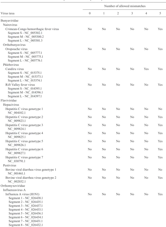
vitro validation. These primers annealed to conserved-re-gions 3 and 7, respectively, amplifying a variable region of 800-806 nt within the NS5 coding sequence. This re-gion enabled the phylogenetic classification of virtually all the flavivirus groups [Supplementary data (Figure)]. The specificity of the primers was validated by in silico PCR analysis using the Geneious software. All sequences used in the testing of specificity are described in Table III. The in silico specificity test was performed at different levels of stringency. It was observed that nonspecific annealing was highly unlikely, occurring only when considering an allowance of five or more mismatches.
The standardisation of the PCR reaction using CRNS5_3F1 and CRNS5_7R6 primers was started with gradient tests in order to determine the best annealing and elongation temperatures (data not show). Once the annealing temperature of 45ºC, and the elongation tem-peratures of 63ºC were determined as optimal, a primer gradient test revealed that 60 pmol of each primer was the ideal amount for the assay, as shown in Fig. 1.
Development of a semi nested-PCR for the detection of DENV1-4 and ZIKV - After standardisation of the first step of the PCR reaction, the next challenge was to determine the regions that allowed for sensitive and spe-cific amplification using a multiplex semi-nested PCR. It was anticipated that this second step would generating amplicons of different sizes, allowing the discrimination between the DENV strains 1-4, as well as ZIKV, using only gel electrophoresis to analyse the PCR results.
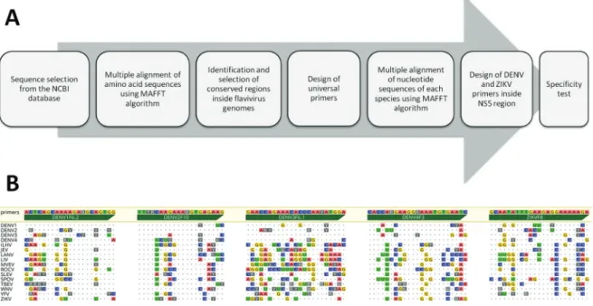
To identify the best target sites for primer design, new CRs were identified from alignment of the 882 ami-no acid sequences of DENV and ZIKV. Subsequently, 14 alignments were performed, one for each sequence set shown in Table I. Conserved regions that existed among the species or serotypes were discarded. Specif-ic CRs located within the NS5 sequence of DENV1-4 and ZIKV genomes were selected as targets for which a new set of 81 candidate primers was designed, based on the following criteria: (1) primer length of 20-25 nu-cleotides; (2) maximum degeneration of eight combi-nations; (3) average Tm from 52-60ºC; (4) absence of degeneration in the three nucleotides at 3’ end; (5) the first two nucleotides of the 3’ end should not be iden-tical (coincidental sites) between the ZIKV and DENV sequences; and (6) secondary structure with ΔG > -5 Kcal/mol. The prediction of the secondary structures was performed with the Geneious software, using the DNA model in the toolkit plugin Vienna. The pipeline used for primer design is summarised in Fig. 2A.
Following primer design, the specificity of each primer was analysed by means of in silico PCR analysis using the complete flavivirus sequence set as a template. This test revealed that most primers would fail to an-neal on a significant number of unspecific targets [Sup-plementary data (Table III)], even when adopting a low level of stringency (i.e. allowing for five mismatches). Among the 81 candidate primers, the five forward prim-ers presented in Table II were selected for in vitro vali-dation. The result of the in silico specificity test for the five selected primers against the sequence set described in Table I is summarised in Fig. 2B.
T A B L E I I P ri m e rs d e si g n e d a n d v al id at e d i n t h is s tu d y P ri me r n a me S e q ue nc e (5 ’→ 3’ ) L e n g ht
Tm (°C
) Tm m e a n (° C ) D ege n e ra cy Se lf -d ime r S p e ci fi ci ty (n º o f s e q s i n w h ic h t h e p ri m e r a n n e al
s / n
º o f s e q s t e st e d ) A m pl ic o n s iz e (b p) C R N S 5 _ 3 F1 A AY T C N A M N S AY G A R A T G T A 20 4 6
.8 - 5
8. 1 52 ,45 32 N ot t e st e d F la v iv ir u se s ( 14 4 2 /14 4 2 ) 80 0 -80 6 C R N S 5 _7 R 6 C C N A R C CA CA T R W A C CA D A T 20 5 2
.1 - 6
0 .0 56 ,0 5 24 N ot t e st e d D E N V1 F 6 .2 A C T C AG C A A A AG A R G C AG T G G 21 5 9.
0 - 6
0 .8 5 9, 9 2 L ow p ro p e n si ty D E N V 1 ( 2 72 /2 72 ) 18 1 D E N V 2 F 1 0 T T Y R C AA G AAA R G T G A G AA G 20 4 8.
8 - 5
6 .0 52 ,4 8 No n e D E N V 2 ( 21 5 /21 5 ) 2 4 5 D E N V 3 F 6 .1 G AA C C A G AAA C A C C C AA Y A T G G A 23 5 8.
7 - 6
TABLE III
Sequences used in the specificity test
Virus taxa
Number of allowed mismatches
0 1 2 3 4 5
Bunyaviridae Nairovirus
Crimean-Congo hemorrhagic fever virus Segment S - NC_005302.1
Segment M - NC_005300.2 Segment L - NC_005301.3
No No No No No Yes
Orthobunyavirus Oropouche virus
Segment S - NC_005777.1 Segment M - NC_005775.1 Segment L - NC_005776.1
No No No No No Yes
Phlebovirus Candiru virus
Segment S - NC_015375.1 Segment M - NC_015373.1 Segment L - NC_015374.1
No No No No Yes Yes
Rift Valley fever virus Segment S - NC_014395.1 Segment M - NC_014396.1 Segment L - NC_014397.1
No No No No No Yes
Flaviviridae Hepacivirus
Hepatitis C virus genotype 1 NC_004102.1
No No No No No No
Hepatitis C virus genotype 2 NC_009823.1
No No No No No Yes
Hepatitis C virus genotype 3 NC_009824.1
No No No No No Yes
Hepatitis C virus genotype 4 NC_009825.1
No No No No No Yes
Hepatitis C virus genotype 5 NC_009826.1
No No No No No Yes
Hepatitis C virus genotype 6 NC_009827.1
No No No No No Yes
Hepatitis C virus genotype 7 NC_030791.1
No No No No No Yes
Pestivirus
Bovine viral diarrhea virus genotype 1 NC_001461.1
No No No No No No
Bovine viral diarrhea virus genotype 2 NC_002032.1
No No No No No Yes
Orthomyxoviridae Influenzavirus A
Influenza A virus (H1N1) Segment 1 - NC_026438.1 Segment 2 - NC_026435.1 Segment 3 - NC_026437.1 Segment 4 - NC_026433.1 Segment 5 - NC_026436.1 Segment 6 - NC_026434.1 Segment 7 - NC_026431.1 Segment 8 - NC_026432.1
Virus taxa
Number of allowed mismatches
0 1 2 3 4 5
Rabdoviridae Vesiculovirus Chandipura virus NC_020805.1
No No No No No Yes
Vesicular stomatitis Indiana virus NC_001560.1
No No No No No Yes
Vesicular stomatitis New Jersey virus NC_024473.1
No No No No No Yes
Vesicular stomatitis Alagoas virus NC_025353.1
No No No No No Yes
Reoviridae Orbivirus
Changuinola virus
Segment 1 - NC_022639.1 Segment 2 - NC_022633.1 Segment 3 - NC_022634.1 Segment 4 - NC_022640.1 Segment 5 - NC_022635.1 Segment 6 - NC_022641.1 Segment 7 - NC_022636.1 Segment 8 - NC_022637.1 Segment 9 - NC_022642.1 Segment 10 - NC_022638.1
No No No No No Yes
Togavidae Alphavirus
Barmah Forest virus NC_001786.1
No No No No No Yes
Chikungunya virus NC_004162.2
No No No No No Yes
Mayaro virus NC_003417.1
No No No No No Yes
Ross River virus NC_001544
No No No No No Yes
Sindbis virus NC_001547.1
No No No No Yes Yes
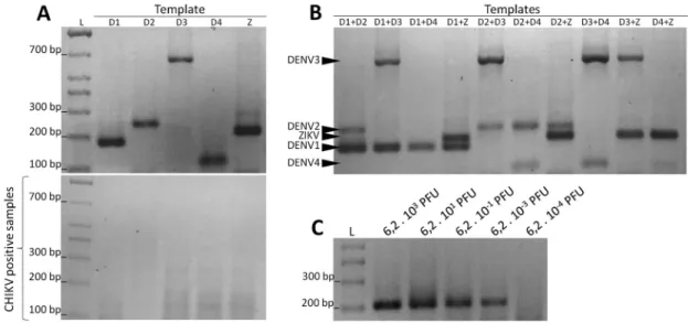
In vitro validation of the semi-nested PCR protocol - The in vitro validation using specific templates con-firmed that the multiplex containing the forward primers DENV1F6.2, DENV2F10, DENV3F6.1, DENV4F3, and ZIKVF8, and the reverse primer CRNS5_7R6 produced specific amplicons that were separated by 2% agarose gel electrophoresis (Fig. 3A top). Separate reactions with each of the forward primers using Chikungunya virus template as a positive control confirmed the specificity of the reactions (Fig. 3A down).
The semi-nested PCR was also tested with heterol-ogous templates containing two different serotypes or species of viruses mixed together. The hexaplex reactions efficiently detected all double-combinations tested of the five viruses. As demonstrated in Fig. 3B, any of the DENV1-4 and ZIKV amplicons in the same electrophore-sis lane were easily separated in a 2% agarose gel. The test
was also able to detect three or more viruses mixed and used as a template, but under these conditions small RNA concentrations could influence the results (data not show).
The sensitivity of the PCR was also assessed using ZIKV as a template. The serial dilution of the ZIKV tem-plate revealed that the nested multiplex reaction was able to detect 6.2 × 10³ plaque forming units (PFU), (Fig. 3C).
In addition, as shown in Fig. 4, the hexaplex reactions efficiently discriminated all serotypes and species using different annealing temperatures. All PCR-generated am-plicons were sequenced by Sanger methodology in order to confirm the specificity of each reaction (data not show).
DISCUSSION
Fig. 1: effect of primer concentration on the reverse transcription-poly-merase chain reaction (RT-PCR) for flavivirus detection. Six reactions were carried out using a temperature of 45ºC for annealing, and 63ºC for extension with Zika virus (ZIKV) cDNA as the template in order to determine the best primer concentration for the use of degenerate primers CRNS5_3F1 and CRNS5_7NR6. The amount of each primer ranged from 10-100 pmol. The amount of each primer used per reaction are presented above each lane (pmol). The black arrow indicates the expected size for the 800-bp amplicon. L = 100-bp size marker.
Fig. 2: pipeline for primer design and results from the specificity test. (A) Schematic of the pipeline used for the design and validation of primers. (B) Sequences of the forward primers targeting the flavivirus NS5 coding region to discriminate dengue virus (DENV)1-4 serotypes and Zika virus (ZIKV) in the semi-nested polymerase chain reaction. Primer sequences are presented in the 5’-3’ orientation above the green arrows. Dot-ted lines correspond to the consensus sequence obtained from the alignment of the sequence sets for each species referenced in Table I. Identities are indicated by dots and mismatches by letters (nucleotide bases or degenerate bases).
sequences allowed for an accurate characterisation of 27 conserved regions. Application of the pipeline with suc-cessive steps involving specific selection criteria identi-fied from the regions CRNS5_3 and CRNS5_7 as the most promising for the designing of universal primers. In agreement, Maher-Sturgess et al. (2008), coinciden-tally identified these regions from the alignment of 257 full-length flavivirus genomes. These regions were set as targets by these investigators for the design of primers that amplify equivalent fragments of NS5, allowing the phylogenetic reconstruction of each flavivirus subgroup. In our study, the alignment of a greater number of se-quences assured the improvement of the universal
prim-ers, mainly by the elimination of potential mismatches, increasing the possibility of amplification of templates with a greater sequence diversity.
A main advantage of the in silico approach was the considerable reduction in the time spent to validate the in vitro protocol. In fact, only the primer concentrations, and the annealing and elongation temperatures required optimising, and without the need for additives or differ-ent mixtures in order to achieve a high specificity. As expected, the concentration of primers was critical to the efficiency of the reaction; for the first step, higher primer concentrations provided the best results. This may have been attributed to the degeneracy of the prim-ers in association with a single type of virus DNA used as a template. In the multiplex reaction, this effect was less pronounced because of the lower degeneration of the primers. In both the first PCR reaction and the nested re-action, the in vitro results corroborated the efficiency of the in silico analyses to predict specificity and the ideal annealing conditions. The nested multiplex reaction pro-duced specific amplicons, even when using templates that contained more than one type of virus, or having different annealing temperatures.
Fig. 4: polymerase chain reaction (PCR) efficiency at different annealing temperatures. The efficiency of the semi-nested PCR reaction containing the six primers was evaluated at different annealing temperatures (45-60ºC) for each of the five viruses (specified on the left). The expected amplicon sizes are indicated (black arrows). L = 100 bp size marker.
Fig. 3: evaluation of the semi-nested polymerase chain reaction (PCR) for identification of dengue virus (DENV)1-4 serotypes and Zika vi-rus (ZIKV). The efficiency of the semi-nested reaction using the primers DENV1F6.2, DENV2F10, DENV3F6.1, DENV4F3, ZIKVF8, and CRNS5_7NR6 was evaluated under different conditions. (A) Size resolution of each amplicon in a 2% agarose gel. The letters above each lane indicate the templates containing RNA of DENV serotypes (D1, D2, D3, and D4) or ZIKV (Z). The results of semi-nested reactions containing each primer individually and a template positive for Chikungunya virus are presented (below). (B) Tests using a mixture of reverse-transcribed RNAs as the template from two types of viruses. All possible combinations among the five viruses have been evaluated and are shown above each their respective gel lanes. The size of each amplicon in the 2% agarose gel are indicated (black arrows). (C) Dilution test to verify the sensitivity of the semi-nested reaction. L = 100 bp size marker.
both interspecific and intraspecific ways. The speci-ficity tests consisted of species from six viral families used for the validation of the primer pair CRNS5_3F1/ CRNS5_7R6, and 1,442 viral genomes for the validation of the DENV and ZIKV primers. Thus, it was possible to evaluate the specificity of primers for sequences that had never been included in previous work.
A final point to be discussed is the technical feasibil-ity of the proposed method. To be effective in a clini-cal setting, the test should be able to detect the pathogen within seven days after the onset of the first symptoms. The method proposed here allowed the investigation of five viruses using a single reaction to discriminate each species by simple gel electrophoresis. In addition, the test was shown to be effective for identifying two different types of virus in the same sample. This is crucial because co-infections are possible, but are rare with more than two types of virus. Another important point is that in the amount of template required for the PCR protocol was small. This is important in the real-world applications be-cause clinical specimens are often limited, for example, volume samples of CSF, or samples from neonates.
The pipeline presented in this work should be considered as an option to reduce the cost and time for the devel-opment of primers to identify other pathogenic species. These features are of value for public health programs, especially in developing countries where DNA sequenc-ing or real time PCR are not feasible.
AUTHORS’ CONTRIBUTION
ARDN - Experimental design and execution, analysis of the in silico and in vitro data, elaboration of figures, and as-sistance in drafting the manuscript; BEBA, HWBP, YMN and ICM - establishment of cell culture, maintenance of viral iso-lates, and nucleic acid extraction; JVF and JMGA - experimen-tal design, provision of samples, contribution with laboratorial knowledge regarding virus culture, provision of reagents, and assistance in drafting the manuscript; DCFL - conception of the general project, experimental design, analysis and discus-sion of the results, assistance in the elaboration of the figures, and preparation of the manuscript.
REFERENCES
Calvet GA, Filippis AM, Mendonça MC, Sequeira PC, Siqueira AM, Veloso VG, et al. First detection of autochthonous Zika virus transmission in a HIV-infected patient in Rio de Janeiro, Brazil. J Clin Virol. 2016; 74: 1-3.
Campos GS, Bandeira AC, Sardi SI. Zika virus outbreak, Bahia, Bra-zil. Emerg Infect Dis. 2015; 21(10): 1885-6.
Chao DY, Davis BS, Chang GJ. Development of multiplex real-time re-verse transcriptase PCR assays for detecting eight medically impor-tant flaviviruses in mosquitoes. J Clin Microbiol. 2007; 45(2): 584-9.
Chow VT, Seah CL, Chan YC. Use of NS3 consensus primers for the polymerase chain reaction amplification and sequencing of dengue viruses and other flaviviruses. Arch Virol. 1993; 133(1-2): 157-70.
DuPont-Rouzeyrol M, O’Connor O, Calvez E, Daurès M, John M, Grangeon J, et al. Co-infection with Zika and dengue viruses in 2 patients, New Caledonia. Emerg Infect Dis. 2015; 21(2): 381-2.
Dyer J, Chisenhall DM, Mores CN. A multiplexed Taq- Man assay for the detection of arthropod-borne flaviviruses. J Virol Methods. 2007; 145(1): 9-13.
Eldadah ZA, Asher DM, Godec MS, Pomeroy KL, Goldfarb LG, Fein-stone SM, et al. Detection of flaviviruses by reverse-transcriptase polymerase chain reaction. J Med Virol. 1991; 33(4): 260-7.
Figueiredo LT, Batista WC, Kashima S, Nassar ES. Identification of Brazilian flaviviruses by a simplified reverse transcription-pol-ymerase chain reaction method using Flavivirus universal prim-ers. Am J Trop Med Hyg. 1998; 59(3): 357-62.
Fulop L, Barrett AD, Phillpotts R, Martin K, Leslie D, Titball RW. Rapid identification of flaviviruses based on conserved NS5 gene sequences. J Virol Methods. 1993; 44(2-3): 179-88.
Guzman MG, Halstead SB, Artsob H, Buchy P, Farrar J, Gubler DJ, et al. Dengue: a continuing global threat. Nat Rev Microbiol. 2010; 8(Suppl. 12): S7-16.
Kumar S, Stecher G, Tamura K. MEGA7: Molecular Evolutionary Genetics Analysis Version 7.0 for bigger datasets. Mol Biol Evol. 2016; 33(7): 1870-4.
Kuno G. Universal diagnostic RT-PCR protocol for arboviruses. J Vi-rol Methods. 1998; 72(1): 27-41.
Lanciotti RS, Kosoy OL, Laven JJ, Panella AJ, Velez JO, Lambert AJ, et al. Chikungunya virus in US travelers returning from India, 2006. Emerg Infect Dis. 2007; 13(5): 764-7.
Mackenzie JS, Gubler DJ, Petersen LR. Emerging flaviviruses: the spread and resurgence of Japanese encephalitis, West Nile and dengue viruses. Nat Med. 2004; 10(Suppl. 12): S98-109.
Maher-Sturgess SL, Forrester NL, Wayper PJ, Gould EA, Hall RA, Barnard RT, et al. Universal primers that amplify RNA from all three flavivirus subgroups. Virol J. 2008; 5: 16.
Nogueira RM, Araujo JM, Schatzmayr HG. Dengue viruses in Brazil, 1986-2006. Rev Panam Salud Publica. 2007; 22(5): 358-63.
Nogueira RMR, Eppinghaus ALF. Dengue virus type 4 arrives in the state of Rio de Janeiro: a challenge for epidemiological surveil-lance and control. Mem Inst Oswaldo Cruz. 2011; 106(3): 255-6.
Pierre V, Drouet M-T, Deubel V. Identification of mosquito- borne flavivirus sequences using universal and reverse transcription/ polymerase chain reaction. Res Virol. 1994; 145(2): 93-104.
Puri B, Henchal EA, Burans J, Porter KR. A rapid method for detection and identification of flaviviruses by polymerase chain reaction and nucleic acid hybridization. Arch Virol. 1994; 134(1-2): 29-37.
Saitou N, Nei M. The neighbor-joining method: A new method for re-constructing phylogenetic trees. Mol Biol Evol. 1987; 4(4): 406-25.
Sánchez-Seco MP, Rosario D, Domingo C, Hernandez L, Valdés K, Guzmán MG, et al. Generic RT-nested-PCR for detection of flaviviruses using degenerated primers and internal control fol-lowed by sequencing for specific identification. J Virol Methods. 2005; 126(1-2): 101-9.
Scaramozzino N, Crance JM, Jouan A, DeBriel DA, Stoll F, Garin D. Comparison of flavivirus universal primer pairs and development of a rapid, highly sensitive heminested reverse transcription-PCR assay for detection of flaviviruses targeted to a conserved region of the NS5 gene sequences. J Clin Microbiol. 2001; 39(5): 1922–7.
Schatzmayr HG, Nogueira RMR, Travassos da Rosa APA. An out-break of dengue virus at Rio de Janeiro-1986. Mem Inst Oswaldo Cruz. 1986; 81(2): 245-6.
Simmonds P, Becher B, Bukh J, Gould EA, Meyers G, Monath T, et al. ICTV virus taxonomy profiles: Flaviviridae. J Gen Virol. 2017; 98(1): 2-3.
Tajima F, Nei M. Estimation of evolutionary distance between nucle-otide sequences. Mol Biol Evol. 1984; 1(3): 269-85.
Tanaka M. Rapid identification of flavivirus using the polymerase chain reaction. J Virol Methods. 1993; 41(3): 311-22.
Villamil-Gómez WE, Rodríguez-Morales AJ, Uribe-García AM, González-Arismendy E, Castellanos JE, Calvo EP, et al.Zika, dengue,and chikungunya co-infection in a pregnant woman from Colombia. Int J Infect Dis. 2016; 51: 135-8.