Leiomyoma of the breast parenchyma: a case report and
review of the literature
Rodrigo Gregório Brandão
I, Simone Elias
II, Afonso Celso Pinto Nazário
III,
Maria do Carmo Guedes Alcoforado Assunção
IV,
Camilla Cirone Esposito Papa
V, Gil Facina
VIDiscipline of Mastology, Department of Gynecology, Universidade Federal de São Paulo (UNIFESP), São Paulo (SP), Brazil
ABSTRACT
INTRODUCTION: Benign tumors are often seen in breast screening examinations. However, the diferen-tial diagnosis is not always simple because of radiological similarity between the diferent benign lesions.
CASE REPORT: We present a rare case report of leiomyoma of the breast parenchyma in a 68-year-old asymptomatic patient. The mammographic and ultrasonographic indings were similar to those observed in benign lesions.
CONCLUSION: The histopathological diagnosis requires careful diferentiation from lesions that have smooth muscle proliferation, especially leiomyosarcoma. The most commonly performed treatment is re-section of the lesion with free margins. Although breast leiomyoma is rare, it should be considered among the diferential diagnoses for breast nodules of benign appearance. Resection with safety margins proved to be the only treatment needed.
IMedical Doctor and Doctoral Student, Discipline
of Mastology, Department of Gynecology, Universidade Federal de São Paulo (UNIFESP), São Paulo (SP), Brazil.
IIMedical Doctor, Doctorate in Medicine and
Assistant Professor, Discipline of Mastology, Department of Gynecology, Universidade Federal de São Paulo (UNIFESP), São Paulo (SP), Brazil.
IIIMedical Doctor, Doctorate in Medicine and
Full Professor of the Department of Gynecology, Universidade Federal de São Paulo (UNIFESP), São Paulo (SP), Brazil.
IVMedical Doctor, Doctorate in Medicine and
Head of the Locus Laboratory, Department of Pathology, Universidade de São Paulo (USP), São Paulo (SP), Brazil.
VUndergraduate Student, Faculdade Santa
Marcelina (FASM), São Paulo (SP), Brazil.
VIMedical Doctor, Doctorate in Medicine,
Full Professor and Head of the Discipline of Mastology, Department of Gynecology, Universidade Federal de São Paulo (UNIFESP), São Paulo (SP), Brazil.
KEY WORDS:
Breast. Ultrasonography. Leiomyoma. Breast neoplasms. Diagnosis.
INTRODUCTION
Leiomyoma is considered to be the rarest non-epithelial tumor of the breast.1 It occurs more
fre-quently in the retroareolar region because of the greater amount of smooth muscle in this location.2
Its presence in the mammary parenchyma is extremely rare, with fewer than 30 cases reported so far in the literature.3 he clinical, radiological and pathological characteristics do not difer
mark-edly from those observed in the most frequent benign lesions. We report a case of leiomyoma in the breast parenchyma that was seen in our service and conducted a review of the literature, with special attention to radiological features that have been described so far.
CASE REPORT
A 68-year-old woman was seen at the Division of Mastology, Department of Gynecology of the Federal University of São Paulo (Universidade Federal de São Paulo, UNIFESP) with a non-palpable tumor that had been detected through screening mammography. he patient presented controlled hypertension and minor degenerative osteoarticular alterations. She reported having had three preg-nancies and two deliveries, with thirty months of breastfeeding. She said that she did not have any other symptoms such as papillary low or cutaneous lesions. She reported having had routine annual mammograms and that she had not had any previous surgery or biopsies. he mammogram per-formed two years earlier did not show any abnormalities. he physical examination was unremark-able, with no evidence of any palpable mass, skin changes or axillary lymphadenopathy.
Imaging indings
was horizontal and parallel to the skin. It had two lobulations and circumscribed margins, and was coincident with the location described through mammography (Figure 1). he lesion did not present any posterior acoustic shadow, hyperechoic halo or other associated abnormal features. he mass was classiied as being in Breast Imaging-Reporting and Data System (BI-RADS) cat-egory 4.
Histopathological indings
An ultrasound-guided breast core biopsy with a 12-gauge needle was performed and ive fragments were obtained. he
Figure 1. Sonographic indings in breast leiomyoma, demonstrating a hypoechoic oval mass that was predominantly circumscribed but sometimes showed microlobulated margins, and which was parallel to the breast skin. It was classiied via ultrasonography in Breast Imaging-Reporting and Data System (BI-RADS) category 4.
A
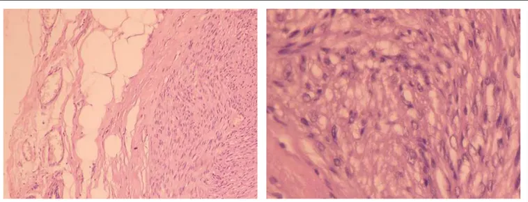
B
Figure 2. Histological sections revealing circumscribed appearance of the lesion, with proliferation of fusiform pattern and lack of atypical forms. Staining with hematoxylin and eosin (10 x and 40 x).
DISCUSSION
Even though Strong’s irst paper on breast leiomyoma was pub-lished in 1913, knowledge of the etiology of this condition remains uncertain.4 It has been taken to originate from
smooth-muscle angiomatous cells, given the “angiocentric” prolifera-tion of smooth muscle that is observed. his theory is rein-forced through the observation that blood vessels are present at locations showing defects or artefacts of histological ixation.5
Current immunohistochemical indings rule out teratogenic ori-gins. Uncertainty remains regarding theories of embryological displacement of smooth muscle cells of the areola, and regard-ing an origin from multipotent mesenchymal cells. hese were proposed in the irst half of the twentieth century by Melnick6
and Shauder.7
Breast leiomyoma occurs predominantly in women, with only one case reported in a man.2 he age of highest incidence is between
40 and 60 years. It presents as an isolated tumor of slow growth, with similar characteristics to the most common benign tumors.8-10
he presence of pain was observed in only three cases, being more frequent in tumors of areolar location due to the contraction of neoplastic muscle cells.11
Physical examination usually reveals a mobile nodule with well-deined limits and ibroelastic consistency, although sometimes it has been reported to have hardened consistency.12-14 Mammographic
images have been described as showing an isodense or hyper-dense oval mass, with outlines that are most oten circumscribed (Table 1).1,13,15-21 Microcalciications relating to leiomyoma have
never been described.15,16,22,23
he efectiveness of mammography is limited in relation to lesions measuring less than 1.0 cm and breasts with predominant glands. Sonography frequently shows a hypoechoic mass with well-deined limits and oval shape.17 Presence of lobulations has
frequently been observed. Growth parallel to the skin has been observed in 100% of the cases. No well-deined posterior acous-tic shadowing has been described.18
Magnetic resonance imaging findings were first reported by Minami et al. They described a circumscribed oval nodule, with hypersignal in T1 and T2, and homogeneous enhance-ment after gadolinium infusion. They pointed out that pres-ence of degeneration can influpres-ence the signal pattern in dif-ferent sequences, as noted in leiomyoma in other regions of the body.16
The differential diagnosis should be done in relation to lesions that have smooth muscle proliferation in the absence of epithelial or ductal structures. In this context, the lesions that comprise the differential diagnosis are angioleiomyoma, fibro-adenoma and malignant phyllodes tumor.24-26 Because mature
adipose tissue is needed to identify cases of hamartoma, this lesion does not provide difficulties in the differential diagnosis.5
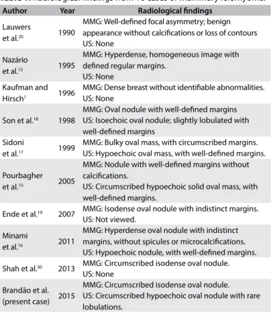
Author Year Radiological indings
Lauwers
et al.20 1990
MMG: Well-deined focal asymmetry; benign appearance without calciications or loss of contours US: None
Nazário
et al.13 1995
MMG: Hyperdense, homogeneous image with deined regular margins.
US: None Kaufman and
Hirsch1 1996
MMG: Dense breast without identiiable abnormalities. US: None
Son et al.18 1998
MMG: Oval nodule with well-deined margins US: Isoechoic oval nodule; slightly lobulated with well-deined margins
Sidoni
et al.17 1999
MMG: Bulky oval mass, with circumscribed margins. US: Hypoechoic oval mass, with well-deined margins.
Pourbagher et al.15 2005
MMG: Nodule with well-deined margins without calciications.
US: Circumscribed hypoechoic solid oval mass, with well-deined margins.
Ende et al.19 2007 MMG: Isodense oval nodule with indistinct margins.
US: Not viewed.
Minami
et al.16 2011
MMG: Hyperdense oval nodule with indistinct margins, without spicules or microcalciications. US: Hypoechoic nodule, with well-deined margins.
Shah et al.30 2013 MMG: Circumscribed isodense oval nodule.
US: None
Brandão et al. (present case) 2015
MMG: Circumscribed isodense oval nodule. US: Circumscribed hypoechoic oval nodule with rare lobulations.
Table 1. Radiological indings from 10 cases of mammary leiomyoma.
MMG = mammogram. US = ultrasonography
In cases of lesions suggestive of leiomyoma, leiomyosarcoma is the main situation that needs to be ruled out.19,27
Presence of 2-16 mitotic igures per 10 high-power igures is the main feature for diagnosing leiomyosarcoma. According to Pourbagher, presence of 1-3 mitotic igures might be considered to represent an intermediate category because of the higher risk of local recurrence and, therefore, treatment that is more radical.15
Boscaino et al. reported local recurrence in two cases initially diag-nosed as leiomyoma. Histological reevaluation of the lesions found presence of increased mitotic activity, and the lesions were reclas-siied as smooth-muscle neoplasms of undetermined prognosis.28
In patients with a conirmed diagnosis of breast leiomyoma, no cases of local recurrence have been reported to date.11-19,22-27
We reviewed the literature in MEDLINE and Lilacs using the English keywords “leiomyoma”, “ibroid tumors”, “benign tumor”, “benign neoplasms”, “breast tumor”, “breast neoplasms” and “ultra-sonography”. We found 30 case reports that described patients with leiomyoma in the breast parenchyma (Table 2).
In reviewing treatments that have been implemented, a wide range of interventions can be identiied, from lumpectomy to radi-cal mastectomy (Table 3).1,2,4-10,13,15-21,24,29 However, since the report
F = female; M = male; W = white; B = black; A = Asian; R = right; L = left; UOQ = upper outer quadrant; UIQ = upper inner quadrant; JIQ = junction of inner quadrants; JOQ = junction of outer quadrants; JUQ = junction of upper quadrants; JLI = junction of lower quadrants; LOQ = lower outer quadrant.
Table 3. Clinical indings from 20 cases of breast leiomyoma
Author Year Sex Race Age (years) Symptoms Location Size (cm) Therapy
Strong4 1913 F W 46 Discomfort UOQ R 6.0
-Schauder7 1927 F W 34 Discomfort UOQ R 3.0 Nodulectomy
Melnick6 1932 F W 45 Pain JLI R “small” Total mastectomy
Leibowich and Lenz8 1940 F W 58 Discomfort Midline 13.8 Total mastectomy
Stein9 1943 F W 54 Discomfort UIQ R 4.0 Radical mastectomy
Craig10 1947 F B 40 Pain LOQ L 10 Nodulectomy
Libcke247 1969 F W 50 Hardening JUQ R 0.5 Nodulectomy
Haagensen29 1971 F - 52 None Midline 2.5 Nodulectomy
Diaz-Arias et al.5 1989 F W 69 None UOQ R 2.0 Nodulectomy
Lauwers et al.20 1990 F B 43 Mammographic inding JUQ L 0.5 Resection with free margins
Nazario et al.13 1995 F B 53 Nodule UIQ L 10 Resection with free margins
Kaufman and Hirsch1 1996 F W 48 Nodule Midline R 1.0 Resection with free margins
Son et al.18 1998 F A 50 Pain UOQ R 1.0 Resection with free margins
Sidoni et al.17 1999 F - 48 Nodule UOQ L 4.0 Resection with free margins
Pourbagher et al. 15 2005 F - 47 Mammographic inding JIQ L 2.5 Resection with free margins
Ende et al.19 2007 F - 48 Mammographic inding JIQ L 1.2 Resection with free margins
Minami et al.16 2011 F A 63 Mammographic inding UIQ R 1.6 Excisional biopsy
Shah et al.21 2013 F W 27 Nodule UIQ L 2.0 Excisional biopsy
Strader et al.2 2013 M W 70 Nodule JOQ L 7.0 Resection with free margins
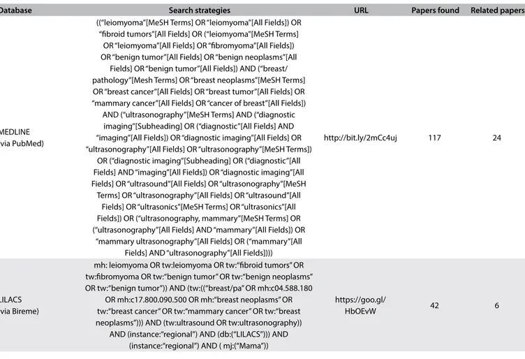
Brandão et al. (current case) 2015 F W 68 Mammographic inding JUQ L 1.4 Resection with free margins Table 2. Search of the literature in medical databases for case reports on leiomyoma in the breast parenchyma. The search was conducted on December 5, 2016
Database Search strategies URL Papers found Related papers
MEDLINE (via PubMed)
((“leiomyoma”[MeSH Terms] OR “leiomyoma”[All Fields]) OR “ibroid tumors”[All Fields] OR (“leiomyoma”[MeSH Terms]
OR “leiomyoma”[All Fields] OR “ibromyoma”[All Fields]) OR “benign tumor”[All Fields] OR “benign neoplasms”[All
Fields] OR “benign tumor”[All Fields]) AND (“breast/ pathology”[Mesh Terms] OR “breast neoplasms”[MeSH Terms] OR “breast cancer”[All Fields] OR “breast tumor”[All Fields] OR “mammary cancer”[All Fields] OR “cancer of breast”[All Fields])
AND (“ultrasonography”[MeSH Terms] AND (“diagnostic imaging”[Subheading] OR (“diagnostic”[All Fields] AND “imaging”[All Fields]) OR “diagnostic imaging”[All Fields] OR “ultrasonography”[All Fields] OR “ultrasonography”[MeSH Terms])
OR (“diagnostic imaging”[Subheading] OR (“diagnostic”[All Fields] AND “imaging”[All Fields]) OR “diagnostic imaging”[All Fields] OR “ultrasound”[All Fields] OR “ultrasonography”[MeSH
Terms] OR “ultrasonography”[All Fields] OR “ultrasound”[All Fields] OR “ultrasonics”[MeSH Terms] OR “ultrasonics”[All Fields]) OR (“ultrasonography, mammary”[MeSH Terms] OR (“ultrasonography”[All Fields] AND “mammary”[All Fields]) OR
“mammary ultrasonography”[All Fields] OR (“mammary”[All Fields] AND “ultrasonography”[All Fields])))
http://bit.ly/2mCc4uj 117 24
LILACS (via Bireme)
mh: leiomyoma OR tw:leiomyoma OR tw:“ibroid tumors” OR tw:ibromyoma OR tw:“benign tumor” OR tw:“benign neoplasms” OR tw:“benign tumor”)) AND (tw:((“breast/pa” OR mh:c04.588.180
OR mh:c17.800.090.500 OR mh:“breast neoplasms” OR tw:“breast cancer” OR tw:“mammary cancer” OR tw:“breast neoplasms”))) AND (tw:ultrasound OR tw:ultrasonography))
AND (instance:“regional”) AND (db:(“LILACS”))) AND (instance:“regional”) AND ( mj:(“Mama”))
https://goo.gl/
CONCLUSION
In conclusion, it can be said that leiomyoma in mammary tissue is an extremely rare condition. he clinical presentation does not difer from that observed in the most common benign tumors of the breast. he radiological indings are characteristically benign, which helps rule out the hypothesis of cancer. In histopathologi-cal evaluations, it is important to pay attention to the diferential diagnosis of leiomyosarcoma. he standard recommended treat-ment is local resection with free margins. In this situation, the risk of local recurrence is practically zero.
REFERENCES
1. Kaufman HL, Hirsch EF. Leiomyoma of the breast. J Surg Oncol. 1996;62(1):62-4.
2. Strader LA, Galan K, Tenofsky PL. Intraparenchymal leiomyoma of the male breast. Breast J. 2013;19(6):675-6.
3. Alawad AA. Multiple parenchymal leiomyomas of the breast in a Sudanese female. Breast Dis. 2014;34(4):165-7.
4. Strong LW. Leiomyoma of the breast. Am J Obstet. 1913;68:53-5. 5. Diaz-Arias AA, Hurt MA, Loy TS, Seeger RM, Bickel JT. Leiomyoma of
the breast. Hum Pathol. 1989;20(4):396-9.
6. Melnick PJ. Fibromyoma of the breast. Arch Pathol. 1932;14:794-8. 7. Schauder H. Über Leiomyome der Brustdrüse. Deutsche Zeitschrift für
Chirurgie. 1927;205(1):58-68. Available from: http://link.springer.com/ article/10.1007/BF02794721. Accessed in 2017 (Apr 26).
8. Leibowich RJ, Lenz G. Primary ibromyoma of the breast: Report of a case and review of the literature. American Journal of Cancer. 1940;38(1):73-5. Available from: http://cancerres.aacrjournals.org/ content/amjcancer/38/1/73.full.pdf. Accessed in 2017 (Apr 26). 9. Stein RJ. Fibroleiomyoma of the breast. Arch Pathol. 1943;33:72-4. 10. Craig JM. Leiomyoma of the female breast. Arch Pathol (Chic).
1947;44(3):314-7.
11. Ku J, Campbell C, Bennett I. Leiomyoma of the nipple. Breast J. 2006;12(4):377-80.
12. Tamir G, Yampolsky I, Sandbank J. Parenchymal leiomyoma of the breast. Report of a case and clinicopathological review. Eur J Surg Oncol. 1995;21(1):88-9.
13. Nazário AC, Tanaka CI, de Lima GR, Gebrim LH, Kemp C. Leiomyoma of the breast. A case report. Sao Paulo Med J. 1995;113(5):992-4. 14. Heyer H, Ohlinger R, Schimming A, Schwesinger G, Grunwald
S. Parenchymal leiomyoma of the breast--clinical, sonographic, mammographic and histological features. Ultraschall Med. 2006;27(1):55-8. 15. Pourbagher A, Pourbagher MA, Bal N, Oguzkurt L, Ezer A. Leiomyoma of the breast parenchyma. AJR Am J Roentgenol. 2005;185(6):1595-7. 16. Minami S, Matsuo S, Azuma T, et al. Parenchymal leiomyoma of the breast: a case report with special reference to magnetic resonance imaging indings and an update review of literature. Breast Cancer. 2011;18(3):231-6. 17. Sidoni A, Lüthy L, Bellezza G, Consiglio M, Bucciarelli E. Leiomyoma of the
breast: case report and review of the literature. Breast. 1999;8(5):289-90.
18. Son EJ, Oh KK, Kim EK, et al. Leiomyoma of the breast in a 50-year-old woman receiving tamoxifen. AJR Am J Roentgenol. 1998;171(6):1684-6. 19. Ende L, Mercado C, Axelrod D, et al. Intraparenchymal leiomyoma of
the breast: a case report and review of the literature. Ann Clin Lab Sci. 2007;37(3):268-73.
20. Lauwers G, de Roux S, Terzakis J. Leiomyoma of the breast. Arch Anat Cytol Pathol. 1990;38(3):108-10.
21. Shah SD, Gupta A, Roy S, Mukhopadhyay S. Intraparenchymal leiomyoma of the breast: a case report. Indian J Surg. 2013;75(Suppl 1):88-9. 22. Manna P, Giuseppetti GM, Latini L, Baldassarre S, Antognoli S. [A case
of leiomyoma of the breast]. Radiol Med. 1993;86(1-2):155-8. 23. Kotsuma Y, Wakasa K, Yayoi E, et al. A case of leiomyoma of the breast.
Breast Cancer. 2001;8(2):166-9.
24. Libcke JH. Leiomyoma of the breast. J Pathol. 1969;98(1):89-90. 25. Roncaroli F, Rossi R, Severi B, Martinelli GN, Eusebi V. Epithelioid
leiomyoma of the breast with granular cell change: a case report. Hum Pathol. 1993;24(11):1260-3.
26. Magro G, Michal M, Bisceglia M. Benign spindle cell tumors of the mammary stroma: diagnostic criteria, classiication, and histogenesis. Pathol Res Pract. 2001;197(7):453-66.
27. Stafyla VK, Gauvin JM, Farley DR. A 53-year-old woman with a leiomyosarcoma of the breast. Curr Surg. 2004;61(6):572-5.
28. Boscaino A, Ferrara G, Orabona P, et al. Smooth muscle tumors of the breast: clinicopathologic features of two cases. Tumori. 1994;80(3):241-5.
29. Haagensen CD. Nonepithelial neoplasms of the breast. In: Haagensen CD (editor). Diseases of the Breast. 2nd ed. Saunders: Philadelphia; 1971.
p. 292-325.
30. Weldon C, Jones B, Daroca P, Beech D. Breast leiomyoma. J La State Med Soc. 1998;150(8):367-70.
Sources of funding: None
Conlict of interest: None
Date of irst submission: November 26, 2016
Last received: December 30, 2016
Accepted: January 4, 2017
Address for correspondence:
Rodrigo Gregório Brandão
Departamento de Ginecologia, Disciplina de Mastologia, Universidade Federal de São Paulo (UNIFESP)
Rua Napoleão de Barros, 608 São Paulo (SP) - Brazil CEP: 040002-233
Tel: (+55 11) 5576-4848 (Ramal 2856) Cel. (+ 55 11) 98161-7511
Fax: (+55 11) 4521-2604