Sao Paulo Med J. 201X; XXX(X):xxx-xxx 1
CASE REPORT
DOI: 10.1590/1516-3180.2016.0351280117Metastatic adenocarcinoma involving the right ventricle and
pulmonary artery leading to right heart failure: case report
Adenocarcinoma metastático envolvendo o ventrículo direito e artéria pulmonar
levando a insuiciência cardíaca direita: relato de caso
Turgut Karabag
I, Caner Arslan
II, Turab Yakısan
III, Aziz Vatan
IV, Duygu Sak
VIstanbul Education and Research Hospital, Istanbul, Turkey
ABSTRACT
CONTEXT: Obstruction of the right ventricular outlow tract due to metastatic disease is rare. Clinical recogni-tion of cardiac metastatic tumors is rare and continues to present a diagnostic and therapeutic challenge.
CASE REPORT: We present the case of a patient who had severe respiratory insuiciency and whose clini-cal examinations revealed a giant tumor mass extending from the right ventricle to the pulmonary artery. We discuss the diagnostic and therapeutic options.
CONCLUSION: In patients presenting with acute right heart failure, right ventricular masses should be kept in mind. Transthoracic echocardiography appears to be the most easily available, noninvasive, cost-efective and useful technique in making the diferential diagnosis.
RESUMO
CONTEXTO: A obstrução da via de saída do ventrículo direito devido a doença metastática é rara. O re-conhecimento clínico de tumores cardíacos metastáticos é raro e continua a apresentar um desaio diag-nóstico e terapêutico.
RELATO DO CASO: Apresentamos o caso de um paciente com insuiciência respiratória grave e cujos exames clínicos revelaram massa de tumor gigante, estendendo-se desde o ventrículo direito até a artéria pulmonar. Discutimos as opções diagnósticas e terapêuticas.
CONCLUSÃO: Em pacientes com insuiciência cardíaca direita aguda, massas do ventrículo direito devem ser mantidas em mente. Ecocardiograia transtorácica parece ser a técnica mais facilmente disponível, não invasiva, custo-efetiva e útil no diagnóstico diferencial.
IMD. Associate Professor, Department of Cardiology, Istanbul Education and Research Hospital, Istanbul, Turkey.
IIMD. Associate Professor, Department of Cardiovascular Surgery, Cerrahpasa Medical School, Istanbul University, Istanbul, Turkey. IIIMD. Resident Physician, Department of Cardiology, Istanbul Mehmet Akif Ersoy Education and Research Hospital, Istanbul, Turkey. IVMD. Resident Physician, Department of Emergency Medicine, Istanbul Education and Research Hospital, Istanbul, Turkey.
VMD. Resident Physician, Department of Internal Medicine, Istanbul Education and Research Hospital, Istanbul, Turkey.
KEY WORDS: Neoplasm metastasis. Adenocarcinoma. Echocardiography. Heart failure. Pulmonary artery.
PALAVRAS-CHAVE: Metástase neoplásica. Adenocarcinoma. Ecocardiograia. Insuiciência cardíaca. Artéria pulmonar.
CASE REPORT | Karabag T, Arslan C, Yakısan T, Vatan A, Sak D
2 Sao Paulo Med J. 201X; XXX(X):xxx-xxx INTRODUCTION
Tumors involving the heart are more commonly metastatic than primary, and the prognosis for metastatic tumors in the heart is extremely poor. Involvement of the right heart is more common than that of the let heart and the clinical course is usually silent in most patients.1 his report presents the case of a 67-year-old
male patient with no previous diseases, in whom a metastatic right cardiac tumor invading the right ventricular outlow tract and the pulmonary artery was detected. We also discuss the diag-nostic and therapeutic techniques.
CASE REPORT
A 67-year-old male patient was admitted to the emergency depart-ment with a 15-day history of progressive fatigue, shortness of breath and respiratory insuiciency. he patient stated that he had not had any complaints until 15 days before admission, and no previous dis-eases had been documented. He reported having progressive short-ness of breath, which irst appeared during exercise 15 days earlier and had then even become apparent at rest. he patient reported having made intermittent use of paracetamol for headache for years. He had a history of smoking and intermittent alcohol use.
On admission, the patient’s general condition was poor, with severe shortness of breath. He was orthopneic and was using acces-sory muscles while breathing. His blood pressure was 80/65 mmHg, and his pulse was 128/minute. His breathing sounds were rough but no rales or rhonchi were heard. Heart sounds were tachycardic, and there was a 2/6 systolic murmur heard in the tricuspid area. He had 1+ edema of both feet. A chest x-ray showed increased vascularization, a dilated pulmonary artery and a cardiothoracic index of > 1. An electrocardiogram revealed an incomplete right bundle branch block and a negative T wave in the V1-3 leads.
Laboratory tests conducted on venous blood sample showed that blood glucose was 124 mg/dl (normal range 74-100); urea, 90.2 mg/dl (normal range 0-50); creatinine, 1.6 mg/dl (normal range 0-1.2); sodium, 134 mg/dl (normal range 132-146); and potassium, 5.2 mg/dl (normal range 3.5-5.5). Liver function tests revealed elevated values: aspartate transaminase, 3418 U/l (nor-mal range 0-50); alanine transaminase, 1204 U/l (nor(nor-mal range 0-50); alkaline phosphatase, 276 U/l (normal range 30-120); and lipase, 92 U/l (normal range 0-67). Total bilirubin and direct bili-rubin levels were slightly elevated: 8.4 g/dl (normal range 6.6-8.3) and 0.24 mg/dl (normal range 0-0.2), respectively. he hemo-gram was unremarkable except for leukocytosis. he prothrom-bin time was 21.2 seconds and was slightly lengthened (normal range 10.4-14.6 seconds). Cardiac markers were also slightly ele-vated: troponin I, 0.45 ng/ml (normal range 0-0.001); and creati-nine kinase-myocardial band, 6.4 ng/ml (normal range 0.6-6.3). An arterial blood gas test revealed pH of 7.49, sO2 of 87.9%, pO2 of 57.2 mmHg and pCO2 of 22.5 mmHg.
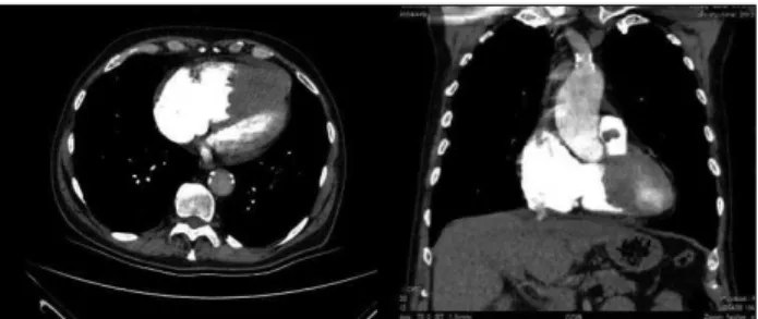
In the light of these data, the patient received an initial diag-nosis of acute toxic hepatitis, taking into consideration the history of pulmonary embolism and recent use of medication. A complete abdominal ultrasound examination revealed a slightly enlarged liver. A contrast computed tomography pulmonary angiogram was performed and did not show any thrombus in the major arter-ies or their branches. On the other hand, a illing defect from the right ventricle to the pulmonary artery, which was interpreted to represent a thrombus, was observed (Figure 1).
herefore, transthoracic echocardiography was performed, revealing a considerably enlarged right ventricle and a lattened interventricular septum, which had shited towards the let ven-tricle. A 6 cm x 5 cm mass, consistent with thrombus echogenicity, was detected inside the right ventricle. It extended to the pulmo-nary artery, with invasion of the pulmopulmo-nary valve, giving rise to to-and-fro motion in each systole (Figure 2).
Right ventricular systolic function was considerably decreased (tricuspid annular plane systolic excursion, TAPSE: 1.6 cm). Let ventricular systolic function was slightly decreased. Severe tricuspid and mitral insuiciency was present. Pulmonary artery systolic pressure was elevated (60 mmHg).
hus, the patient was referred for consultation in the depart-ment of cardiovascular surgery and was immediately scheduled for emergency surgery. During surgery, it was found that the mass was a tumor. he tumor had invaded the anterior wall of the right ven-tricle, interventricular septum and right ventricular outlow tract. he tumor was removed from the interventricular septum and the
Figure 1. Contrast computed tomography pulmonary
angiogram showing a dilated right ventricle and a illing defect, both in the right ventricle and in the pulmonary artery.
Figure 2. Echocardiogram showing the mass inside the right ventricle, which extended to the pulmonary artery and invaded the pulmonary valve, in apical four-chamber view (A) and parasternal short-axis view (B).
A B
Karabag T, Arslan C, Yakısan T, Vatan A, Sak D
Metastatic adenocarcinoma involving the right ventricle and pulmonary artery leading to right heart failure: case report | CASE REPORT
Sao Paulo Med J. 201X; XXX(X):xxx-xxx 3
anterior wall of the right ventricle. he resulting ventricular septal defect was closed with a patch (Figure 3a) and the tricuspid valve was replaced (Figure 3b). Intraoperative transesophageal echocar-diography revealed only moderate mitral insuiciency, and there-fore mitral valve replacement was not considered.
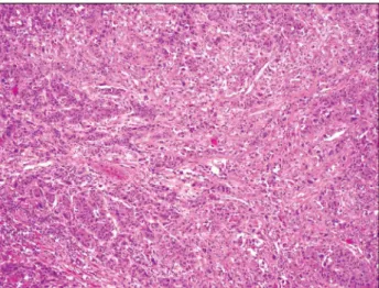
he pathology report showed that the mass was a poorly dif-ferentiated metastatic adenocarcinoma, which had probably origi-nated from the lungs. It exhibited partial neuroendocrine difer-entiation (Figure 4).
he patient’s postoperative hemodynamic condition did not improve, despite inotropic support. He developed cardiopulmo-nary arrest on postoperative day 3 and died, despite resuscita-tion attempts.
DISCUSSION
Tumors that are metastatic to the heart are rare. Cardiac involve-ment at autopsy has been described in 6% to 20% of patients with malignant neoplasms.2 he lungs have been reported to be
the most common primary origin of metastatic neoplasms, fol-lowed by nonsolid neoplasms such as lymphoma or leukemia and tumors of the liver and colon, respectively.3 he epicardium is the
most commonly involved site, followed by the myocardium and the endocardium.4
he systematized results from searching the literature through the main databases are presented in Table 1. he data in the litera-ture show that diagnosing and treating tumors located in the right ventricular outlow tract is extremely challenging. he symptoms usually depend on the size and location of the tumor. he most common symptoms include shortness of breath, syncope and cya-nosis.5 Some patients may present with nonspeciic symptoms and
tumors are usually detected incidentally. Inability to measure blood pressure with the patient in the seated position is likely to indicate occlusion of the pulmonary artery by the tumor.5 hese tumors may
reach huge sizes, thus resulting in occlusion of the right ventricular outlow tract and right ventricular volume overload and dilation, which is likely to result in severe right ventricular failure.6
In addi-tion, neoplasms may cause fatal clinical consequences including arrhythmia, acute heart failure and sudden death. Tumors may disintegrate into fragments, thus embolizing the pulmonary vas-cular bed. Accordingly, patients may present with clinical signs and symptoms of pulmonary embolism. Emergency surgery should be considered upon detection of right ventricular outlow tract obstruction.4 Early diagnosis enables timely surgical treatment,
thus increasing survival. In addition, the data in the literature indi-cate that postoperative adjuvant chemotherapy may be useful in patients with adenocarcinomas of gastrointestinal origin that are metastatic to the heart.7,8
In the case presented here, the patient presented with a 15-day history of progressive shortness of breath. He showed clear signs and symptoms of right ventricular failure. A diagnosis of pul-monary embolism was initially considered, but irstly contrast computed tomography and then transthoracic echocardiography revealed an intracardiac tumor. Upon detection of obliteration of the right ventricular outlow tract and invasion of the pulmonary valve by the tumor, the patient was sent for emergency surgery. Despite successful excision of the mass, the patient’s postoperative hemodynamic condition did not improve, even though inotropic support was provided, and he subsequently died.
Figure 4. Adenocarcinoma iniltration to the myocardium. Tumor cells form coarse solid trabecular structures (100 x; hematoxylin and eosin).
Table 1. Systematic search of the literature performed in April 2016
Database Search strategies Found Used
MEDLINE (via PubMed)
(metastatic adenocarcinoma)
and (heart) 1,388 4
LILACS (via Bireme)
(metastatic adenocarcinoma)
and (heart) and (pulmonary) 0 0
Cochrane Library
(metastatic adenocarcinoma)
and (heart) and (failure) 30 0 Figure 3. Resection of tumor from the heart, with closure
of the ventricular septal defect using a patch (A) and replacement of the tricuspid valve (B).
A
B
Metastatic adenocarcinoma involving the right ventricle and pulmonary artery leading to right heart failure: case report
CASE REPORT | Karabag T, Arslan C, Yakısan T, Vatan A, Sak D
4 Sao Paulo Med J. 201X; XXX(X):xxx-xxx
Although the tumor was diagnosed pathologically, the origin of the tumor could not be identiied. Pathologically, the tumor was likely to have originated from the lungs, gastrointestinal tract or pancreaticobiliary system. However, this could not be conirmed because the patient’s family did not give permission for an autopsy.
CONCLUSION
Right ventricular metastatic tumors are relatively rarer than other types. In patients presenting with shortness of breath with rapid progression and right heart failure, right ventricular obstruc-tion should be kept in mind, along with other possible diagno-ses. Transthoracic echocardiography appears to be the most eas-ily available, noninvasive, cost-efective and useful technique in making the diferential diagnosis.
REFERENCES
1. Patel SA, Herfel BM, Nolan MA. Metastatic colon cancer involving the right atrium. Tex Heart Inst J. 2012;39(1):79-83.
2. Ewald FW Jr, Scherf AH. A 60-year-old man with a malignant tumor of the upper airway and unexplained respiratory failure. Chest. 1997;111(1):239-41.
3. Abraham KP, Reddy V, Gattuso P. Neoplasms metastatic to the heart:
review of 3314 consecutive autopsies. Am J Cardiovasc Pathol. 1990;3(3):195-8.
4. Hayashi S, Isobe I, Hayashi M, et al. Successful resection of intracardiac invasive thymoma with right ventricular inlow tract occlusion. Intern
Med. 2000;39(2):138-42.
5. Soejima Y, Niva A, Tanaka M, et al. Large right ventricular myxoma in a 79-year-old male. Intern Med. 1996;35(5): 380-2.
6. Bouzas-Mosquera A, Flores-Rios X, Aldama G. Primary cardiac
rhabdomyosarcoma causing obstruction to the right ventricular outlow. Eur J Echocardiogr. 2007;8(5):406-7.
7. Koizumi J, Agematsu K, Ohkado A, Shiikawa A, Uchida T. Solitary cardiac metastasis of rectal adenocarcinoma. Jpn J Thorac Cardiovasc Surg.
2003;51(7):330-2.
8. Makhija Z, Deshpande R, Desai J. Unusual tumours of the heart: diagnostic and prognostic implications. J Cardiothorac Surg. 2009;4:4.
This case was presented at EuroEcho-Imaging 2016 in Leipzig, Germany.
Sources of funding: None
Conlict of interest: None
Date of irst submission: December 22, 2016
Last received: January 14, 2017
Accepted: January 28, 2017
Address for correspondence:
Turgut Karabag
Department of Cardiology, Istanbul Education and Research Hospital Kasap İlyas Mah., Org. Abdurrahman Naiz Gürman Cd., PK: 34098 Istanbul — Turkey
Tel. +90 (212) 459 62 30
E-mail: [email protected]
Karabag T, Arslan C, Yakısan T, Vatan A, Sak D