J of Evolution of Med and Dent Sci/ eISSN- 2278-4802, pISSN- 2278-4748/ Vol.4/ Issue 12/Feb 09, 2015 Page 1970
ROLE OF ULTRASONOGRAPHY & COLOR DOPPLER IN THE EVALUATION OF
THYROID NODULES WITH HISTOPATHOLOGICAL CORRELATION
K. J. S. S. Raghu Teja1, Anil Kumar Sakalecha2, Purnima Hedge3, T. N. Suresh4, P. N. Sreeramulu5,
B. N. Kishore Kumar6, Haritha. P7
HOW TO CITE THIS ARTICLE:
K. J. S. S. Raghu Teja, Anil Kumar Sakalecha, Purnima Hegde, T. N. Suresh, P. N. Sreeramulu, B. N. Kishore Kumar, Haritha. P. Role of Ultrasonography & Color Doppler in the Evaluation of Thyriod Nodules with Histo-pathological Correlation. Journal of Evolution of Medical and Dental Sciences 2015; Vol. 4, Issue 12,
February 09; Page: 1970-1985, DOI: 10. 14260/jemds/2015/285
ABSTRACT: PURPOSE OF THE STUDY: 1. To identify morphologic patterns on sonography, those are predictive of benign thyroid nodules and malignant thyroid nodules. 2. To evaluate the efficacy of sonography in differentiating benign and malignant thyroid nodules in comparison with histopathology. MATERIALS AND METHODS: HRS of thyroid was performed in 60 patients with clinically palpable STN, referred to Department of Radiodiagnosis, R. L. Jssalappa Hospital and Research Centre, Tamaka, Kolar, Karnataka over a period of 24months, using SIEMENS ACCUSON X 300 and SIEMENS G 50 with 5-10 MHz transducers. Thyroid sonographic findings relevant to benign or malignant thyroid nodules were recorded and these findings were compared with histopathology reports. RESULTS: Out of 60 cases of solitary thyroid nodules evaluated at sonography, 41 were diagnosed to be benign thyroid nodules. 19 were malignant thyroid nodules. After histopathological evaluation, 43 cases were found to be benign thyroid nodules and 17 were malignant thyroid nodules. Among benign thyroid nodules, follicular adenoma 55.8% (24 cases)was most common followed by nodular hyperplasia 27.9% (12 cases)and colloid nodule 16.3%(7 cases). Among malignant nodules, papillary carcinoma 88.2% (15 cases) was most common followed by follicular carcinoma 11.8% (2 cases). CONCLUSION: Thyroid nodules were more common in the females of age group 31 – 45 years. Sonography is a safe, fairly accurate investigation to differentiate benign from malignant thyroid nodules with sensitivity of 82.3 % and specificity of 88.3 %.
KEYWORDS: solitary thyroid nodule; high resolution sonography; histopathology.
INTRODUCTION: Thyroid nodule is a discrete lesion and a common clinical condition. Solitary thyroid nodules are commonly being present in up to 50% of the elderly population. Thyroid nodules are found in up to 20% clinically by palpation and in up to 70% on sonographic studies.1 The thyroid
gland is unique among endocrine glands, in that it is the first endocrine gland to appear in the fetus. It is the largest of all endocrine glands (Weighing about 25 gm) and is the only one which is amenable to direct physical examination because of its superficial location.2 One or more additional
non-palpable thyroid nodules may be found by sonography in about 50% of patients with a clinically palpable solitary nodule and they are also incidentally detected by imaging studies performed for various reasons.3
STN is one of the commonest thyroid disorders. STN is common in females than males with a ratio of 5:1 and prevalence mainly depends on age, sex, iodine intake, diet and environmental exposure, though STN is common in women, malignancy in STN is common in men.4
J of Evolution of Med and Dent Sci/ eISSN- 2278-4802, pISSN- 2278-4748/ Vol.4/ Issue 12/Feb 09, 2015 Page 1971 clinically. Many sonographic features have been described to differentiate benign nodules from malignant nodules.5 For each thyroid nodule, gray scale and color Doppler ultrasound are used to
evaluate the sonographic features, which include shape, echogenicity (Hypoechoic or isoechoic or hyperechoic), composition (Solid, purely cystic, cyst with thin septa, mixed and spongiform), margin, halo, as well as presence or absence of coarse / microcalcification, vascularity of the nodule and presence or absence of regional lymphnodes.6
Sonographic features which may be highly predictive of benign nodule on the basis of certain characteristics which includes wider than tall in shape, hyperechoic or isoechoic, solid nodule or purely cystic / cystic with thin septa / mixed / spongiform, well-defined margins, complete peripheral halo, coarse or egg shell peripheral calcification and perinodular vascularity.7
Sonographic features which may be highly predictive of malignant nodule are taller than wide shape, predominantly hypoechoic or hyperechoic, solid nodule, ill-defined margins, incomplete peripheral halo, microcalcification, intranodular vascularity, regional lymphadenopathy and local invasion of adjacent structures.8
Prediction of malignancy using sonography still remains difficult. Since there is overlap of sonographic features between benign and malignant thyroid nodules, as it is not possible to distinguish a benign follicular adenoma from follicular carcinoma by sonography, FNAC, core biopsy and frozen sections, as vascular and capsular invasion can only be evaluated on histological specimen, so sonographic features are usually corroborated with histopathology results in differentiating various thyroid nodules.9
The goal in evaluating a thyroid nodule is to determine whether it is benign nodule or malignant nodule so that patients can undergo treatment at an earlier stage to reduce possible morbidity and mortality due to the disease, while avoiding unnecessary tests and surgery in patients with benign nodules.11
MATERIALS AND METHODS: All patients referred for HRS of the thyroid with clinically suspected STN to department of Radiodiagnosis at R. L. Jalappa Hospital and research centre, Kolar are taken for the study.
Total 60 cases who were diagnosed clinically with solitary thyroid nodule in R. L. Jalappa Hospital and research centre, Kolar, over a 24 months study period from December 2010 to November 2012.
INCLUSION CRITERIA:
All patients with clinical diagnosis of solitary thyroid nodule who are 18 yrs of age and above.
EXCLUSION CRITERIA:
Patient with diffuse thyroid disease. Patients with multinodular goiter.
Method of Performing Examination: Sonography of neck was performed by using SIEMENS ACCUSON X 300 & SIEMENS G 50 with 5-10 MHz transducers.
J of Evolution of Med and Dent Sci/ eISSN- 2278-4802, pISSN- 2278-4748/ Vol.4/ Issue 12/Feb 09, 2015 Page 1972 away from sternal notch area. The entire thyroid was scanned from upper to the lower pole and the isthmus in the longitudinal and transverse planes.
Sonographic features of thyroid nodule were noted in the prepared proforma, with these sonographic features the thyroid nodule was predicted as benign or malignant, later these patients underwent surgery.
The data collected from these patients was analyzed using descriptive tools like specificity, sensitivity and predictive value of sonographic diagnosis on comparison with histopathological diagnosis.
RESULTS: The present study deals with results of sonography of the thyroid nodule regarding prediction of benign and malignant nodule in comparison with histopathological diagnosis.
Number of cases (n=60)
Percentage (%)
Benign 41 68.3%
Malignant 19 31.7%
Total 60 100
TABLE 1: INCIDENCE OF NODULES ON SONOGRAPHY
Age (years)
Number of cases (n= 60)
Percentage (%)
21-30 13 21.7%
31-40 24 40%
41-50 14 23.3%
51-60 8 16%
61& above 1 2%
Total 60 100%
TABLE 2: AGE INCIDENCE OF STN ON SONOGRAPHY
Sex Number of cases(n=50) Percentage (%)
Female Male
51 9
85% 15%
Total 60 100%
J of Evolution of Med and Dent Sci/ eISSN- 2278-4802, pISSN- 2278-4748/ Vol.4/ Issue 12/Feb 09, 2015 Page 1973
Location Number of cases(n=50) Percentage (%)
Right lobe 39 65%
Left lobe 19 31.7%
Isthmus 2 3.3%
Total 60 100%
TABLE 4: LOCATION OF NODULES ON SONOGRAPHY
SL. No.
Morphologic Pattern
No. of
cases Benign Malignant
1. Spongiform 2 2 -
2. Cyst with colloid clot 3 3 -
3. Giraffe - - -
4. White knight 13 12 1
5. Red light 4 1 3
6. Hypoechoic 13 1 12
7. Isoechoic without halo 8 2 6
8. Isoechoic with halo 9 7 2
9. Ring of fire 3 2 1
10. Mixed 2 1 1
TABLE 5: MORPHOLOGIC PATTERNS
ON SONOGRAPHY AS DESCRIBED(By waters et al)5
Category AP/TRANS ratio Total (%) < 1cm > 1cm
Benign 35(85.4%) 6(14.6%) 41(100%)
Malignant 4(21.1%) 15(78.9%) 19(100%)
TABLE: 6 DISTRIBUTION OF AP/TRANS RATIO ACCORDING TO SONOGRAPHIC DIAGNOSIS
Category Echotexture Total
(%) Hyperechoic Isoechoic Hypoechoic Anechoic
Benign 14 (34%) 17(41.6%) 3(7.3%) 7(21.2%) 41(100%)
Malignant 1(5.2%) 4(21.2%) 14(73.6%) - 19(100%)
J of Evolution of Med and Dent Sci/ eISSN- 2278-4802, pISSN- 2278-4748/ Vol.4/ Issue 12/Feb 09, 2015 Page 1974
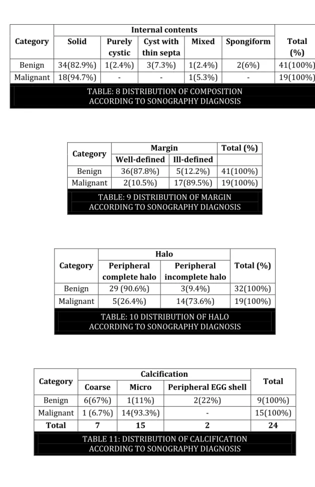
Category
Internal contents
Total (%) Solid Purely
cystic
Cyst with thin septa
Mixed Spongiform
Benign 34(82.9%) 1(2.4%) 3(7.3%) 1(2.4%) 2(6%) 41(100%)
Malignant 18(94.7%) - - 1(5.3%) - 19(100%)
TABLE: 8 DISTRIBUTION OF COMPOSITION ACCORDING TO SONOGRAPHY DIAGNOSIS
Category Margin Total (%) Well-defined Ill-defined
Benign 36(87.8%) 5(12.2%) 41(100%) Malignant 2(10.5%) 17(89.5%) 19(100%)
TABLE: 9 DISTRIBUTION OF MARGIN ACCORDING TO SONOGRAPHY DIAGNOSIS
Category
Halo
Total (%) Peripheral
complete halo
Peripheral incomplete halo
Benign 29 (90.6%) 3(9.4%) 32(100%)
Malignant 5(26.4%) 14(73.6%) 19(100%)
TABLE: 10 DISTRIBUTION OF HALO ACCORDING TO SONOGRAPHY DIAGNOSIS
Category Calcification Total Coarse Micro Peripheral EGG shell
Benign 6(67%) 1(11%) 2(22%) 9(100%)
Malignant 1 (6.7%) 14(93.3%) - 15(100%)
Total 7 15 2 24
J of Evolution of Med and Dent Sci/ eISSN- 2278-4802, pISSN- 2278-4748/ Vol.4/ Issue 12/Feb 09, 2015 Page 1975
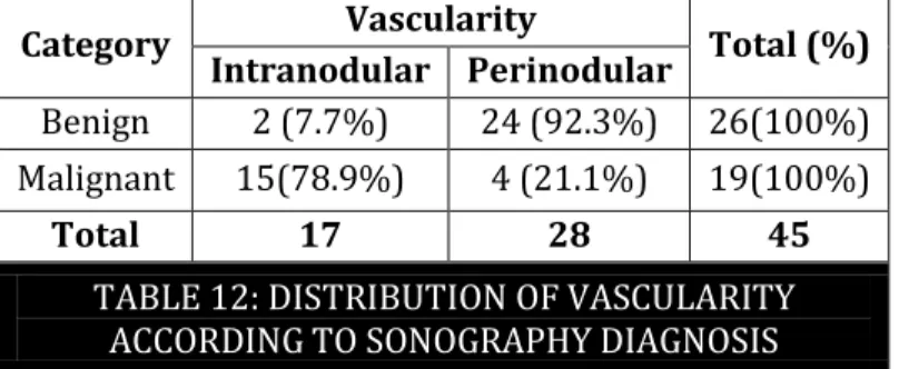
Category Vascularity Total (%) Intranodular Perinodular
Benign 2 (7.7%) 24 (92.3%) 26(100%)
Malignant 15(78.9%) 4 (21.1%) 19(100%)
Total 17 28 45
TABLE 12: DISTRIBUTION OF VASCULARITY ACCORDING TO SONOGRAPHY DIAGNOSIS
HISTOPATHOLOGICAL DIAGNOSIS
Benign nodules Number of cases (35) Percentage (100%)
Follicular adenoma 24 55.8%
Nodular hyperplasia 12 27.9%
Colloid nodule 7 16.3%
TOTAL 43 100%
TABLE 13: TYPES OF BENIGN NODULES
Malignant lesions Number of cases(15) Percentage (100%)
Papillary carcinoma 15 88.2%
Follicular carcinoma 2 11.8%
Medullary carcinoma - -
TABLE 14: TYPES OF MALIGNANT NODULES
H R S
HISTOPATHOLOGY
Benign Malignant TOTAL
Benign 38 3 41
Malignant 5 14 19
TOTAL 43 17 60
TABLE 15: COMPARISION OF SONOGRAPHY WITH HISTOPATHOLOGY
SENSITIVITY- 82.3% SPECIFICITY- 88.3%
J of Evolution of Med and Dent Sci/ eISSN- 2278-4802, pISSN- 2278-4748/ Vol.4/ Issue 12/Feb 09, 2015 Page 1976
SONOGRAPHY MORPHOLOGIC FEATURES OF BENIGN NODULES
Case 1: 45 year male with colloid clot. Transverse sonographic image of the left thyroid lobe shows cyst with colloid clot (arrow) which does not shows vascularity on color Doppler image and also showing comet- tail artifacts (arrow head).
Case 2: 30 year female. Transverse gray scale sonographic image of the right thyroid lobe shows mixed nodule (Both cystic and solid) suggestive of benign nodule.
Case 1
J of Evolution of Med and Dent Sci/ eISSN- 2278-4802, pISSN- 2278-4748/ Vol.4/ Issue 12/Feb 09, 2015 Page 1977
Case 3: 32 year female with follicular adenoma. Transverse Doppler sonographic image of the right thyroid lobe shows hypervascular nodule.
Case 4: 21 year female with nodular hyperplasia. Transverse gray scale sonographic image of the right thyroid lobe shows circumscribed homogenous isoechoic nodule with complete hypoechoic peripheral halo.
Case 3
J of Evolution of Med and Dent Sci/ eISSN- 2278-4802, pISSN- 2278-4748/ Vol.4/ Issue 12/Feb 09, 2015 Page 1978
Case 5: 35 year female. Transverse gray scale sonographic image of the left thyroid lobe shows peripheral egg shell calcification (Arrow) suggestive of benign nodule.
Case 6: 42 year male. Longitudinal gray scale sonographic image of the left thyroid lobe shows circumscribed homogenous isoechoic nodule with coarse calcifications (arrow) suggestive of benign nodule.
Case 5
J of Evolution of Med and Dent Sci/ eISSN- 2278-4802, pISSN- 2278-4748/ Vol.4/ Issue 12/Feb 09, 2015 Page 1979
Case 7: 55 year female with hyperplastic nodule. Longitudinal Doppler image of the left thyroid lobe shows peripheral hypervascularity.
SONOGRAPHIC MORPHOLOGIC FEATURES OF MALIGNANT NODULES
Case 8: 41 years female with papillary carcinoma. Transverse gray scale sonographic image of the right thyroid lobe shows a circumscribed hypoechoic nodule with incomplete peripheral halo (arrow).
Case 7
J of Evolution of Med and Dent Sci/ eISSN- 2278-4802, pISSN- 2278-4748/ Vol.4/ Issue 12/Feb 09, 2015 Page 1980
Case 9: 62 year male with papillary carcinoma. Longitudinal gray scale sonographic image of the right thyroid lobe shows homogenous hypoechoic nodule with ill-defined borders and shows internodular vascularity.
Case 10: 45 year female with papillary carcinoma. Longitudinal gray scale sonographic image of the left thyroid lobe shows micro-calcifications and refractive shadows from edge of a solid nodule (arrow).
Case 9
J of Evolution of Med and Dent Sci/ eISSN- 2278-4802, pISSN- 2278-4748/ Vol.4/ Issue 12/Feb 09, 2015 Page 1981
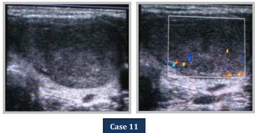
Case 11: 25 year female with follicular neoplasm. Longitudinal sonographic image of the left thyroid lobe shows solid homogenous egg shaped nodule with thin capsule with intranodular vascularity on color Doppler image which proved to be follicular carcinoma on histopathology.
HISTOPATHOLOGY IMAGES
FIG. 1: Microphotography showing Nodular Hyperplasia.
FIG. 2: Microphotography showing Follicular adenoma. Case 11
Fig. 1
J of Evolution of Med and Dent Sci/ eISSN- 2278-4802, pISSN- 2278-4748/ Vol.4/ Issue 12/Feb 09, 2015 Page 1982
FIG. 3: Microphotography showing Papillary Carcinoma.
DISCUSSION: Sonography is a choice of investigation in evaluation of thyroid nodules. The high resolution of ultrasound has resulted in discovery of large number of thyroid nodules which are obscured clinically. Many sonographic features have been described to differentiate benign and malignant nature of the nodule. The present study was done on 60 patients.
The study done by Burch HB et al showed that highest age incidence thyroid nodule is between 21-50 years, the maximum being 31-40 years.12 In the present study the commonest age
group affected was 31-40 years.
The study done by Tsegaye et al showed that females are more commonly affected than males with male to female ratio was about 1:4.1.13 In present study female predominance (85%) was noted
with a male to female ratio of 1:5.6 and correlates with the above study.
The study done by Horwath E et al showed that 71% of nodules commonly occur in right thyroid lobe.14 In the present study 65% of nodules occurred in right thyroid lobe and followed by
31.7% in left thyroid lobe.
The study done by Watters et al showed that four specific morphologic features such as spongiform configuration, Cyst with colloid clot, Giraffe pattern and diffuse hyperechogenicity are predictive of benign thyroid nodules and had 100% specificity for benignity that do not require biopsy.5 In the present study, nodules with spongiform configuration, cyst with colloid clot and
diffuse hyperechogenicity were diagnosed as benign lesions on HRS and were confirmed by histopathology reports which correlates with the above study. There is no giraffe pattern in the present study.
The study done by Grabe SK et al showed that AP/TRANS ratio < 1was noted in 95.3 %of benign nodules and AP/TRANS ratio > 1 was noted in 83.7 % of malignant nodules.15 In present study
AP/TRANS ratio < 1 was noted in 85.4 % of benign nodules and AP/TRANS ration > 1 was noted in 78.9 % of malignant nodules which correlates with the above study.
The study done by Kim et al showed that 55% of hyperechoic, 42% isoechoic nodules and 3% of hypoechoic are benign nodules and 80% of hypoechoic nodules and 20% of isoechoic nodules are malignant nodules.16 In the present study, 34% of hyperechoic nodules, 41.6% of isoechoic nodules
and 7.3% of hypoechoic nodules were benign.73.6%of hypoechoic nodules and 21.2% of isoechoic nodules were malignant.
J of Evolution of Med and Dent Sci/ eISSN- 2278-4802, pISSN- 2278-4748/ Vol.4/ Issue 12/Feb 09, 2015 Page 1983 The study done by Hegde A et al showed that most benign and malignant nodules are solid, making it difficult to use this criterion for differentiating the two.17 In present study shows 82.9% of
benign nodules were solid, whereas 94.7% of malignant nodules were solid.
The study done by Papini E et al showed that nodule had well defined margin were benign and nodule had ill-defined margin were mostly malignant nodules. 18 In the present study, 87.8% of
nodules had well-defined margins and 5% of nodule had ill-defined margins were benign nodules and 89.5% of the nodules had ill-defined margins and 10.5% well-defined margins were malignant nodules.
The study done by Wienke JR et al showed that 93.7% had peripheral complete halo in benign nodules and 83.3 % had peripheral incomplete halo in malignant nodules.19 In the present study
90.6% of the nodules which had peripheral complete halo were benign nodules and 73.6% of the nodules which had peripheral incomplete halo were malignant nodules.
The study done by Wienke JR et al showed that coarse calcification seen in 72% and peripheral egg shell calcification seen in 35% of benign nodules, whereas microcalcification seen in 16% of malignant nodules.19 In present study coarse calcification were seen in 67 % and peripheral
egg shell calcification in 22 % of benign nodules and microcalcification was seen in 93.3 % of malignant nodules.
The study done by Propper RA et al showed that perinodular vascularity is seen in benign nodules and intranodular vascularity is seen in malignant nodules.20 In the present study perinodular
vascularity was seen in 92.3 % of benign nodules and 21.1% of malignant nodules, whereas intranodular vascularity was seen in 78.9 % of malignant nodules.
In the present study of 60 cases of solitary thyroid nodules, Sonographic diagnosis was made as benign in 41 cases, 38 cases were confirmed as benign by histopathology. Remaining 3 cases were diagnosed as malignant by histopathology. Sonologically these 3 cases showed features of benign nodule such as AP/TRANS ratio < 1, isoechoic nodule, well defined, peripheral thick complete halo.
In the present study of 60 cases, Sonographic diagnosis was made as malignant in 19 cases, 14 cases were confirmed as malignant by histopathology. Remaining 5 cases were diagnosed as benign by histopathology. Sonologically these 5 cases showed features of malignant nodule such as AP > 1, isoechoic nodule, peripheral complete halo and intranodular vascularity.
The sonographic mismatch of predicting benign and malignant nodules in the present study was mainly noted in cases of follicular adenoma and follicular carcinoma. They both differ only in the vascular and capsular invasion which is very difficult to diagnose on sonography.Hence in such cases histopathological examination only gives the correct diagnosis.
The study done by Jones et al showed that sonography has sensitivity of 75 % and specificity of 83 % in differentiating benign from malignant nodules.21 In present study, sonography was able to
differentiate benign from malignant nodules with sensitivity of 82.3% and specificity of 88.3 % which correlates with above study.
CONCLUSION: The incidence of STN was more common in female population. The peak incidence of STN was found in the age group of 31-40 years.
J of Evolution of Med and Dent Sci/ eISSN- 2278-4802, pISSN- 2278-4748/ Vol.4/ Issue 12/Feb 09, 2015 Page 1984 Sonographic features such as taller than wide shape, markedly hyperechogenicity / hypoechogenecity, ill-defined margins, peripheral incomplete halo, microcalcification and intranodular vascularity with or without perinodular vascularity are highly predictive of a malignant nodule.
Sonography is specific (88.3%) and sensitive (82.3 %) in differentiating benign from malignant nodule.
BIBLIOGRAPHY:
1. Baier ND, Hahn PF, Gervais DA, Samir A, Halpern EF, Mueller PR, et al. Fine needle aspiration biopsy of thyroid nodules: Experience in a cohort of 944 pts. AJR 2009; 193:1175-1179.
2. Park K. Nutrition and Health, Park’s textbook of Preventive and Social Medicine 18th Edn: 2005; 10: 419-420.
3. Cappelli C, Castellano M, PirolaI, Cumetti D, Agosti B, Gandossi E, et al. The predictive value of ultrasound findings in the management of thyroid nodules. QJ Med 2007; 100:29-35.
4. Welker MG, Orlow D. Thyroid nodules. AmFam physician 2003; 67: 559-667.
5. Bonavita JA, Mayo J, Babby J, Bennett G, Oweity T, Macari M, et al. Pattern recognition of benign nodules at Ultrasound of the thyroid: which nodules can be left alone. AJR 2009; 193:207-223. 6. Frates MC, Benson CB, Charboneau JW, Cibas ES, Clark OH, Coleman BG, et al. Management of
Thyroid Nodules Detected at US: Society of Radiologists in Ultrasound Consensus Conference Statement. Radiology 2005; 237(3):794-800.
7. Hegedüs L. Clinical practice. The thyroid nodule. N Engl J Med. 2004; 351: 1764–71.
8. Polpi MB, Rastogi A, Bhalla PJS, Solanki Y. Utility of gray-scale ultrasound to differentiate benign from malignant thyroid Nodules. Indian J RadiolImaging.2012; 22:63-68.
9. Ahn SS, Kim EK, Kang DR, Lim SK, kwak JY, Kim MJ.Biopsy of thyroid nodules: Comparison of three sets of Guidelines. AJR 2010; 194:31-37.
10.Erik K, Alexander, Jenny P, Heering, Carol B, Mary C, et al.Assessment of Nondiagnostic Ultrasound-Guided FineNeedle Aspirations of Thyroid Nodules. J ClinEndocrinolMetab2002; 87(11):4924-4927.
11.John B, Hanks. Thyroid anatomy.Textbook of Surgery 17thEdn.2004; 1: 947-983.
12.Burch HB, Jose RM, Smile SR, lyengar KR. The role of imprint cytology in intraoperative diagnosis of thyroid swelling. Indian J Pathol Microbiol 2002; 45(4):393-396.
13.Tsrgaye B, Ergete W. Histopathologic pattern of thyroid disease. Med J 2003; 80(10): 525-528. 14.Horvath E, Majlis S, Rossi R, et al. An ultrasonogram reporting system for thyroid nodules
stratifying cancer risk for clinical management. J Clin Endocrinol Metab. 2009; 94:1748–51. 15.Grebe SK, Hay ID. Follicular cell–derived thyroid carcinomas. Cancer Treat Res 1997; 89: 91-140. 16.Kim EK, Park CS, Chung WY, Kim DI, Lee JT. New sonographic criteria for recommending
fine-needle aspiration biopsy of non palpable solid nodules of the thyroid. AJR 2002; 178:687–91. 17.Hegde A, Gopinathan A, Abu Bakar R, Ooi CC, Koh YY, Lo RH.A method in the madness in
ultrasound evaluation of thyroid nodules. Med J 2012; 53(11): 766–773.
J of Evolution of Med and Dent Sci/ eISSN- 2278-4802, pISSN- 2278-4748/ Vol.4/ Issue 12/Feb 09, 2015 Page 1985 19.Wienke JR, Chong WK, Fielding JR, Zou KH, Mittelstaedt CA. Sonographic features of benign
thyroid nodules: interobserver reliability and overlap with malignancy. J Ultrasound Med. 2003; 22:1027–31.
20.Propper RA, Skolnick ML, Weinstein BJ, Dekker A. The non-specificity of the thyroid halo sign. J ClinUltrasound.1980; 8: 129–32.
21.Som PM, Curtin HD, Mancuso AA. An imaging-based classification for the cervical nodes designed as an adjunct to recent clinically based nodal classifications" Arch. Otolaryngol. Head Neck Surg. 1999; 125 (4): 388–96.
AUTHORS:
1. K. J. S. S. Raghu Teja 2. Anil Kumar Sakalecha 3. Purnima Hegde 4. T. N. Suresh 5. P. N. Sreeramulu 6. B. N. Kishore Kumar 7. Haritha P.
PARTICULARS OF CONTRIBUTORS:
1. Post Graduate, Department of Radiology, Sri Devaraj Urs Medical College, Tamaka, Kolar, Karnataka.
2. Professor, Department of Radiology, Sri Devaraj Urs Medical College, Tamaka, Kolar, Karnataka.
3. Professor & HOD, Department of Radiodiagnosis, Sri Devaraj Urs Medical College, Tamaka, Kolar, Karnataka. 4. Professor, Department of Pathology, Sri
Devaraj Urs Medical College, Tamaka, Kolar, Karnataka.
5. Professor, Department of Surgery, Sri Devaraj Urs Medical College, Tamaka, Kolar, Karnataka.
6.
6. Professor of Radiology, department of Radiodiagnosis, Sri Devaraj Urs Medical College, Tamaka, Kolar, Karnataka. 7. Post Graduate, Department of
Radiodiagnosis, Sri Devaraj Urs Medical College, Tamaka, Kolar, Karnataka.
NAME ADDRESS EMAIL ID OF THE CORRESPONDING AUTHOR:
Dr. K. J. S. S. Raghu Teja, Post Graduate in Radiology, Department of Radiodiagnosis, Sri Devaraj Urs Medical College, Tamaka, Kolar, Karnataka.
E-mail: [email protected].