Experimental use of a cellulosic biopolymer as a new
material for suburethral sling in the treatment of stress
urinary incontinence
_______________________________________________
Roberto G. Lucena
1, Salvador V. C. Lima
1, José L. de A. Aguiar
1, Rogerson T. Andrade
1, Flávia C. M.
Pinto
1, Fabio O. Vilar
11 Núcleo de Cirurgia Experimental do Departamento de Cirurgia da Universidade Federal de Pernambuco,
Recife, Pernambuco, Brasil
ABSTRACT
ARTICLE
INFO
______________________________________________________________ ______________________
Purpose: To analyze the interaction between the cellulose exopolysaccharide (CEC) and urethral tissue when used as a pubovaginal sling.
Materials and Methods: Forty Wistar rats were divided into four groups. In groups A and B the cellulose exopolysaccharide (CEC) was implanted around the urethral tissue (bladder neck below the upper margin) and the rats were sacrificed at 30 and 90 days. Similar procedure was used in groups C and D using a polypropylene mesh. After sacri-fice bladder and urethra were sent for histological analysis. The histological parameters (inflammatory reaction) by evaluated by quantitative analysis. For collagen deposition analysis it was used stereological method.
Results: The cellulose exopolysaccharide (CEC) was inert and well preserved at the implanted region at the time of examination. Morphologic alterations were not found at the CEC implant but some reactions of foreign body type were observed at the ad-jacent structures. In some areas a process of neovascular formation was observed. Ste-reological analysis at the suburethral area showed a significant difference in collagen presence in favor of CEC.
Conclusions: The CEC implant showed adequate results when used as a suburethral sling with good integration to the host tissue, preserving its architecture.
Key words:
Suburethral Slings; Urinary Incontinence; Cellulose; Biopolymers
Int Braz J Urol. 2015; 41: 1148-53
_____________________
Submitted for publication: May 30, 2014
_____________________
Accepted after revision: January 28, 2015
INTRODUCTION
Urinary incontinence affects millions of wo-men in the World. It is estimated a prevalence ran-ging from 10 to 52% of the adult population (1).
Many surgical procedures have been pro-posed for the treatment of female SUI. Pubovaginal slings have shown good efficacy with satisfactory re-sults in the long term follow-up (2). Autologous fas-cia has been widely used and became the treatment of choice for many women suffering from SUI (2, 3).
The use of tension–free vaginal tape (TVT) started a new era of the use of heterologous materials which simplifies the procedure and decreases morbidity (4).
Several synthetic materials including poly-propylene and polytetrafluorethylene (PTFE) have been used with this purpose despite the higher risk of infection and erosion (5, 6).
In the present study we evaluated the inte-raction of the cellulose exopolysaccharide (CEC) with urethral tissue when used as a pubovaginal sling.
MATERIALS AND METHODS
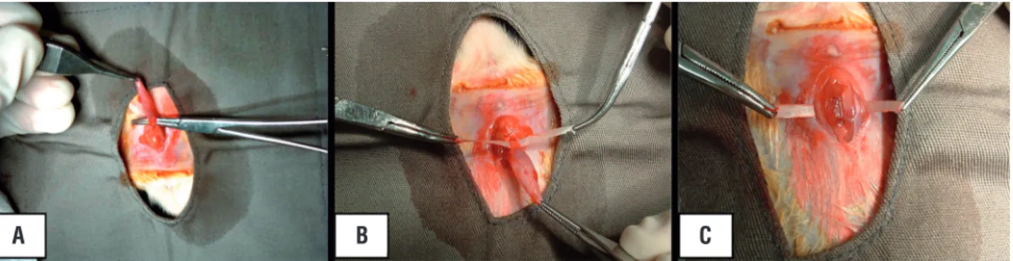
Forty female Wistar rats with weight ran-ging from 215 to 297g were used in the present study. They were divided into 4 groups of 10. Two groups (A and B) were submitted to the implant of CEC that had 5cm in length and 0.3cm in width and groups C and D received the polypropylene mesh with the same dimensions. In both cases the surgical implantation occurred below the bladder neck with tension free fixation at the rectus mus-cle fascia (Figure-1).
Animals in subgroups A and C were sa-crificed 4 weeks after the implant and animals in subgroups B and D after 12 weeks in order to eva-luate tissue reaction and integration to the host. Bladder, urethra and abdominal musculature were removed as a single block keeping their anato-mic relationship. The whole block was preserved in buffered 10% formalin for 15 days after which was processed for histological analysis.
Histological slides (5 microns) were pro-cessed using Haematoxilin-Eosin (HE) staining to evaluate inflammatory reaction. The following criteria were used for this purpose: (1) Absent: no inflammation or less than 5% at the analyzed area; (2) Slight: inflammatory reaction between 5 and 25%; (3) Moderate: Inflammatory reaction betwe-en 25 and 70%; (4) Intbetwe-ense: over 70% of the stu-died area. Collagen quantification was performed
through stereological analysis and the slides were stained by Picrosirius red. Samples were analyzed by the same pathologist using 40X magnification.
Numeric variables were expressed by central tendency and dispersion. Kruskal-Wallis test was used to express continuous variables and Chi Square for categorical variables. The significance level to re-ject the nullity hypothesis was 5% (p<-0.05).
This research followed the rules of the expe-rimental ethics code and the animal protecting laws practiced in Brazil. This study was approved by the Ethics Committee for animal study of the Institution.
RESULTS
No postoperative surgical complication was found in all animals. During sacrifice no atypical tissue reaction was observed as a conse-quence of the use of both materials. No fistula or abscess was found. Macroscopically, a good incor-poration was observed (Figure-2).
Microscopically the subaponeurotic area showed intense inflammatory reaction in both groups with a significant presence of polymor-phonuclear neutrophils in the analysis after 4 we-eks. No atypical formation was observed.
The same analysis performed in animals sacrificed at 12 weeks showed a slight increase in collagen deposition at the subaponeurotic area in animals receiving CEC as compared to polypropyle-ne mesh (25.90% vs 22.30), p=0.21 (Figure-3).
Stereological analysis at the suburethral area showed a significant difference of collagen presence in favor of CEC (58.60% vs 28.10), p=0.003.
Figure 1 - Surgical technique of implantation showing dissection of the urethra (A), positioning of the CEC at the suburethral space (B) and fixation to the abdominal wall (C).
A B C
The inflammatory reaction induced by CEC sling showed an intense proliferation of polymorphonuclear cells even in the late phase. Some angiogenesis with discrete neovasculari-zaton penetrating the new material was found. No capsule formation was found around the CEC implant (Figure-4).
The qualitative analysis concerning in-flammation and the degree of necrosis in groups B and D in both suburethral and subaponeurotic
regions showed similar results. The polypropyle-ne group showed less inflammatory reaction than CEC in the suburethral area.
DISCUSSION
The inflammatory reaction from a synthe-tic material in an animal model represents an im-portant step in the evaluation of its future use in clinical trials. Several materials have been used as Figure 2 - Demonstration of polypropylene (A) and CEC (B) in situ at 120 days sacrifice. Adequate incorco-poration to the implanted area.
Figure 3 - Analysis of collagen deposition in the suburethral space. Comparison between the biomaterials.
A B
slings in the treatment of SUI and at the present polypropylene mesh is incorporated to various in-dustrial mechanisms that are currently in clinical use (3, 5, 6, 9). The main reason to study a new material such as CEC is its physical properties and biocompatibility as well as the fact of being produced from a renewable material such as su-garcane sub product.
The cellulosic exopolysaccharide (CEC) has already been tested concerning cytotoxicity which has been evaluated through the adhesion index, nitric acid production and cellular viabili-ty of alveolar macrophages. These tests have been done in rats and no toxic reaction was found in the cell culture. There was also a high compa-tibility level in three cytotoxicity studies (10) which follow the recommendations of the Food and Drug Administration (FDA) that states that implantable medical devices and their consti-tuents should not produce adverse reactions both locally and systemically, have no carcinogenic effect and produce no harm to the reproductive system and development (11).
CEC presents satisfactory physical pro-perties such as elasticity, resistance to traction
and flexibility allowing easy modulation in di-fferent shapes including the possibility of using as a sling. In an independent study in rats poly-propylene mesh and CEC were implanted into the peritoneum and demonstrated that there was incorporation of both bio prosthesis without the presence of exudates and fistulas (12).
There is growing concern among surge-ons using different types of industrial materials specially related to the risks of erosion, infection and biocompatibility status (5, 6, 13). In the ear-ly phase of the present study it was observed a high level of granulocytes infiltration as well as the presence of fibroblasts. These findings re-present typical aspects of tissue repair involving the vascularization process. In this phase there is activation of the coagulation, platelets aggre-gation that involves its invasion by neutrophils, monocytes, macrophages and fibroblasts. This phenomenon is known as chemotaxis (13). The centripetal migration of endothelial cells in the area of the CEC implant interferes in the angio-genesis process justifying tissue remodeling and the integration of this new material in the subu-rethral area.
Microscopic analysis of the suburethral area showed a significantly elevated amount of collagen (30.5%) in the group that used CEC im-plant as compared to the control group. Erosion related to the use of the material was not found. A greater collagen deposition was observed in the suburethral region for both groups of mate-rials when compared to subaponeurotic area.
This significant increase in collagen den-sity is probably related to the interaction be-tween cytokines and cells related to the healing process. Early research conducted to evaluate the biomechanics characteristics of CEC membrane demonstrated that after implantation there is an increase of its resistance to traction associated to the tissue integration. This phenomenon may represent a relevant point in favor of the incre-ase of maintenance of continence rates when in clinical use (14).
In the present study a more intense in-flammatory reaction was found at the subure-thral area when compared to the control group in the 12 weeks group but this was not associated with urethral erosion.
Tissue reaction of polypropylene sling was studied in three different types of mesh which are different in relation to the structure and size of the pores (15). Other studies emphasize the role of these pores in allowing the filling with connective tissue, migration of immunocompetent cells and angiogenesis (16). Macroporous materials present greater molecular permeability and consequently allow quicker fibrinous fixation. They work as a biologic glue preventing the accumulation of se-cretions (2).
Histological analysis demonstrates a de-crease in inflammatory reaction and fibrosis when a larger porous material is used as a sling. On the other hand lower porosity materials tend to facili-tate capsule formation. This phenomenon did not occur with the CEC membrane.
More recently, small intestine submucosa (SIS) has been introduced as an option to be used as a sling in the treatment of SUI. This material is an acellular matrix that is produced from the intestinal submucosa of pigs. It is composed of collagen, gro-wth factors, glycosaminoglycan and glycoprotein. The porosity is described as microscopic (17, 18).
Due to its composition SIS is infiltrated by the host cells that quickly proliferate and result in regeneration of local tissue in a well organized way. The authors concluded that there was com-plete absorption of the sling material due to the exceptional biocompatibility (18). Incontinence recurred in all patients probably due to the ma-terial absorption and loss of urethral support. Al-though no clinical study has proven this idea it is theoretically hypothesized that materials to be used with this purpose must stay in situ for a mi-nimum of 12 weeks.
CEC was not affected by the degradation process and no acute complications such as abscess formation or extrusion or fistulas related to the im-plantation of this material. We believe that the pre-sence of collagen coating the suburethral mucosa may be responsible for the preservation of this tis-sue architecture and viscoelastic characteristics (12, 14). This suburethral support probably reduces the risks of erosion which is one of the main compli-cations associated with the use of synthetic slings.
It is important to emphasize that the ob-jective of the present research was not to evalu-ate the functional properties of the CEC implant since no tension was applied over it. The absence of suburethral erosion and reduced inflammatory reaction as well as the tendency for incorporation in the long term as demonstrated by the angioge-nesis process may be the most important features to be taken in account.
CONCLUSIONS
The CEC implant was shown to be appro-priate when used as a suburethral sling in rats. Adequate integration to the host tissue and preser-vation of its architecture were the main findings of the study. The utilization of CEC may be con-sidered as an ideal material to be used as a sling but the impact of its use in clinical practice still has to be tested.
ACKNOWLEDGEMENTS
To Sidney Pratt, native speaker that revi-sed the English version of this text, Canadian, BA, MAT (The Johns Hopkins University), RSA diplo-ma (TEFL).
FINANCIAL SOURCE
Federal government (Ministry of Science and Technology, MCT): FINEP (Studies and Projects Financing Agency) and CNPQ (National Counsel of Technological and Scientific Development).
CONFLICT OF INTEREST
None declared.
REFERENCES
1. McCormick KA, Newman DK, Colling J, Pearson BD. Urinary incontinence in adults. Am J Nurs. 1992;92:75-88.
2. Chaikin DC, Rosenthal J, Blaivas JG. Pubovaginal fascial sling for all types of stress urinary incontinence: long-term analysis. J Urol. 1998;160:1312-6
3. Cross CA, Cespedes RD, McGuire EJ. Our experience with pubovaginal slings in patients with stress urinary incontinence. J Urol. 1998;159:1195-8.
4. Petros PE, Ulmsten UI. An integral theory of female urinary incontinence. Experimental and clinical considerations. Acta Obstet Gynecol Scand Suppl. 1990;153:7-31.
5. Norris JP, Breslin DS, Staskin DR. Use of synthetic material in sling surgery: a minimally invasive approach. J Endourol. 1996;10:227-30.
6. Bent AE, Ostergard DR, Zwick-Zaffuto M. Tissue reaction to expanded polytetrafluoroethylene suburethral sling for urinary incontinence: clinical and histologic study. Am J Obstet Gynecol. 1993;169:1198-204.
7. Vilar F, Aguiar JL, Lima SV, Machado M, Pontes F, Lucena R, et al. Doença de Peyonie: estudo de um novo material com perspectiva de aplicação clínica. Int Braz J Urol. 2006; 32: 167-7.
8. Lima SV, Aguiar JLA, Pereira LA, Machado M, Andrade R, Lima R. A new dressing for hypospadias surgery. Int Braz J Urol. 2006; 32: 189-9.
9. Kobashi KC, Govier FE. Perioperative complications: the first 140 polypropylene pubovaginal slings. J Urol. 2003;170:1918-21
10. US Food and Drug Administration. Electronic and information technology accessibility standards economic assessment. [serial Internet]. Nov 2000. [cited in 2007 Out 11]. Available at: www.fda.gov/accessibility.html
11. Lima FR, Lima JRA, Hirakawa P, Medeiros Jr. MD, Lima FMT, Aguiar JLA. Resposta inflamatória a membrana de biopolímero de cana-de-açúcar e telas de Polipropileno implantas no peritôneo parietal de ratos. An Fac Méd Univ Fed Pernamb. 2005; 50: 37-40.
12. Castro CM, Aguiar JL, Melo FA, Silva WT, Marques E, Silva DB. Citotoxicidade de biopolímero de cana-de-açúcar. An Fac Med Univ Fed Pernamb. 2004;49:119-23.
13. Yildirim A, Basok EK, Gulpinar T, Gurbuz C, Zemheri E, Tokuc R. Tissue reactions of 5 sling materials and tissue material detachment strength of 4 synthetic mesh materials in a rabbit model. J Urol. 2005;174:2037-40.
14. Slack M, Sandhu JS, Staskin DR, Grant RC. In vivo comparison of suburethral sling materials. Int Urogynecol J Pelvic Floor Dysfunct. 2006;17:106-10.
15. Rutner AB, Levine SR, Schmaelzle JF. Processed porcine small intestine submucosa as a graft material for pubovaginal slings: durability and results. Urology. 2003;62:805-9. 16. Wiedemann A, Otto M. Small intestinal submucosa for
pubourethral sling suspension for the treatment of stress incontinence: first istopathological results in humans. J Urol. 2004;172:215-8.
17. Silveira AA, Dantas ML, Almeida YM, Aguiar JL. Estudo biomecânico de membranas de biopolímero de cana-de-açúcar perfuradas e contínuas implantadas no celular subcutâneo da parede abdominal de ratos. An Fac Med Fed Pernamb. 2007;52:55-9.
18. Thiel M. Análise quantitativa da fibrose e semiquantitativa da reação inflamatória de quatro diferentes slings sintéticos. [Tese Doutorado]. Campinas-SP: Universidade Estadual de Campinas; 2006. Available at: http://www.bibliotecadigital. unicamp.br/document/?code=vtls000386571