INTRODUCTION
In the literature, several studies point out that 10% to 20% of sexually active adults have HPV infection, although only 1% presents clas-sic condyloma and 2% visible lesions after acetic acid application (1). According to world litera-ture, it is rational to expect the existence of 3 to 6 million males infected with HPV (2).
The relationship between HPV infection and cervical cancer is well established and there are strong evidences that it may also be implicat-ed in the etiology of anal and genital cancer (3).
The studies related to the determination of prevalence of HPV infection in males are very important, since they can present a subclinical and asymptomatic infection and become poten-tial source of infection of HPV to their male or female sexual partners (4).
In view of all these facts, our study was designed to determine the prevalence of precur-sor lesions of penile cancer, to establish the con-cordance among different diagnostic techniques (PCR, Hybrid Capture (HC) and Peniscopy with acetic acid 5%) in the diagnosis of infection with the Human Papilloma Virus (HPV) of the penis of Objectives: To determine the prevalence of precursor lesions of penile cancer, to
establish the concordance of diagnostic techniques (PCR, Hybrid Capture (HC) and peniscopy with acetic acid 5%) in the diagnosis of Human Papilloma Virus (HPV) of the penis of men infected with HIV and to evaluate the infl uence of the immune status.
Patients, Methods and Results: 276 men were studied, with a median age of 34.6 years. Prevalence of High Risk HPV, Low Risk HPV and infection with both, ac-cording to HC, was 43%, 32% and 22%, respectively. PCR showed 50% of posi-tivity for HPV DNA. Peniscopy was positive in 27% of individuals. Peniscopy showed good specifi city and low sensitivity for the detection of penile HPV, and low concordance with PCR. Men with white lesions had a 3.6 higher relative risk of positivity for HPV. The most common clinical lesion observed was vegetation, identifi ed in 29% of patients. PCR and HC techniques showed high sensitivity for HPV DNA and there was an excellent correlation between them. Immunosup-pressed individuals with CD4< 200 cells/mm3 had the highest prevalence of pre-malignant lesions that were observed in 10% of the studied individuals.
Conclusions: Peniscopy was important for identifi cation and treatment of sub-clinical lesions. PCR and HC techniques were sensitive methods for the detection of HPV DNA with high concordance. Severely immunosuppressed individuals showed a higher prevalence of pre-malignant lesions of the penis.
Clinical and laboratorial study of HPV infection in men
infected with HIV
_______________________________________________
Giuseppe Figliuolo, Jusimara Maia, Alex P. Jalkh, Angelica E. Miranda, Luiz C.L. Ferreira
Fundação da Medicina Tropical Dr. Heitor Vieira Dourado, Manaus, Brazil
ABSTRACT ARTICLE INFO
_______________________________________________________________ _____________________
Key words:
DNA Probes; HPV; Men; HIV
Int Braz J Urol. 2012; 38: 411-18
________________
Submitted for publication: January 01, 2011
________________
HIV positive males and to evaluate the influence of the immunologic status on the occurrence of the lesions.
MATERIAIS AND METHODS
This is a cross-sectional descriptive study of men affected with HIV attended at the Funda-ção de Medicina Tropical Dr. Heitor Vieira Doura-do (FMT-HVD). Data including demographic, epi-demiologic and clinical characteristics of patients were collected. Physical examination (urological inspection and peniscopy with acetic acid 5%), molecular biological tests (PCR in house and Hy-brid Capture II, Digene & Co®) and conventional
histopathology study were also performed. Crite-ria for inclusion were: HIV positive males, with ≥ 18 years old, who provided written informed consent to join the study. Criteria for exclusion were: HIV negative males, Indians, psychiatric patients and those that didn’t complete all steps of the study. Data were collected at Epi Info® version 6.04 platform and the statistical analysis was made through Statistical Package for Social Sciences® (SPSS) version 16.0 for Windows.
Patients lied in supine position in order to be submitted to the urological and scrotal in-spection. A surgical brush with saline was rubbed against the foreskin, balanopreputial sulcus, glans and navicular fossa of the penis. The brush was immersed in an Eppendorf vial containing 1 mL of T1 buffer (commercial kit for nuclear extrac-tion Spin Tissue-Macherey-Nagel®). The vial was tightly closed and sent to the laboratory, where was maintained at -70oC until PCR analysis.
Another brush was used to Hybrid Cap-ture (HC) for High and Low Risk HPV, and it was stored in an appropriate kit.
After the cytological collection, we pro-ceeded with peniscopy and penile and scrotal inspection. A gauze soaked with acetic acid 5% was placed around the penis for 10 minutes. Positive lesions (white lesions) were biopsied, except in patients with previous histopathologi-cal or laboratorial diagnosis or that didn’t allow the procedure. The samples were fixed in buff-ered formalin 10% and were sent to histopatho-logical studies.
For statistical analysis, it was used Pear-son’s Chi-Square test with Yates correction whenever necessary; Fisher’s exact test was used to categorical variables for values under 5 and significance analysis including Odds Ratio (OR) and 95% confidence intervals was performed. Significance was established for p < 0.05 (5%).
For concordance analysis, it was used the kappa (k) associative test.
RESULTS
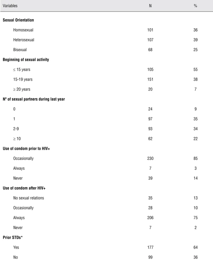
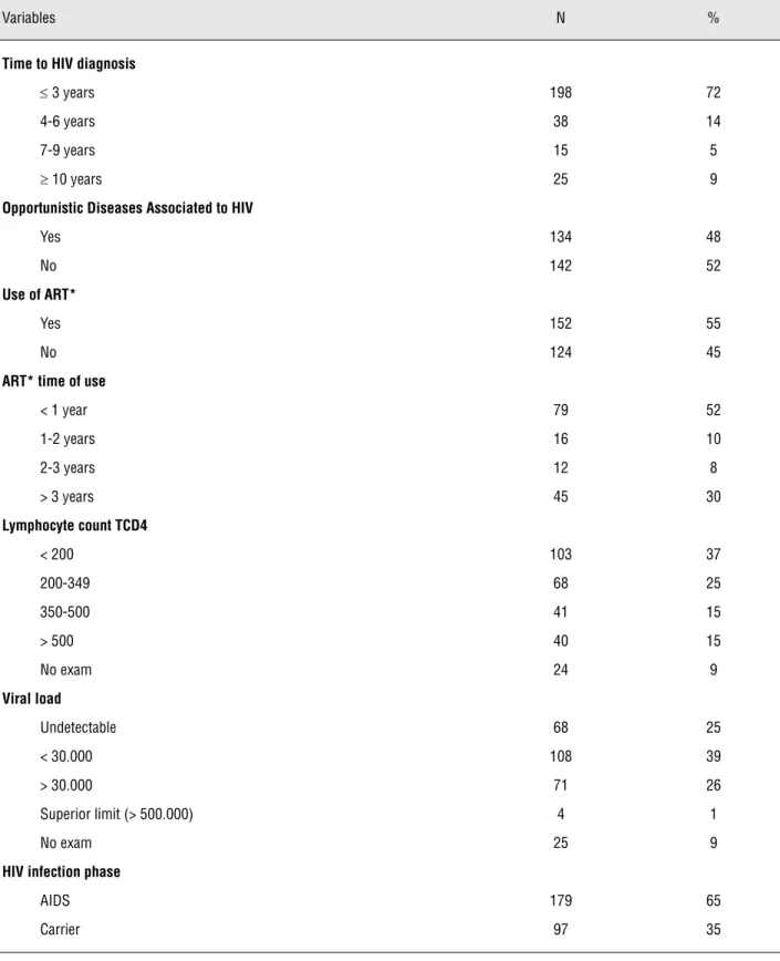
Two hundred and seventy six HIV-posi-tive male patients older than 18 years old were included. Median age was 34.6 years. Table-1 depicts socio-demographics characteristics of patients. Table-2 shows the variables related to sexual behavior, use of condoms and previous STDs and Table-3 those related to HIV virus.
Peniscopy was positive for white lesions in 27% of patients. The most frequent lesion was vegetation (29%) (Table-4). Biopsy was obtained in 22% of participants; some of them had more than one lesion and a total of 75 fragments of skin were collected for conventional histopatho-logical study (Table-5). Pre-malignant lesions were observed in 10% of patients (Table-6), and most of them (59%) had CD4 < 200 cells/mm3.
According to HC, the prevalence for High Risk, Low Risk and both High and Low Risk HPV infection were 43%, 32% and 22%, respectively. PCR had 50% of positivity for HPV DNA.
The concordance between peniscopy and PCR was observed in 62% of samples, revealing a “weak concordance” according to kappa associa-tive test (k = 0.2317). Patients with white lesions observed at peniscopy had a 3.6 higher risk of HPV infection.
Peniscopy was considered a diagnostic test with high specificity (86%) and low sensitiv-ity (37%). Positive and negative predictive values were 73% and 58%, respectively. Exam accuracy was 62%.
Table 1 - Socio-demographic variables of 276 men with HIV+/AIDS.
Demographic indices N %
Age
18-29 years 101 36
30-49 years 151 55
≥ 50 years 24 9
Race/Colour
Brown 207 75
White 47 17
Black 18 7
Yellow 4 1
Marital status
Single 158 57
Married 53 19
Fixed partner 56 20
Divorced 7 3
Widow 2 2
Education
Illiterate 2 1
Primary 97 35
Secondary 125 45
Terciary 52 19
Use of unlawfully drugs
Yes 66 24
No 210 76
Smoking
Yes 75 27
Table 2 - Distribution of sexual behavior variables, use of condoms and previous STDs of 276 HIV+/AIDS males.
Variables N %
Sexual Orientation
Homosexual 101 36
Heterosexual 107 39
Bisexual 68 25
Beginning of sexual activity
≤ 15 years 105 55
15-19 years 151 38
≥ 20 years 20 7
Nº of sexual partners during last year
0 24 9
1 97 35
2-9 93 34
≥ 10 62 22
Use of condom prior to HIV+
Occasionally 230 85
Always 7 3
Never 39 14
Use of condom after HIV+
No sexual relations 35 13
Occasionally 28 10
Always 206 75
Never 7 2
Prior STDs*
Yes 177 64
No 99 36
Table 3 - Variables associated to the presence of HIV virus.
Variables N %
Time to HIV diagnosis
≤ 3 years 198 72
4-6 years 38 14
7-9 years 15 5
≥ 10 years 25 9
Opportunistic Diseases Associated to HIV
Yes 134 48
No 142 52
Use of ART*
Yes 152 55
No 124 45
ART* time of use
< 1 year 79 52
1-2 years 16 10
2-3 years 12 8
> 3 years 45 30
Lymphocyte count TCD4
< 200 103 37
200-349 68 25
350-500 41 15
> 500 40 15
No exam 24 9
Viral load
Undetectable 68 25
< 30.000 108 39
> 30.000 71 26
Superior limit (> 500.000) 4 1
No exam 25 9
HIV infection phase
AIDS 179 65
Carrier 97 35
DISCUSSION
The prevalence of HPV in our study was higher than of the study of Goldstone et al. They evaluated 602 HIV negative males who were en-gaged in sex with other males and observed a prevalence of 18.2% of HPV infection of the penis using also PCR (6).
Peniscopy showed high specificity and low sensitivity. However, most studies showed a weak specificity of the exam and also a good sensitivity (7-9). We believe that our results were biased due to the high prevalence of HPV in the studied population (around 50%), explaining the good specificity, and that most of the patients had subclinical or latent infection with HPV, that im-paired the identification through peniscopy, only with biomolecular techniques, explaining the low sensitivity.
Some risk characteristics for HPV infec-tion of penis of HIV positive males were identi-fied in our study, including heterosexual behavior (higher rate of penile HPV infection compared to
bisexuals and homosexuals, probably due to the high rate of female infection in our population, demonstrated in several studies done in Manaus (10-12).
PCR and HC techniques had high concor-dance and sensitivity for the detection of HPV. Rodrigues et al. (13) demonstrated that HC and PCR techniques for the detection of HPV in clini-cal samples had a fair concordance, including conventional and real time techniques (k = 0.338). When they compared conventional PCR with real time PCR they observed an almost perfect concor-dance (k = 0.818).
There are very few studies related to in-traepithelial neoplasms or penile cancer of HIV positive men. Kreuter et al. studied 263 HIV-pos-itive homosexual men and found penile intraep-ithelial neoplasms of penis in 11 (4.2%) and of anus in 156 (59.3%) (14).
The limitations of our study included the small size of sample that prevented a strong as-sociation among the techniques. The study was conducted in an AIDS ambulatory. However, most
Table 4 - Distribution of dermatological lesions observed in 75 positive peniscopies. After: Rook’s 2010 (5).
Peniscopy Lesion N (%)
Vegetation 25 29
White lesion 13 15
Ulcer 10 12
Papule normochromic 13 15
Crust 1 1
Papule Hypochromic 4 5
Papule Hyperchromic 4 5
Macula hypochromic 9 10
Macula hyperchromic 3 4
Eritema 1 1
Hyperchromic Plate 2 2
Table 5 - Distribution of histopathological findings of patients with clinical lesions detected.
Histopathological Findings N (%)
Angioceratoma 1 1
Chronic inespecifc balanitis 2 2
Inespecific ulcerated balanitis 1 1
Inespecific chronic balanopostitis 2 2
Condyloma Acuminata 19 27
Flat Condyloma 1 1
Epidermodysplasia 2 2
Nonspecific chronic eczema 1 1
Hypermelanose 1 1
High grade intra-epitelial lesion (HSIL) 4 6
Fungal infection 1 1
Scleroathrofic lichen 2 2
Lichen planus 1 1
Low grade Intra-epitelial lesion (LSIL) 18 25
Molluscum Contagiosum 6 9
Melanocytic nevi 1 1
Bowenoid papulosis 5 7
Chronic inespecific postitis 1 1
No significative alterations 6 9
Total 75 100
Table 6 – Distribution of histopathological findings of 27 pre-malignant lesions.
Pre-malignant lesions
Epidermodysplasia Verruciformis Like 02
High grade intra-epitelial lesion (HSIL) 04
Low grade Intra-epitelial lesion (LSIL) 17
Bowenoid papulosis 03
Bowenoid papulosis+ LSIL+Bowenoid papulosis 01
HIV+/AIDS patients from Amazonas are attend-ed at FMT-HVD, which we believattend-ed allowattend-ed the study of a significant sample of patients.
We believe that the present results can be used to delineate preventive programs for early detection of penile cancer, in individuals with higher risk, including immunosuppressed pa-tients. Diagnosis and treatment of male partners infected with HPV would also allow a reduction of sexually transmitted diseases.
CONCLUSIONS
Prevalence of DNA HPV was approxi-mately 50%.
Peniscopy proved to be a high specific and low sensitive exam.
Concordance of peniscopy and PCR for the detection of HPV was low.
Concordance of PCR and HC for HPV de-tection was excellent.
We observed a prevalence of 10% of pa-tients with pre-malignant lesions determined by histopathological studies and that most of them were severely immunosuppressed (TCD4 < 200 cells/mm3).
CONFLICT OF INTEREST
None declared.
REFERENCES
1. Gollnick H, Barasso R, Jappe U, Ward K, Eul A, Carey-Yard M, Milde K: Safety and efficacy of imiquimod 5% cream in the treatment of penile genital warts in uncircumcised men when applied three times weekly or once per day. Int J STD AIDS. 2001; 12: 22-8.
2. Silverberg MJ, Ahdieh L, Munoz A, Anastos K, Burk RD, Cu-Uvin S, et al.: The impact of HIV infection and immunodefi-ciency on human papillomavirus type 6 or 11 infection and on genital warts. Sex Transm Dis. 2002; 29: 427-35.
3. Internacional Agency for Research on Cancer (IARC). Working Group on the Evaluation of Carcinogenic Risks to Humans. Human papillomaviruses. IARC Monogr Eval Carcinog Risk Hum 2008; 1-636.
4. Olsson SE, Kjaer SK, Sigurdsson K, Iversen OE, Hernandez-Avila M, Wheeler CM, et al.: Evaluation of quadrivalent HPV 6/11/16/18 vaccine efficacy against cervical and anogenital disease in subjects with serological evidence of prior vaccine type HPV infection. Hum Vaccin. 2009; 5: 696-704.
5. Rook’s textbook of dermatology. Nomeclatura do Comitê da liga Internacional da Sociedade de Dermatologia; 2010. 6. Goldstone S, Palefsky JM, Giuliano AR, Moreira ED Jr, Aranda
C, Jessen H, et al.: Prevalence of and risk factors for human papillomavirus (HPV) infection among HIV-seronegative men who have sex with men. J Infect Dis. 2011; 203: 66-74. 7. Carvalho JJM. Prevalência e padronização diagnóstica da
infecção genital pelo HPV em homens atendidos em clínica urológica. São Paulo. Tese (mestrado). FMSCSP; 1999, 109p. 8. Carvalho JJM. Identificação do grupo de risco em pacien-tes com infecção pelo HPV com diagnóstico pela peniscopia confirmado pelo teste de biologia molecular. São Paulo. Tese (Doutorado). FMSCSP; 2002, 63p.
9. Chaves JHB, Vieira TKB, Ramos JS, Bezerra AFS. Peniscopia no rastreamento daslesões induzidas pelo papilomavirus hu-mano. Rev Bras Clin Med. 2011; 9: 30-5.
10. Souza PM. Detecção do Papilomavírus Humano (HPV) na Cérvice Uterina de Pacientes HIV positivas e em portadoras de AIDS. Manaus. Tese (Mestrado). Fundação de Medicina Tropical do Amazonas; 2004.
11. Brock MF. Alterações Colpocitológicas em pacientes portado-ras do Vírus HIV atendidas na Fundação de Medicina Tropical do Amazonas. Tese (Mestrado). Fundação de Medicina Tropi-cal do Amazonas; 2005.
12. Corrêa GJ. Prevalência do Papilomavírus Humano (HPV) em mulheres portadoras de lesões intra-epiteliais escamosas de alto grau e carcinoma epidermóide invasor de colo uterino. Tese (Mestrado). Fundação de Medicina Tropical do Amazo-nas; 2005.
13. Rodrigues AD, Cantarelli VV, Frantz MA; Pilger DA, Pereira FS. Comparação das técnicas de captura de híbridos e PCR para a detecção de HPV em amostras clínicas. J Bras Patol Med Lab 2009; 45 (6):457-62.
14. Kreuter A, Brockmeyer NH, Weissenborn SJ, Gambichler T, Stücker M, Altmeyer P, et al.: Penile intraepithelial neoplasia is frequent in HIV-positive men with anal dysplasia. J Invest Dermatol. 2008; 128: 2316-24.
______________________
Correspondence address
Dr. Giuseppe Figliuolo Fundação de Medicina Tropical Dr. Heitor Vieira Dourado, Manaus, Brazil
Parque Tropical, Rua 08, 28-B / 1104, Edifício: Nápoles. Bairro: Parque 10