Copyright © 2008 by Sociedade Brasileira de Pediatria
O
RIGINALA
RTICLEPeriventricular leukomalacia in very low birth weight
preterm neonates with high risk for neonatal sepsis
Rita C. Silveira,1 Renato S. Procianoy,2 Juliana C. Dill,3 Cristine S. da Costa3
Abstract
Objective:To investigate the association between periventricular leukomalacia (PVL) and neonatal sepsis in very
low birth weight infants (VLBWI).
Methods:We studied VLBWI with a clinical suspicion of infection who had been born at our institution between the
1st of August, 2005 and the 31st of July, 2007. Children were excluded if they died before reaching 14 days, had malformations of the central nervous system or congenital infections. Ultrasound brain scans were carried out on the third day and weekly up until the sixth week of life or discharge. Periventricular leukomalacia was diagnosed by persistent diffuse periventricular hyperechogenecity for more than 7 days, or by periventricular cysts. The VLBWI were separated into two groups on the basis of the presence or absence of PVL. Sepsis was defined as clinical manifestation plus a positive culture. The Mann-Whitney, chi-square andttests were applied followed by logistic regression.
Results:A total of 88 VLBWI were studied. Of these, 62 (70.5%) survived and 51 (57.8%) had PVL. Both groups
were similar in terms of birth weight, gestational age, Apgar score, type of delivery, SNAPPE-II score, presence of necrotizing enterocolitis, persistent ductus arteriosus and deaths. Sepsis and mechanical ventilation were more common in the group with PVL (23.5 and 2.7%, p = 0.005; 86 and 59%, p = 0.004, respectively). Both of these were identified as, independent risk factors for PVL by logistic regression (p = 0.027 and 0.015, respectively).
Conclusions:Chorioamnionitis has been defined as a risk factor for PVL. We have demonstrated that neonatal
sepsis is also an important risk factor. We believe that the systemic inflammatory response is the principal factor involved in the etiopathogenesis of PVL among VLBWI.
J Pediatr (Rio J). 2008;84(3):211-216:Prematurity, neonatal sepsis, periventricular leukomalacia, very low birth weight.
Introduction
The increased survival of progressively more premature newborn infants has resulted in a significant number of pre-term infants who grow up with neurocognitive abnormali-ties.1,2Currently, periventricular leukomalacia (PVL) is the principal neurological problem affecting children born extremely premature. Approximately 25% of newborn infants with birth weights below 1,500 g who survive to discharge exhibit moderate to severe permanent motor deficits, such as spastic diplegia. At school age, 25 to 50% of children who
had been diagnosed with PVL manifest cognitive and learning deficits.3-5
The presentation of PVL is subclinical and so diagnosis is made by neuroimaging examinations (cerebral ultrasound and/or magnetic resonance). An early diagnosis of PVL can be achieved using transfontanelar cerebral ultrasound during the neonatal period used as screening for brain injuries. On the ultrasound brain scan, areas of periventricular hyperecho-genecity can be observed which will later develop into periven-tricular cysts and/or diffuse hyperechogenic areas in the white
1. Doutora. Professora adjunta, Universidade Federal do Rio Grande do Sul (UFRGS), Porto Alegre, RS, Brazil.
2. Chefe, Serviço de Neonatologia, Hospital de Clínicas de Porto Alegre (HCPA), Porto Alegre, RS, Brazil. Professor titular, UFRGS, Porto Alegre, RS, Brazil. 3. Residente de 3º ano (R3), Neonatologia.
Financial support: CNPQ-DECIT/MS and FIPE-HCPA.
No conflicts of interest declared concerning the publication of this article.
Suggested citation:Silveira RC, Procianoy RS, Dill JC, da Costa CS. Periventricular leukomalacia in very low birth weight preterm neonates with high risk for neonatal sepsis. J Pediatr (Rio J). 2008;84(3):211-216.
Manuscript received Nov 23 2007, accepted for publication Feb 14 2008.
doi:10.2223/JPED.1777
matter.6,7This diffuse component of PVL is common among extreme preterm newborns, and the focus appears to be asso-ciated with the inflammatory response resulting from ischemia or infection.3,7,8In these two situations, the factors involved in etiopathogenesis are related to prematurity.
The critical period of cerebral myelination is between 23 and 32 weeks’ postconceptional age, when pre-oligodendrocytes differentiate into immature oligoden-drocytes, responsible for initiating myelination of the white matter.9
The reduction of cerebral perfusion in very small preterm neonates with deficient cerebrovascular autoregulation is a high risk for the occurrence of injuries to the periventricular white matter.8This being so, preterm newborn infants exposed to intrauterine infections are vulnerable to pre-oligodendrocyte death in the face of ischemic insults, even when mild and insufficient to cause injuries by themselves.2,8-10
Many experimental and clinical studies have demon-strated the link between intrauterine infection and increased occurrence of PVL.11-13A meta-analysis of 26 studies, relat-ing chorioamnionitis to PVL, concluded that, among the new-born infants of mothers with chorioamnionitis, the chances of developing cystic PVL were three times greater.14 Notwith-standing, studies relating the presence of neonatal sepsis in extreme preterms with increased occurrence of PVL are rare and do not deal with this high risk population specifically.
The objective of our study was to investigate the pres-ence of PVL and the possible risk factors associated with its development, particularly neonatal sepsis, in a population that is especially vulnerable to these conditions: very low birth weight preterm newborn infants (VLBWI).
Methods
This was a prospective cohort study of a population of new-born infants with birth weights between 500 and 1,500 g, new-born at the obstetrics center at our hospital and admitted to the neonatal intensive care unit (NICU) during the period between 1st August, 2005 and 31st July, 2007. Inclusion was based on the assessment of the duty physician in whose opinion the infants enrolled exhibited clinical signs suggestive of infec-tion during the first week of life.
Exclusion criteria were death before 14 days of life, pres-ence of major congenital malformations or malformation of the central nervous system, congenital STORCH infections (syphilis, toxoplasmosis, rubella, cytomegalovirus or her-pes) or HIV+ mother.
An Informed Consent Form was read and signed by par-ents or guardians before the first cerebral ultrasound scan. The study was approved by Research Ethics Committee at the institution, protocol no. 04/446.
All newborn infants enrolled underwent a first cerebral ultrasound at 3 days of life; and then weekly until the sixth week of life or hospital discharge, if this took place before 6 weeks of life. This screening made it possible to diagnose PVL. Observational clinical monitoring while in NICU allowed for assessment of possible risk factors for PVL. VLBWI were dis-charged from hospital when they reached a weight of 2,000 g, were able to receive full oral enteral feeding and were clini-cally stable.
After the serial ultrasound brain scans, patients were divided in two groups:
Group 1: VLBWI with diagnosis of PVL based on diffuse or cerebral cystic white matter injury.
Group 2: VLBWI without diagnosis of PVL, with cerebral echography negative for this condition.
Images were acquired via the anterior fontanelle, with a LOGIQ 500 Scanner (G. E. Medical Systems, United States) and an 11 MHZ transducer. The researchers who carried out the scans were unaware of the patients’ clinical status in terms of presence or absence of sepsis; images were recorded on the machine and reviewed by two researchers who were quali-fied in cerebral ultrasound scanning.
The criteria for diagnosing PVL were the presence of dif-fuse periventricular hyperechogenecity that persisted for a period of more than 7 days, without forming cysts (diffuse periventricular white matter injuries) or the presence of cys-tic lesions of at least 0.5 cm in diameter, distributed bilater-ally and located close to the external angles of the lateral ventricles.6,15
The presence of ventricular dilatation without cerebral hemorrhage during any of the serial ultrasound scans was considered to be secondary to necrosis of the white matter present in the diffuse PVL component.3
The following data were collected prospectively and later compared between the two groups: sex, birth weight, gesta-tional age, fifth minute Apgar score, SNAPPE-II score, ante-natal corticosteroid use, neoante-natal sepsis, respiratory distress syndrome (RDS), peri-intraventricular hemorrhage (PIVH), non-hemorrhagic periventricular dilatation and pressure posi-tive mechanical ventilation for more than 24 hours. Mortality was also compared between groups.
Gestational age was determined according to the moth-er’s obstetric history (date of last menstruation) and con-firmed by obstetric ultrasound, at 12 weeks’ pregnancy at the latest. In the absence of any reliable maternal data, gesta-tional age was determined by somatic physical and neurologi-cal examination of the newborn, using the New Ballard method.16
Infants were diagnosed with RDS if they exhibited expira-tory grunting, nostril flaring, sternum retraction, a need for ≥ 40% oxygen and a chest X ray with diffuse ground-glass appearance and a need for exogenous surfactant.
Presence or absence of PIVH was determined from the first ultrasound brain scan and during weekly monitoring scans, according to the classification defined by Papile et al.18
The criteria for diagnosing neonatal sepsis were clinical signs of infection in the presence of microorganism in blood or cerebrospinal fluid cultures. Serum samples for blood cul-ture were obtained using the aseptic collection technique and according to the recommendations laid out by Schelonka et al.19
Clinical signs of infection were the presence of at least three of the findings referred to below, or two clinical findings plus one or more maternal risk factors20,21: thermal instability; apnea, bradypnea, grunting, tachypnea, sternum and subcostal retraction, nostril flaring and cyanosis; hypo-tonia and convulsions; irritability and lethargy; gastrointes-tinal symptoms, such as abdominal distension, vomiting, gastric residues and difficulty accepting food; idiopathic jaundice; cutaneous pallor, cold skin, perspiration, hypoten-sion and capillary refill time longer than 3 s; signs of bleed-ing, with clinical status suggestive of disseminated intravascular coagulation; subjective assessment: the new-born “does not look well”.
Maternal risk factors were obtained from their obstetric records and their medical and perinatal histories. The events most often described were maternal fever, urinary infection and more than 18 hours of rupture of membranes.
The sample size was calculated from published data reporting the occurrence of PVL at 7 to 26% of infants born preterm and weighing less than 1,500 g using proportions. Based on a statistical power of 90% with a level of signifi-cance of 5% (α = 0.05) and an estimated PVL incidence of 33% in high risk populations (VLBWI with presumed or con-firmed sepsis), the resulting sample size was 30 newborn infants in each group.10
Statistical analysis
Data are presented in the form of mean and standard deviation or median and interquartile range (from the 25th to the 75th percentiles), depending on the behavior of the vari-able in question. Differences between means were analyzed using Student’sttest, and differences between medians were subject to the Mann-Whitney test. Categorical variables were analyzed using the chi-square test. Variables were included in the logistic regression model is they exhibited statistical sig-nificance (p < 0.05) in the comparison between groups, and their odds ratios as risk factors for PVL were calculated. The level of significance was p < 0.05.
Results
A total of 180 VLBWI were born during the 2 years of this study, 88 of whom met the inclusion criteria and were fol-lowed up until their sixth week of life, hospital discharge or later death (after 14 days of life). A total of 26 VLBWI (29.5%) died while in NICU, and 62 (70.5%) were discharged from hos-pital. Nasal Continuous Positive Airway Pressure (CPAP) was used in 85 (96.6%) newborn infants, since it is routine at our service to install nasal CPAP for all VLBWI as soon as they exhibit any type of respiratory distress. Antenatal corticoster-oids were given to 51 patients (58% of the population) and nine of these had PIVH, in contrast with 25 out of 37 newborn infants whose mothers had not been given antenatal corticos-teroids (p < 0.001).
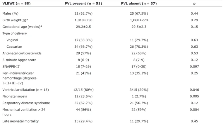
The exposure group (group 1) comprised 51 VLBWI with PVL, and was compared with 37 VLBWI in the group without PVL (group 2). Type of delivery, presence of perinatal asphyxia, necrotizing enterocolitis and persistent ductus arte-riosus were similar between groups. With the exception of five newborn infants who exhibited ventricular dilatation which could not be defined as either secondary to PVL or post-hemorrhagic, ventricular dilatation was significantly more frequent among VLBWI with PVL than among those with-out PVL. Three newborn infants withwith-out PVL exhibited ven-tricular dilatation secondary to cerebral hemorrhage diagnosed by means of serial ultrasound scans (Table 1). Neo-natal sepsis and the use of mechanical ventilation with inter-mittent positive pressure were significantly more frequent among newborn infants with PVL (Table 1).
The logistic regression model employed took the pres-ence of PVL as dependent factor, and as independent factors just those variables that had exhibited statistical significance in the univariate analyses (p < 0.05) and were not secondary to PVL. Therefore, just neonatal sepsis and mechanical ven-tilation were analyzed as independent factors, and both proved significantly associated with PVL (Table 2).
Twelve newborn infants with PVL had positive blood cultures: eight Staphylococcus coagulase negative, two
Streptococcus agalactiae, one Streptococcus α-hemolitico
and oneEscherichia coli. Just one newborn without PVL had a blood culture positive for Staphylococcus coagulase nega-tive. Staphylococcus coagulase negativewas present in 69.2% of cases of sepsis and was the most prevalent germ.
Discussion
Shalak & Perlman have written about the importance of antenatal corticosteroid therapy for reducing severe cerebral hemorrhage, since it accelerates maturation of the germinal matrix, increases systemic arterial pressure with improved cerebral perfusion and appears to be associated with less severe cases of RDS.22Our data demonstrate that more than half of our VLBWI (58%) had been given antenatal corticos-teroid, and that 66% of those did not present hemorrhage or exhibit level I PIVH with later reabsorption, demonstrating the efficacy of this preventative measure for reducing the occur-rence of cerebral hemorrhage inpremature newborn infants, since those who were given antenatal corticosteroid suffered significantly fewer cerebral hemorrhages.
It has been suggested that antenatal corticosteroid may be protective against injuries to cerebral white matter in
VLBWI due to its anti-inflammatory function.23In our study, a complete course of antenatal corticosteroid was given to 57% of the mothers whose newborn infants developed PVL and to 60% of the mothers of newborn infants without PVL; its administration did not result in a reduced incidence of PVL among VLBWI.
Neonatal sepsis is a clinical condition that activates a cas-cade of acute inflammatory response mediators. Kadhim et al., in two of their studies, found evidence of significant expression of tumor necrosis factor alpha (TNF-α) and, to a lesser extent, of interleukin-1 beta (IL-1β) in cerebral tissue with PVL, with increased cytokines production in cases where PVL was associated with infection. The inflammatory reaction was detected at an early stage of PVL and extended up to the last phase, cavitation.24,25
Table 1- Periventricular leukomalacia in very low birth weight infants and possible risk factors
VLBWI (n = 88) PVL present (n = 51) PVL absent (n = 37) p
Males (%) 32 (62.7%) 25 (67.5%) 0.44
Birth weight(g)* 1,010±250 1,068±270 0.29
Gestational age (weeks)* 29.2±2.5 29.5±2.3 0.15
Type of delivery
Vaginal 17 (33.3%) 11 (29.7%) 0.63
Caesarian 34 (66.7%) 26 (70.3%) 0.63
Antenatal corticosteroids 29 (57%) 22 (60%) 0.53
5-minute Apgar score 8 (6-9) 8 (7-9) 0.12
SNAPPE-II† 18 (7-29) 17 (0-30) 0.097
Peri-intraventricular hemorrhage (degrees I+II+III+IV)
21 (41%) 13 (35.1%) 0.25
Ventricular dilatation (n = 15) 12/15 (80%) 3/15 (20%) 0.046
Neonatal sepsis 12 (23.5%) 1 (2.7%) 0.005
Respiratory distress syndrome 32 (62.7%) 21 (56.7%) 0.12
Mechanical ventilation > 24 hours
44 (86%) 22 (59%) 0.004
Late neonatal mortality 15 (29.4%) 11 (29.7%) 0.45
PVL = periventricular leukomalacia; VLBWI = very low birth weight infants.
Data presented as mean ± standard deviation, median (interquartile range), n (%), chi-square test or Fisher’s exact test when not indicated otherwise. * Student’sttest.
†Mann-Whitney test.
Table 2- Neonatal sepsis and mechanical ventilation as risk factors for periventricular leukomalacia in very low birth weight infants (logistic regression)
OR CI p
Confirmed sepsis 11.613 (1.42-94.9) 0.027
Mechanical ventilation 4.3 (1.32-14.43) 0.015
Prematurity is a considerable neonatal risk factor for PVL, especially for white matter injuries with a diffuse component (non-cystic). There are speculations that other neonatal con-ditions are associated with diagnosis of PVL, such as perinatal asphyxia, hypovolemia and early neonatal sepsis. Many of these factors cause a reduction in systemic blood pressure, although clinical studies have not demonstrated an associa-tion between PVL and postnatal systemic hypotension.22,26,27 It was recently suggested that alterations in cerebral blood flow are a necessary condition for PVL, but are not enough to cause it in isolation. The preference for premature infants is related to the timing of the insult and the distribution of sus-ceptible pre-oligodendrocytes around the brain.8A question that remains is the influence of the cascade of inflammatory events that occurs in neonatal sepsis. The concern of this study, however, was to exclusively select a population at greater risk of suffering PVL, i.e. sick VLBWI, although not nec-essarily with a definite diagnosis of neonatal sepsis.
The presence of ischemia, inflammation or systemic infec-tion appears to be etiopathogenic for PVL. It is possible that PVL in preterm newborns is associated with an altered inflam-matory response equilibrium within the central nervous sys-tem, and that proinflammatory cytokines act directly on the brain, and not as part of a larger systemic response.10 Stud-ies that have assessed the association between maternal chorioamnionitis and PVL have not examined the occurrence of sepsis in premature newborns.11-14Our study assessed neonatal sepsis as an important risk factor for PVL in VLBWI. We did not asses the presence of chorioamnionitis because we did not have histology for all placentas and, consequently, lacked diagnoses of subclinical chorioamnionitis, which is a risk factor for PVL that has already been described.28
We avoided possible sample selection bias, defining neo-natal sepsis as present when newborn infants had a positive culture. For this reason we can safely say that the occurrence of neonatal sepsis in VLBWI increases their chances of PVL by 11 times. In the majority of cases, PVL has a subclinical pre-sentation, and it is therefore of fundamental importance to understand its risk factors, which, together with serial ultra-sound brain scans, make an early diagnosis possible. It is pos-sible to detect PVL by means of transfontanelar cerebral ultrasound, as long as the newborn infant is monitored weekly and up to hospital discharge. This is also useful to predict the development of cerebral palsy.6,29-31Vollmer et al. studied preterm newborns at 24 to 32 weeks’ gestational age and assessed the efficacy of cerebral ultrasound for detection of cerebral lesions and their relation with delayed neuropsycho-motor development at 8 years of age. These authors found that poor prognosis depended fundamentally on the pres-ence and type of lesion found with cerebral ultrasound.26It is possible that serial cerebral ultrasound scans, in addition to clinical findings from VLBWI in neonatal intensive care units, such as neonatal sepsis, are important prognostic factors.
One possible limitation of our study was that we did not use magnetic resonance imaging to confirm the diagnosis of PVL, especially the diffuse component of white matter inju-ries. Although magnetic resonance imaging has demon-strated greater diagnostic sensitivity and specificity for PVL, the elevated costs and technical difficulties involved, such as the need for transportation from NICU to radiology depart-ment, limits its use as a routine,7,30whereas cerebral ultra-sound scans are accessible, practical and well-adapted to bedside use.15We therefore believe that our study makes an additional contribution by demonstrating that it is possible to reach a diagnosis of PVL using ultrasound and following an appropriate follow-up protocol.
We already know that maternal chorioamnionitis is a risk factor for PVL. In this study we demonstrated that neonatal sepsis is also an important risk factor for PVL in VLBWI with clinical suspicion of neonatal infection. It is probable that the systemic inflammatory response to perinatal and neonatal infections is the principal factor involved in the etiopathogen-esis of PVL, which is a disease with significant morbidity, in common with cerebral palsy. Further studies attempting to block this inflammatory response may be promising for reduc-ing the magnitude of the PVL and, as a consequence, improve the future quality of life of these VLBWI.
Acknowledgements
The authors would like to thank Vânia Naomi Hirakata and Daniela Benzano, for their help with the statistical analysis.
References
1. Vohr B, Wright LL, Dusick AM, Mele L, Verter J, Steichen JJ, et al. Neurodevelopmental and functional outcomes of extremely low birth weight infants in the National Institute of Child Health and Human Development Neonatal Research Network, 1993-1994. Pediatrics. 2000;105:1216-26.
2. Wood NS, Marlow N, Costeloe K, Gibson AT, Wilkinson AR. Neurologic and developmental disability after extremely preterm birth. EPICure Study Group.N Engl J Med. 2000;343:378-4.
3. Volpe JJ. Cerebral White matter injury of the preterm infant-more common than you think (commentaries). Pediatrics. 2003;112: 176-80.
4. Msall ME.Neurodevelopmental surveillance in the first 2 years after extremely preterm birth:evidence,challenges, and guidelines.Early Hum Dev. 2006;82:157-66.
5. Pagliano E, Fedrizzi E, Erbetta A, Bulgheroni S, Solari A, Bono R. Cognitive profiles and visuoperceptual abilities in preterm and term spastic diplegic children with periventricular leukomalacia. J Child Neurol. 2007;22:282-8.
6. Leijser LM, de Vries LS, Cowan FM.Using cerebral ultrasound effectively in the newborn infant.Early Hum Dev. 2006;82: 827-35.
8. Back SA, Riddle A, McClure MM. Maturation-dependent vulnerability of perinatal white matter in premature birth.Stroke. 2007;38:724-30.
9. Back SA, Luo NL, Borenstein NS, Volpe JJ, Kinney HC. Arrested oligodendrocyte lineage progression during human cerebral white matter development: dissociation between the timing of progenitor differentiation and myelinogenesis. J Neuropathol Exper Neurol. 2002;61:197-211.
10. Ellison VJ, Mocatta TJ, Winterbourn CC, Darlow BA, Volpe JJ, Inder TE.The relationship of CSF and plasma cytokine levels to cerebral white matter injury in the premature newborn.Pediatr Res. 2005;57:282-6.
11. Yoon BH, Romero R, Park JS, Kim M, Oh SY, Kim CJ, et al.The relationship among inflammatory lesions of the umbilical cord (funisitis), umbilical cord plasma interleukin-6 concentration, amniotic fluid infection, and neonatal sepsis.Am J Obstet Gynecol. 2000;183:1124-9.
12. Dammann O, Phillips TM, Allred EN, O’Shea TM, Paneth N, Van Marter LJ, et al.; Elgan Study Investigators.Mediators of fetal inflammation in extremely low gestational age newborns. Cytokine. 2001;13:234-9.
13. Baud O, Emilie D, Pelletier E, Lacaze-Masmonteil T, Zupan V, Fernandez H, et al. Amniotic fluid concentrations of interleukin-1β, interleukin-6 and TNF-α in chorioamnionitis before 32 weeks of gestation: histological associations and neonatal outcome.Br J Obstet Gynaecol. 1999;106:72-7.
14. Wu YW, Colford JM Jr.Chorioamnionitis as a risk factor for cerebral palsy (a meta-analysis).JAMA. 2000;284:1417-24.
15. Leijser LM, de Vries LS, Rutherford MA, Manzur AY, Groenendaal F, de Koning TJ, et al.Cranial ultrasound in metabolic disorders presenting in the neonatal period: characteristic features and comparison with MR Imaging.AJNR Am J Neuroradiol. 2007;28:1223-31.
16. Ballard JL, Khoury JC, Wedig K, Wang L, Eilers-Walsman BL, Lipp R.New Ballard Score, expanded to include extremely premature infants.J Pediatr. 1991;119:417-23.
17. Richardson DK, Corcoran JD, Escobar GJ, Lee SK.SNAP-II and SNAPPE-II: Simplified newborn illness severity and mortality risk scores.J Pediatr. 2001;138:92-100.
18. Papile L, Burstein J, Burstein R, Koffler H. Incidence and evaluation of subependymal haemorrhage: a study of children with a birthweight less than 1,500 gm. J Pediatr. 1978;92: 529-34.
19. Schelonka RL, Chai MK, Yoder BA, Hensley D, Brockett RM, Ascher DP.Volume of blood required to detect common neonatal pathogens.J Pediatr. 1996;129:275-8.
20. Buck C, Bundschu J, Gallati H, Bartmann P, Pohlandt F. Interleukin-6: a sensitive parameter for the early diagnosis of neonatal bacterial infection.Pediatrics. 1994;93:54-8.
21. de Bont ES, Martens A, Van Raan J, Samson G, Fetter WP, Okken A, et al.Tumor necrosis factor-alpha, interleukin-1 beta, and interleukin-6 plasma levels in neonatal sepsis.Pediatr Res. 1993;33:380-3.
22. Shalak L, Perlman JM.Hemorrhagic-ischemic cerebral injury in the preterm infant: current concepts.Clin Perinatol. 2002;29: 745-63.
23. Agarwal R, Chiswick ML, Rimmer S, Taylor GM, McNally RJ, Alston RD, et al.Antenatal steroids are associated with a reduction in the incidence of cerebral white matter lesions in very low birthweight infants. Arch Dis Child Fetal Neonatal Ed. 2002;86: F96-F101.
24. Kadhim H, Tabarki B, Verellen G, De Prez C, Rona AM, Sébire G. Inflammatory cytokines in the pathogenesis of periventricular leukomalacia. Neurology. 2001;56:1278-84.
25. Kadhim H, Tabarki B, de Prez C, Rona AM, Sébire G.Interleukin-2 in the pathogenesis of perinatal white matter damage. Neurology. 2002;58:1125-8.
26. Vollmer B, Roth S, Baudin J, Stewart AL, Neville BG, Wyatt JS. Predictors of long-term outcome in very preterm infants: gestational age versus neonatal cranial ultrasound.Pediatrics. 2003;112:1108-14.
27. Volpe JJ.Neurobiology of periventricular leukomalacia in the premature infant.Pediatr Res. 2001;50:553-62.
28. Perlman JM, Risser R, Broyles S.Bilateral cystic periventricular leukomalacia in the premature infant: associated risk factors. Pediatrics. 1996;97:822-7.
29. De Vries LS, van Haastert IC, Rademaker KJ, Koopman C, Groenendaal F.Ultrasound abnormalities preceding cerebral palsy in high-risk preterm infants.J Pediatr. 2004;144:815-20.
30. Inder TE, Anderson NJ, Spencer C, Wells S, Volpe JJ.White matter injury in the premature infant: a comparison between serial cranial sonographic and MR findings at term.AJNR Am J Neuroradiol. 2003;24:805-9.
31. Silveira RC, Procianoy RS. Lesões isquêmicas cerebrais no recém-nascido pré-termo de muito baixo peso. J Pediatr (Rio J). 2005;81:S23-32.
Correspondence: Renato S. Procianoy Rua Silva Jardim, 1155/701
CEP 90450-071 - Porto Alegre, RS - Brazil Tel.: +55 (51) 3331.5726