ISSN 1806-3713 © 2016 Sociedade Brasileira de Pneumologia e Tisiologia
http://dx.doi.org/10.1590/S1806-37562016000000280
Tumor seeding along the needle track after
percutaneous lung biopsy
Leonardo Guedes Moreira Valle1, Rafael Dahmer Rocha1,
Guilherme Falleiros Mendes1, José Ernesto Succi2, Juliano Ribeiro de Andrade1
1. Departamento de Radiologia Intervencionista, Hospital Israelita Albert Einstein, São Paulo (SP) Brasil. 2. Departamento de Cirurgia Torácica, Hospital Israelita Albert Einstein, Albert Einstein, São Paulo (SP) Brasil.
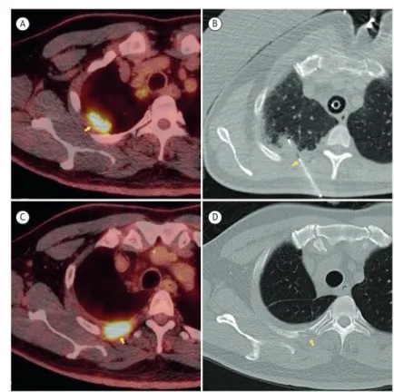
A 56-year-old male patient underwent percutaneous biopsy of a nodule in the right lung apex (Figure 1A). The tip of a 19-gauge coaxial needle was positioned in the posterior chest wall (Figure 1B), and six samples of the lesion were obtained with a 20-gauge core needle. The pathological analysis revealed squamous cell carcinoma. Using an anterior approach, we performed right upper lobectomy with tumor-free margins. At 6 months of follow-up, a positron emission tomography-CT scan of the chest showed an 18F- luorodeoxyglucose-avid soft
tissue mass (Figure 1C) in the T3-4 interspace, along the biopsy tract, as well as bone erosion of the right third rib posteriorly (Figure 1D), suggesting tumor seeding. A subsequent CT scan of the chest, obtained
two months later, conirmed local disease progression.
We then performed en bloc resection with disease-free
pleural margins, and the pathological analysis conirmed
that tumor seeding had occurred.
Tumor seeding along the biopsy route is exceedingly rare. Certain factors, such as the use of large-bore cutting needles, increase the risk of such tumor cell dissemination, that risk also being greater when the tumor is an adenocarcinoma.
RECOMMENDED READING
Kim JH, Kim YT, Lim HK, Kim YH, Sung SW. Management for chest wall implantation of non-small cell lung cancer after ine-needle aspiration biopsy; Eur J Cardiothorac Surg. 2003;23(5):828-32. http://dx.doi. org/10.1016/S1010-7940(03)00095-2
Figure 1. Nodule in the right lung apex and percutaneous biopsy of the same: in A, positron emission tomography-CT (PET-CT) scan showing the nodule (arrow); in B, CT scan showing the point of insertion of the coaxial needle (arrow); in C, PET-CT scan after 6 months of follow-up, showing an 18F-luorodeoxyglucose-avid soft tissue mass; and in D, CT scan after 6 months of follow-up, showing bone erosion of the right third rib posteriorly (arrow).
A
C
B
D
J Bras Pneumol. 2016;42(1):71-71