Wegener Granulomatosis: Otologic Manifestation
as First Symptom
Carla Fabiane da Costa
1Jose Fernando Polanski
11Department of Otorhinolaryngology, Hospital Universitario
Evangelico de Curitiba, Curitiba, PR, Brazil Int Arch Otorhinolaryngol 2015;19:266–268.
Address for correspondenceCarla Fabiane da Costa, Resident Doctor, Department of Otorhinolaryngology, Hospital Universitário Evangélico de Curitiba, Rua Pedro Viriato Parigot de Souza 3288, Curitiba, PR 81200100, Brazil (e-mail: [email protected]).
Introduction
Wegener granulomatosis (WG) is a rare multisystem autoim-mune disease of unknown etiology.1,2It is a systemic vasculi-tis, affecting small and medium-sized vessels of the upper and lower respiratory tract and kidneys, including necrotizing granulomatous inflammation.2,3WG has a spectrum of clinical presentations that includes recurrent respiratory infection, renal manifestations, and nonspecific systemic symptoms. Otologic manifestations are occasionally thefirst to appear in 20 to 60% of cases of WG, and serous otitis media is the most common presentation in ear disease.4–6However, WG is not a
common diagnosis in ear, nose, and throat (ENT) practice. We describe a case of a 50-year-old woman with severe bilateral hearing loss as thefirst symptom of the disease.
Differential Diagnosis
The differential diagnosis of WG with otologic manifestations may include tuberculous otitis media, cholesteatoma, Langer-hans cell histiocytosis, neoplastic diseases, and other forms of vasculitis, sarcoidosis, and systemic lupus erythematosus.
Tuberculous otitis media is characterized by painless otorrhea that fails to respond to the usual antimicrobial
treatment in a patient with evidence of tubercle infection elsewhere, followed by multiple tympanic membrane perfo-rations, abundant granulation tissue, bone necrosis, and preauricular lymphadenopathy. Deafness is out of proportion with the apparent degree of development of disease seen in the otoscopy.
An ear exam may show a pocket or perforation in the eardrum often with drainage when cholesteatoma occurs.
Sarcoidosis is an idiopathic disease that presents in ana-tomic areas of concern to otorhinolaryngologists. It can cause dysfunction of both auditory and vestibular systems. In patients known previously to have sarcoidosis, this disease should be seriously considered. In patients presenting with otologic disorders and associated facial nerve paralysis or other neuropathies, uveitis, granulomatous meningitis, or diabetes insipidus, sarcoidosis should be suspected. An ex-amination of the eyes as well as a chest X-ray is imperative. Sudden andfluctuating neurosensory hearing loss has been reported.
Case Report
The patient presented with complaints of severe and pro-gressive bilateral hearing loss with onset 6 months Keywords
►
Wegener
granulomatosis
►
otitis media
►
hearing loss
Abstract
Introduction
Wegener granulomatosis is a systemic vasculitis affecting small and
medium-sized vessels of the upper and lower respiratory tract and kidneys.
Objective
To describe a case of Wegener disease with atypical manifestation.
Resumed Report
We describe the case of a 50-year-old woman with chronic otitis media
and sensorineural hearing loss as the primary symptoms, without other manifestations.
Conclusion
In cases of acute ear manifestations with or without hearing loss and with
poor response to usual treatments, Wegener granulomatosis should be included among
the possible etiologies. After adequate diagnoses and treatment of this rare disease,
there was favorable evolution.
received May 9, 2014
accepted after revision July 1, 2014
published online November 11, 2014
DOI http://dx.doi.org/ 10.1055/s-0034-1387164. ISSN 1809-9777.
Copyright © 2015 by Thieme Publicações Ltda, Rio de Janeiro, Brazil
Case Report
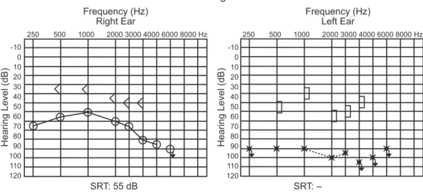
previously. Symptoms evolved with otorrhea and pain refractory to broad-spectrum antibiotics and nonsteroidal anti-inflammatory drugs. During ENT examination, oto-scopy showed bilateral thickening and retraction of the tympanic membrane andfluid in the middle ear, with no other clinical findings. After carrying out the audiometry (►Fig. 1) and emittanciometry, ventilation tubes were placed bilaterally.
After 1 month, the patient complained of weight loss (7 kg/mo), lack of appetite, fevers, night sweats, asthenia, and dry cough. Laboratory findings revealed mild normo-chromic normocytic anemia, Westergren erythrocyte sedi-mentation elevated rate, elevated C-reactive protein, nonreactive Mantoux tuberculin skin test, negative sputum smear, and negative HIV antibody.
Computed tomography of the temporal bone (►Fig. 2) and audiometry were performed. Chest computed tomography showed the presence of pulmonary nodules and masses with excavation areas in the left upper and lower lobes, with moderate pericardial effusion (►Fig. 3). Bronchoscopy with transbronchial biopsy showed inconclusive pathologic result, with the presence of discretefibrous thickening and septal fibrosis nodular focus.
The patient was also referred to the Department of Rheu-matology for antineutrophil cytoplasmic antibodies testing, which was positive, and then antiproteinase 3 antibody testing, which was also positive, confirming the diagnosis. 250 500 1000 2000 3000 4000 6000 8000 Hz 250 500 1000 2000 3000 4000 6000 8000 Hz
-10 0 10 20 30 40 50 60 70 80 90 100 110 120
-10 0 10 20 30 40 50 60 70 80 90 100 110 120
Audiogram
Frequency (Hz)Right Ear
Frequency (Hz) Left Ear
Hearing Level (dB) Hearing Level (dB)
SRT: 55 dB SRT: –
Fig. 1 Audiogram before treatment. Abbreviation: SRT, speech reception threshold.
Fig. 2 Computed tomography of the temporal bone.
Fig. 3 Chest computed tomography showed the presence of pul-monary nodules and masses with excavation areas in the left upper and lower lobes and moderate pericardial effusion.
International Archives of Otorhinolaryngology Vol. 19 No. 3/2015
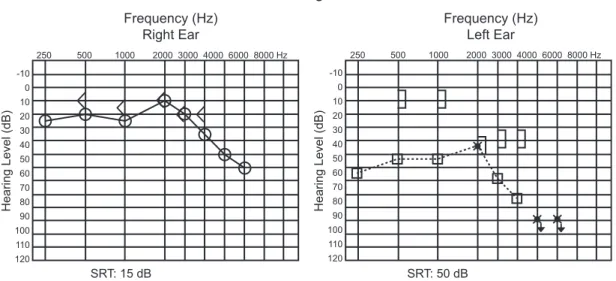
With the onset of steroid (prednisone 60 mg/d), the patient’s symptoms improved and hearing thresholds recov-ered significantly after 1 week of treatment (►Fig. 4). The patient currently uses cyclophosphamide monthly and cor-ticosteroids daily with good clinical and laboratory control.
Discussion
WG is a rare idiopathic disease, immunologically mediated, which affects the small arteries of the upper and lower respiratory tracts and the kidneys, causing inflammation with necrosis, granuloma formation, and vasculitis in these organs.1–4The average age of diagnosis is usually between 20
and 40 years, and males are affected more than females (1.5:1.0).5 Otologic manifestation varies widely, occurring in20 to 60% of the affected patients, and includes serous otitis media, chronic otitis media, sensorineural hearing loss, vertigo, tinnitus, and facial palsy.4,6 Treatment should be started early for a better hearing outcome.7 Pulmonary manifestations occur in 45% of cases at initial presentation and 87% during the course of the disease.8,9Ocular involve-ment in WG can be part of the initial presentation of the disease in 8 to 16% of cases. The majority of studies have reported initial manifestations in ENT, followed by lung, skin, and kidneys.3,10Immunosuppressive drugs are thefirst-line therapy.1,2,5
Final Comments
In cases of acute ear manifestations with or without hearing loss and with poor response to usual treatments, WG should be included among the possible etiologies. As the prognosis
depends on early diagnosis and appropriate treatment, the otolaryngologist plays a decisive role in reducing the mor-bidity and mortality rate in this disease.
References
1 Rossini BAA, Bogaz EA, Yonamine FK, Testa JRG, Penido NdeO. Refractory otitis media as thefirst manifestation of Wegener’s granulomatosis. Braz J Otorhinolaryngol 2010;76(4):541
2 Scalcon MRR, Pereira IA, Rachid Filho A, Paiva ES. Manifestação otológica localizada em paciente com granulomatose de Wegener. Rev Bras Reumatol 2008;48(4):253–255
3 Antunes T, Barbas CSV. Granulomatose de Wegener. J Bras Pneu-mol 2005;31(Suppl 1):S21–S26
4 Pires APBA, Sousa NJA, Sousa RCA, et al. Wegener’s granulomatosis presenting with bilateral facial nerve palsy. Acta ORL 2008;26(4): 209–259
5 Rezende CEB, Rodrigues REC, Yoshimura R, Uvo IP, Rapoport PB. Wegener’s granulomatosis: a case report. Rev Bras Otorrinolar-ingol 2003;69:261–265
6 Cahali S, Souza MMA, Silveira MC, Cabali MB, Cahali RB. Wegener’s granulomatosis—case report with otological manifestation asfirst symptom. Braz J Otorhinolaryngol 1997;63(1):72–74
7 Takagi D, Nakamaru Y, Maguchi S, Furuta Y, Fukuda S. Otologic manifestations of Wegener’s granulomatosis. Laryngoscope 2002; 112(9):1684–1690
8 Fauci AS, Haynes BF, Katz P, Wolff SM. Wegener’s granulomatosis: prospective clinical and therapeutic experience with 85 patients for 21 years. Ann Intern Med 1983;98(1):76–85
9 Cordier JF, Valeyre D, Guillevin L, Loire R, Brechot JM. Pulmonary Wegener’s granulomatosis. A clinical and imaging study of 77 cases. Chest 1990;97(4):906–912
10 Correa JC, Azevedo AG, Rubens J, Rocha G. Granulomatose de Wegener: análise de dois casos. J Bras Med 1985;48(6): 34–38
Audiogram
Frequency (Hz) Right Ear
Frequency (Hz) Left Ear
250 500 1000 2000 3000 4000 6000 8000 Hz 250 500 1000 2000 3000 4000 6000 8000 Hz
-10 0 10 20 30 40 50 60 70 80 90 100 110 120
-10 0 10 20 30 40 50 60 70 80 90 100 110 120
SRT: 50 dB SRT: 15 dB
Hearing Level (dB) Hearing Level (dB)
Fig. 4 Audiogram performed after treatment. Abbreviation: SRT, speech reception threshold.
International Archives of Otorhinolaryngology Vol. 19 No. 3/2015
Wegener Granulomatosis Costa, Polanski