Catheter-associated bloodstream infections (CA-BSI) in
wards: a prospective comparative study between subclavian
and jugular access
Infecção de corrente sanguínea relacionada a cateter venoso central (ICSRC) em enfermarias:
estudo prospectivo comparativo entre subclávia e jugular interna
Gustavo Lopes Gomes Siqueira1, Walkiria Hueb2, Rodrigo Contreira3, Maria Aparecida Nogueron4, Daniela Muniz Cancio4,
Roberto Augusto Cafaro5
Abstract
Background: Positive hemoculture associated with central venous catheters has been studied in intensive care units (ICU), but is still controversial if the internal jugular vein access has a higher incidence of infection than subclavian or femoral vein access.
Objective: To compare catheter-related bloodstream infection (CABSI) rates between internal jugular and subclavian vein access in patients admitted to surgical wards.
Methods: his is a prospective, descriptive and comparative study of 114 central venous catheters placed in 96 patients admitted to the surgical wards of a tertiary-care hospital. he following parameters were studied: local of insertion of the catheter (internal jugular versus subclavian), number of lumens (single versus double) and duration of use (longer or shorter than 14 days), in order to determine their inluence in CABSI rates.
Results: he CABSI rate was 9,64% (11 catheters), with no signiicant statistical diferences regarding the number of lumens (p=0.274), and duration of use (p=0.156). he CABSI rate was higher in the subclavian vein than in the internal jugular vein access (OR 11.2, 95%CI 1.4–90.8; p=0.023).
Conclusions: he internal jugular vein access has a lesser incidence of CABSI than subclavian vein access in patients admitted to surgical wards.
Keywords: catheterization, central venous; cross infection; bacteremia.
Resumo
Contexto: Hemocultura positiva associada a cateter venoso central tem sido estudada em unidades de terapia intensiva (UTI), mas ainda é controverso se o acesso jugular tem maior incidência de complicações infecciosas que o acesso na veia subclávia.
Objetivo: Comparar índice de infecção entre os acessos na jugular interna e os na veia subclávia em pacientes internados nas enfermarias de cirurgia.
Métodos: Estudo prospectivo, descritivo e comparativo com 114 cateteres em 96 pacientes admitidos nas enfermarias de cirurgia de um Hospital Quaternário, tendo como variáveis o local de inserção, número de lumens, tempo de uso, comparando-os com o índice de complicações infecciosas.
Resultados: O índice de infecção foi de 9,64% (11 cateteres), sem signiicância estatística quando comparados o número de lumens (mono versus duplo) e infecção (p=0,274); também sem signiicância estatística a comparação entre o tempo de uso (≥14 dias) e infecção (p=0,156). Comparando os acessos jugular e subclávia, encontramos signiicância estatística tendo infecção em 17,2% na subclávia e 1,8% na jugular, com p=0,005. Índice de Hemocultura positivo associado a cateter venoso central foi maior no acesso subclávia quando comparado com jugular interna, com OR 11,2, IC95% 1,4–90,9; p=0,023.
Conclusões: O acesso venoso central na jugular interna tem menor risco de infecção se comparado com subclávia em enfermarias.
Palavras-chave: cateterismo venoso central; infecção hospitalar; bacteriemia.
Study carried out at Irmandade Santa Casa de Misericórdia de São Paulo – São Paulo (SP), Brazil.
1 Staf physician, Sector of Vascular and Endovascular Surgery, Hospital João XXIII – Campina Grande (PB), Brazil; Ex-Resident of the Department of Vascular Surgery at Santa Casa de São Paulo
– São Paulo (SP), Brazil.
2 Staf physician, Department of Vascular Surgery; Head of Vascular Access Group (GAV), Santa Casa de São Paulo – São Paulo (SP), Brazil; Member of the Vascular Access Society. 3 Assistant Staf Physician, Sector of Infectology, Santa Casa de São Paulo – São Paulo (SP), Brazil.
4 Nurse of Grupo de Acessos Venosos (GAV), Santa Casa de São Paulo – São Paulo (SP), Brazil. 5 Chief of Surgery, Department of Vascular Surgery, Santa Casa de São Paulo – São Paulo (SP), Brazil.
Introduction
The development of multiple techniques and the technological advances in vascular access procedures have prolonged and saved the lives of innumerable pa-tients. However, vascular access procedures are not free of complications that can sometimes be catastrophic. For this reason, great care should be taken in the care of such patients from the moment of catheter insertion through catheter removal.
In the United States, it is estimated that about 5 million central venous catheters (CVC) are inserted in patients every year. Catheter-related bloodstream infection (CABSI) is the third most common cause of
nosocomial infection (14%)1. As a consequence,
hospi-tal length of stay is increased from 7 to 19 days, with
mortality rates as high as 25%2,3.
Risk factors for this complication are: a) duration of catheter use; b) type of catheter; c) the number of lumens; d) type of infusion solution; e) insertion technique; f )
insertion site4,5. It is well established that, for central
ve-nous catheters, the infection rate is higher when the fem-oral access, rather than the jugular or subclavian access is used. There is no consensus in the literature, however, when the latter two insertion sites are compared. Some papers have shown higher infection rates for the internal
jugular access, but only in ICU patients5,6.
There are few published studies of patients admitted to settings other than the ICU. Marshall et al. performed a search on the National Library Medicine in 2007 us-ing the key words “bloodstream”, “infection” and “non-ICU”, but were able to find only four papers. The first large observational and descriptive study on this subject was conducted in Germany and published in 2006. The authors found an infection rate of 4.3/1000 days of use, which is higher than the rate of 1.8/1000 days of use in
the ICU8.
The first large observational study carried out in the USA in 2007, showed an infection rate in ICUs
sim-ilar to that from Germany7 — a controversial result, for
it was expected that the infection rate would be lower in patients with less severe disease outside the ICU. A
cross-sectional study published by Trick et al.9 in 2004
showed that 83% of all questionable catheter insertions
were performed in the ward (1.8% in ICU vs 8.5% in
ward), which suggests that one of the factors predis-posing to catheter-related infection in wards would be the large number of questionable insertions. There have been no studies in Brazil or in the international
literature addressing the issue of insertion sites as the cause of (CABSI).
Thus, due to the high frequency of CVC insertion in surgical patients, the severe complications of such pro-cedures, the economic impact of bacteremia or sepsis in the treatment of those patients, and the dearth of studies on catheter insertion in the ward, we decided to study which venous insertion sites are most frequently associ-ated with (CABSI).
Patients and Methods
From March 11 through June 11 2009, a prospective, descriptive and comparative study was carried out in the
surgical wards of Hospital Central da Santa Casa de São
Paulo. A total of 96 patients had 114 catheters, evaluated by filling a protocol and evaluated from the day of inser-tion to the day of removal.
Inclusion criteria: all patients admitted to a ward who required central venous access, had the catheter in-serted through the jugular or subclavian veins and had the protocol filled correctly.
Exclusion criteria: inadequate filling of the protocol and catheter insertion by venous accesses other than the subclavian and jugular veins.
Catheter insertion was performed by anesthesiology and surgery resident physicians in operating rooms or in the surgical wards, always using aseptic technique. There was no distinction in our protocol between catheters in-serted in either setting. The choice of jugular or subcla-vian access was random, and the conversion from one site to the other during the procedure was not reported in the protocol.
Information on the outcome and complications of each CVC was collected and reported on the protocol by two nurses of the Vascular Access Group (GAV) of the
Santa Casa de São Paulo.
The criteria for the diagnosis of CABSI were: paired blood cultures collected from a peripheral vein and from the CVC, with the culture from the CVC turning positive earlier and with a higher number of micro-organisms than the peripheral blood; and the growth of the same micro-organism from both samples; clinical signs of in-fection such as fever and/or chills; and the exclusion of other causes of bloodstream infection. Our criteria were
based on the CDC Guidelines 20026. The time lag
be-tween the positive culture from the CVC to the periph-eral blood sample was 120 minutes. According to Bouza
CABSI diagnosis, this scheme presents higher sensitivity and negative predictive values. The onset of symptoms of sepsis more than 48 hours after the patient’s admission to the ward was also a diagnosis criterion, according to the
infection protocol described by Marschall et al.7.
Blood culture collection was performed according to the institution’s protocol: 10 mL of blood collected from the catheter after discarding the initial 10 mL and 10 mL of blood collected from a peripheral vein were immedi-ately sent to analysis in separate test tubes.
All cases of infection received a final evaluation from an infectologist of GAV.
All catheters were evaluated by the medical team, but the patient’s attending physician made the decision of removing or not the catheter. All catheters that were removed had positive blood cultures.
Regarding statistical analysis, all variables were sub-mitted to descriptive analysis. Quantitative variables were assessed according to minimum and maximum values, calculation of mean, standard deviation and median. For qualitative variables, absolute and relative frequencies
were calculated. The Student t test was used to compare
means. The chi-square test was used to evaluate propor-tion homogeneity, and when expected frequencies were less than 5, the Fisher Exact test was used. To obtain pre-dictive factors of death, the model for multivariate analy-sis was adjusted. Sensitivity and specificity values were determined by the Receiver Operating Characteristic (ROC) curve, while Odds Ratio was obtained by logistic regression. The software used for statistical analysis was the SPSS 15.0 software, with significance level set at 5%.
The study protocol was approved by the Ethics
Committee of Irmandade Santa Casa de Misericórdia de
São Paulo (protocol 030/10).
Results
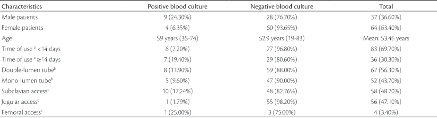
During the study, 114 catheters from 96 patients were analyzed, with results depicted in Table 1. The group of patients was comprised of 35 females (36.4%) and 61 males (63.5%), with mean age of 59 years, and mean hospital stay of 18 days. Catheter-related infection was identified in 9.64% of the cases (11 catheters).
Mean catheter duration of use was 12.3 days (1 to 69 days), and 31.3% of all catheters were used for more than 15 days. No protocol of catheter removal or exchange
(CDC guidelines) was necessary6. The statistical
analy-sis on days of use and infection did not show significant correlations (p=0.156), although 19.4% of catheters (7 out of 36) used for more than 14 days had to be removed for infection.
Regarding the number of lumens, one case had to be excluded from the sample because the number of cathe-ter lumens was not informed. Out of the total, 54.8% (62) were double-lumen catheters and 45.1% (51) were sin-gle-lumen catheters. In the comparative analysis, 4 out of the 51 single-lumen catheters (7.84%) and 7 out of the 62 double-lumen catheters (11.9%) presented infection, without statistically significant differences (p=0.274).
The micro-organisms found in blood cultures were similar to those reported in literature, that is, polymi-crobial in 2 cases 918.1%) and monomipolymi-crobial in 9 cases
(81.8%). Negative-coagulase staphylococcus was the most
common finding, with 5 positive cultures, followed by
Pseudomonas aeruginosa, identified in 4 cultures. The subclavian vein was the access in 58 cases (50.9%) and the jugular vein in 56 cases (49.1%). Regarding the catheter insertion site, statistical analysis showed infec-tion rates of 17.2% in the subclavian vein and 1.8% in
Characteristics Positive blood culture Negative blood culture Total
Male patients 9 (24.30%) 28 (76.70%) 37 (36.60%)
Female patients 4 (6.35%) 60 (93.65%) 64 (63.40%)
Age 59 years (35-74) 52.9 years (19-83) Mean: 53.46 years
Time of use a <14 days 6 (7.20%) 77 (96.80%) 83 (69.70%)
Time of use a ≥14 days 7 (19.40%) 29 (80.60%) 36 (30.30%)
Double-lumen tubeb 8 (11.90%) 59 (88.00%) 67 (56.30%)
Mono-lumen tubeb 5 (9.60%) 47 (90.00%) 52 (43.70%)
Subclavian accessc 10 (17.24%) 48 (82.76%) 58 (48.70%)
Jugular accessc 1 (1.79%) 55 (98.20%) 56 (47.10%)
Femoral accessc 1 (25.00%) 3 (75.00%) 4 (3.40%)
Table 1. Characteristics comparison between infected and non-infected catheters.
CA–BSI– Catheter-related bloodstream infections; a Non signiicant p value (p=0.18); b Non signiicant p value (p=0.246); c p=0.008; One of the 13 medical reports did not specify what the site
the jugular vein (p=0.005) (Figure 1). The Relative Risk (Odds Ratio) of infection in subclavian vein compared to jugular vein was 11.2 times higher (95%CI 1.4–92.8; p=0,023).
Discussion
Central venous catheter-related infections have been extensively studied in the literature Most studies, however, have been carried out at ICUs, with severely ill patients, who are difficult to manage and often in
venti-latory support. Climo et al.11 published a study of 2,459
patients in 2003. Central venous access was used in 29% of the patients; 55% of the ICU patients and 24% of the ward patients had central venous catheters. However, the absolute number of patients in the ward was higher
than in the ICU (506 vs 212), which reflects on the
im-portance of care and preparation for catheter insertion. The objective of our study was to evaluate patients from the surgical wards, not comparing them to those from the ICU.
Studies have shown a 1 to 13% prevalence rate of
positive blood cultures related to CVCs12-15; In our
pa-tients we have found a rate of 9.64% (11 catheters). This is a value in the upper normal range high and concern-ing value, but we should be taken into account that most CVCs were inserted by inexperienced first-year
residents. According to Bernard and Stahl13, the risk of
mechanical and infectious complications of CVC inser-tions depend on the operator’s experience (cutoff num-ber: 50 procedures). A significant difference has been observed in the infectious complication rate of proce-dures performed by experienced (25%) compared to
in-experienced operators (56%)16.
There is a scant number of studies in the literature on catheter-related infections in settings other than the ICU. The first study conducted in the USA, published
by Marschall in 20077, showed an infection rate similar
to that found in ICUs, leading to believe that in settings other than the ICU the risk of catheter-related infection is higher, probably because more unnecessary proce-dures are performed and the level of care is lower than in the ICU. .
Regarding the mean duration of catheter use of 12.3 days, even though most literature reports
recom-mend removing the catheter after 14 days of use17-19,
we decided to follow CDC guidelines and remove only the catheters with clear evidence of infection. The wide variation of duration of catheter use on our series (from 1 to 69 days), with 30% used for longer than 14 days, should not change the recommendation of removing or exchanging the catheter after 14 days of use, for, after that period of time, the catheter becomes more
suscep-tible to infection18,19. Despite the fact that the infection
rate was 19.4% for the catheters used longer than 14 days and only 5.1% for catheters used for less time, no statisti-cal significance was observed.
he insertion site that carries the lowest risk of infec-tion has long been a subject of controversy in the litera-ture. Until the controversy is resolved, each hospital has to establish its own protocol. According to CDC Guideline
20026, the preferred insertion site regarding the risk of
infection is the subclavian vein, but no deinitive clinical trial has been conducted to clarify this issue.
Deshpande et al.20 did not find statistically
signifi-cant differences on the rates of catheter colonization or infection between three different insertion sites (jugu-lar, subclavian and femoral) in ICU patients. Merrer et
al.5 conducted a more specific trial and showed that the
femoral catheters had a higher risk of infection than
subclavian catheters (19.8% vs 4.5%, p<0.001), but a
comparison between the jugular and subclavian veins was not made. In a study published in 2005, Lorente
et al.21 evaluated 2,595 catheters and found a
statisti-cally significant difference of infection rate between three sites: femoral access was associated with higher incidence of infection compared to the other accesses, and the jugular access was associated with a signifi-cantly higher incidence of infection, compared to the subclavian access (RR: 3.1; p=0,005).
The femoral access tends to have a higher infec-tion rate, probably from the fact that the groin skin has a dense bacterial flora. The higher prevalence of
Figure 1. Jugular vein versus subclavian vein infection.
20.0
In
fe
ct
io
n (%)
18.0 16.0 14.0 12.0 10.0 8.0 6.0 4.0 2.0 0.0
J SC
Local p=0.005
1.8
17.2
infection in the jugular access compared to the subcla-vian access remains under investigation. It is probably due to two factors: (1) proximity to the oral cavity; (2) higher density of the local bacterial flora due to the high local temperature and difficulty of keeping
occlu-sive bandages21. The limitation of all studies is that they
were carried out in the ICU setting, with sicker patients with fever, some of them on ventilatory support with endotraqueal tube or thacheostomy and difficulty to clear oral secretions.
The present study found a lower incidence of jugu-lar access infection compared to the subclavian access. This finding, which differs from the literature, is prob-ably explained by the fact that ward patients do not pres-ent the difficult clinical problems seen in ICU patipres-ents. One cannot state however that the jugular access has less risk of infection, for further studies with larger samples would be necessary to prove such statement. Our pa-tients were not randomized according to the admission diagnosis, and even though it has been published that
critical patients present lower rates of infection22, several
yet unidentified factors may be responsible for catheter-related infections.
Conclusion
Our results show superiority of the jugular access over the subclavian access regarding the incidence of CABSI in settings other than the ICU.
Acknowledgment
he authors thank Raisa Zoraide Cunha de Melo for the original text revision and contextual structuring.
References
1. Richards M, Edwards J, Culver D, Gaynes R. Nosocomial infections in combined medical-surgical intensive care units in the United States. Infect Control Hosp Epidemiol. 2000;21(8):510-15.
2. Rosenthal V, Guzman S, Pezzotto S, Crnich C. Efect of an infec-tion control program using educainfec-tion and performance feedback on rates of intravascular device-associated bloodstream infec-tions in intensive care units in Argentina. Am J Infect Control. 2003;31(7):405-9.
3. Orsi G, Di Stefano L, Noah N. Hospital-acquired, laboratory-con-irmed bloodstream infection: increased hospital stay and direct costs. Infect Control Hosp Epidemiol. 2002;23(4):190-7.
4. Safdar N, Kluger DM, Maki DG. A review of risk factors for catheter-related bloodstream infection caused by percutaneously inserted, noncufed central venous catheters: implications for preventive strategies. Medicine (Baltimore). 2002;81(6):466-79.
5. Merrer J, De Jonghe B, Golliot F, Lefrant JY, Rafy B, Barre E, et al. Complications of femoral and subclavian venous catheteriza-tion in critically ill patients: a randomized controlled trial. JAMA. 2001;286(6):700-7.
6. O’Grady NP, Alexander M, Dellinger EP, Gerberding JL, Heard SO, Maki DG et al. Guidelines for the prevention of intravascular cath-eter-related infections. Am J Infect Control. 2002;30(8):476-89.
7. Marschall J, Leone C, Jones M, Nihill D, Fraser VJ, Warren DK. Catheter-associated bloodstream infections in general medical patients outside the intensive care unit: a surveillance study. Infect Control Hosp Epidemiol. 2007;28 (8):905-9.
8. Vonberg RP, Behnke M, Gefers C, Sohr D, Ruden H, Dettenkofer M, et al. Device-associated infection rates for non-intensive care unit patients. Infect Control Hosp Epidemiol. 2006;27(4):357-61.
9. Trick WE, Vernon MO, Welbel SF, Wisniewski MF, Jernigan JA, Weinstein RA. Unnecessary use of central venous catheters: the need to look outside the intensive care unit. Infect Control Hosp Epidemiol. 2004;25(3):266-8.
10. Bouza E, Alvarado N, Alcala L, Perez MJ, Rincón C, Muñoz P. A randomized and prospective study of 3 procedures for diagnosis of Catheter-Related bloodstream infection without catheter with-drawal. Clinical Infectious Diseases 2007;44:820-6.
11. Climo M, Diekema D, Warren DK, Herwaldt LA, Perl TM, Peterson L, et al. Prevalence of the use of central venous access devices with-in and outside of the with-intensive care unit: results of a survey among hospitals in the prevention epicenter program of the Centers for Disease Control and Prevention. Infect Control Hosp Epidemiol. 2003;24(12):942-5.
12. Goetz AM, Wagener MM, Miller JM, Muder RR: Risk of infection due to central venous catheters: efect of site of placement and catheter type. Infect Control Hosp Epidemiol. 1998,19(11):842-5.
13. Richet H, Hubert B, Nitemberg G, Andremont A, Buu-Hoi A, Ourbak P, et al. Prospective multicenter study of vascular-cath-eter-related complications and risk factors for positive central-catheter culture in intensive care unit patients. J Clin Microbiol. 1990;28(11):2520-5.
14. Moro ML, Vigano EF, Cozzi Lepri A. Risk factors for central venous catheter-related infections in surgical and intensive care units. he Central Venous Catheter Related Infections Study Group. Infect Control Hosp Epidemiol. 1994;15(4 Pt 1):253-64.
15. Sadoyama G, Gontijo Filho PP. Comparison between the jugular and subclavian vein as insertion site for central venous catheters: microbiological aspects and risk factors for colonization and infec-tion. Braz J Infect Dis. 2003;7(2):142-8.
16. Bernard RW, Stahl WM. Subclavian vein catheterizations: A prospective study. I. Noninfectious complications. Ann Surg. 1971;173(2):184-90.
17. Richet H, Hubert B, Nitemberg G, Andremont A, Buu-Hoi A, Ourbak P, et al. Prospective multicenter study of vascular-catheter-related complications and risk factors for positive central-catheter cultures in intensive care unit patients. J Clin Microbiol. 1990; 28(11):2520-25.
19. Corona ML, Peters SG, Narr BJ, hompson RL. Infections related to central venous catheters. Mayo Clin Proc. 1990;65(7):979-86.
20. Deshpande KS, Hatem C, Ulrich HL, Currie BP, Aldrich TK, Bryan-Brown CW, Kvetan V. he incidence of infectious complications of central venous catheters at the subclavian, internal jugular, and femoral sites in an intensive care unit population. Crit Care Med. 2005;33(1):13-20; discussion 234-5.
21. Lorente L, Henry C, Martin MM, Jimenez A, Mora ML. Central ve-nous catheter-related infection in a prospective and observational study of 2,595 catheters. Crit Care. 2005;9(6):R631-5.
22. Mnatzaganian G, Galai N, Sprung CL, Zitser-Gurevich Y, Mandel M, Ben-Hur D, et al. Increased risk of bloodstream and urinary in-fections in intensive care unit (ICU) patients compared with pa-tients itting ICU admission criteria treated in regular wards. J Hosp Infect. 2005;59(4):331-42.
Correspondence Gustavo Lopes Gomes Siqueira Rua Aureliano Coutinho, 77, apto 93 CEP 01224-020 – São Paulo (SP), Brazil E-mail: gustavomed@yahoo.com