Abstract
Objectives: To assess the impact of increased thoracic kyphosis on pulmonary function and functional capacity in children and adolescents with cystic ibrosis (CF) and to verify the inluence of disease severity, age and nutritional status on this deformity.
Method: This was a cross-sectional, analytical study conducted at a university hospital. It included CF patients with conirmed diagnosis and without pulmonary exacerbation. The sample was submitted to postural assessment, spirometry (FEV1, FVC and FEV1/FVC) and 6-minute walk test distance (6-MWT distance). Data were analyzed using the Mann Whitney test, Spearman correlation and logistic regression.
Results: Forty-two patients were enrolled, 61.9% presented increase of thoracic kyphosis. There was no difference in values of FEV1, FVC, FEV1/FVC and 6-MWT distance between the groups with or without thoracic kyphosis (p = 0.407; p = 0.756; p = 0.415; p = 0.294). In the group without alteration, patients with more disease severity had a mean FEV1 of 74.1±21.9% and FVC of 79.8±18.7% while in those of lesser severity higher values were found (95.6±12.2% and 97.6±13.2%, respectively) (p = 0.027 and p = 0.027). The presence of kyphosis was correlated with age (p = 0.048) but not with severity (p = 0.151) and body mass index (p = 0.088).
Conclusions: There was a high prevalence of increased thoracic kyphosis in children and adolescents with CF. The deformity did not affect pulmonary function and functional capacity and there was no relationship with disease severity. Regardless of posture, worsening of disease severity determined worsening of pulmonary function.
J Pediatr (Rio J). 2012;88(4):310-6: Posture, cystic ibrosis, pulmonary function.
O
riginala
rticleCopyright © by Sociedade Brasileira de Pediatria
310 1. MSc in Sciences. Universidade Estadual de Campinas (UNICAMP), Campinas, SP, Brazil. 2. Master’s candidate, Saúde da Criança e do Adolescente. UNICAMP, Campinas, SP, Brazil. 3. MSc, Saúde da Criança e do Adolescente. UNICAMP, Campinas, SP, Brazil.
4. Associate tenured professor, Departamento de Pediatria, Faculdade de Ciências Médicas (FCM), UNICAMP, Campinas, SP, Brazil. Laboratório de Fisiologia Pulmonar, Centro de Investigação em Pediatria da FCM, UNICAMP, Campinas, SP, Brazil.
5. PhD, Saúde da Criança e do Adolescente. UNICAMP, Campinas, SP, Brazil. Coordinator, Serviço de Fisioterapia em Pediatria, FCM, Hospital de Clínicas, UNICAMP, Campinas, SP, Brazil. Coordinator, Pós-Graduação em Fisioterapia Pediátrica, UNICAMP, Campinas, SP, Brazil.
6. PhD, Saúde da Criança e do Adolescente, UNICAMP, Campinas, SP, Brazil. Professor Graduação e Pós-Graduação em Fisioterapia, Universidade do Estado de Santa Catarina (UDESC), Florianópolis, SC, Brazil.
No conflicts of interest declared concerning the publication of this article.
Suggested citation: Okuro RT, Côrrea EP, Conti PB, Ribeiro JD, Ribeiro MA, Schivinski CI. Influence of thoracic spine postural disorders on cardiorespiratory parameters in children and adolescents with cystic fibrosis. J Pediatr (Rio J). 2012;88(4):310-6.
Manuscript submitted Jan 17 2012, accepted for publication Apr 25 2012. http://dx.doi.org/10.2223/JPED.2206
Inluence of thoracic spine postural disorders
on cardiorespiratory parameters in children
and adolescents with cystic ibrosis
Renata Tiemi Okuro,1 Ester Piacentini Côrrea,2 Patrícia Blau Margosian Conti,3
José Dirceu Ribeiro,4 Maria Ângela Gonçalves Oliveira Ribeiro,5 Camila Isabel Santos Schivinski6
Introduction
The increase in life expectancy in cystic ibrosis (CF) involves the development of secondary complications related to the musculoskeletal system.1 The occurrence of
changes in body posture that can harm functions related to the cardiopulmonary system is among them.2
This is because postural abnormalities have a strong inluence on the respiratory effort, as this determines
a permanent stimulus on the musculoskeletal support, potentially aggressive in age groups in development.3,4
In CF, pulmonary disease exerts a positive pressure, repeated over and over the thoracic framework, resulting in a thoracic kyphosis. This deformity is the result of the obstructive process and the constant coughing episodes caused by the hypersecretion characteristic of the disease. Its presence may contribute to declining respiratory function.6
It is recommended that interventions be performed in an attempt to prevent and/or minimize emerging postural abnormalities. Early treatment is crucial in order to minimize the impairment of respiratory function. The pre-puberty phase (8-12 years) is described as the best period to begin this closer attention, because the growth phase is the most suitable for any intervention.6
Therefore, the objective of this study was to assess the impact of increased thoracic kyphosis on respiratory function and on the functional capacity of children and adolescents with CF and to assess the inluence of disease severity, age and nutritional aspects on the presence of this deformity.
Method
A cross-sectional analytical study was carried out at the Cystic Fibrosis Clinic of the Department of Pediatrics, Hospital de Clinicas da Universidade Estadual de Campinas (UNICAMP), Brazil. Included subjects were children and adolescents from 7 to 19 years, followed in regular service, with a diagnosis of CF conirmed by clinical history, altered sweat test (chloride greater than 60 mmol/L) in at least two samples and molecular genetic study. The excluded patients were those dependent on oxygen and those in acute pulmonary exacerbation, according to the Cystic Fibrosis Clinical Score7 and Cystic Fibrosis Foundation scores8. These are instruments routinely used in outpatient consultations.9
The CF outpatient service provides assistance to approximately 150 children and adolescents. All patients followed up by the service within the range deined by the study group were invited to participate in the study, which was performed from January to December, 2011.
The sample was submitted to postural assessment through the New York test (NYT),10,11 examination of
pulmonary function by spirometry and the 6-minute walk test (6MWT). Height and weight anthropometric data, as well as the computation of the body mass index (BMI) were also recorded.
NYT is an objective method of assessing posture that includes 13 body segments. It has a scoring system that allows quantitative analysis and the rating of the assessed postural disorder. Each body segment is scored according to the suitability of their alignment (5 points - normal pattern; 3 - moderate change; and 1 - severe). At the end
of the assessment the points of all segments are added up, and the individual’s posture is classiied as normal (56-65 points), moderate (40-55) or severe (1-39).11 This study
speciically examined the position of the thoracic spine in lateral view, which determines the degree of curvature of this segment (normal, with a slight increase or a high degree of kyphosis). This change was also classiied as normal, moderate or severe, according to the NYT, and patients were grouped according to presence (NYT moderate or severe) or absence (normal NYT) of increased thoracic kyphosis. They were labeled as group with thoracic kyphosis and group without it, respectively. For data analysis, this variable was considered as qualitative.
Pulmonary function was assessed by spirometry (Medgraphics CPFS/D spirometer) in the Pulmonary Function Laboratory, School of Medical Sciences, UNICAMP, following regulations by the American Thoracic Society (ATS, 1995).12
The percentage of the predicted value of the variables of forced expiratory volume in one second (FEV1) and forced
vital capacity (FVC) was considered. A cutoff point of 80% was considered to divide the sample below the predicted value (less than 80%) and above it (80%).
After a 15 minutes rest, the 6MWT was performed to assess the functional capacity of patients with CF. 6MWT was also performed following ATS’ recommendations (ATS, 2002).13 The distance covered (DC) was recorded. The predicted distance for each participant was calculated according to Prienitz et al.14 For data analysis, patients
were grouped according to test performance and DC (DC: below and above the predicted).
The severity of cystic ibrosis was also considered using the Shwachman score (SS) modiied by Doershuk.15
The SS covers items related to general activity, physical examination, nutrition and radiological indings, with a range from 20 to 100 points. Patients were classiied as severe (score below 40), moderate (between 40 and 55), medium (from 56 to 70), good (between 71 and 85) and excellent (from 86 to 100 points). For data analysis, the sample was divided into two groups: the groups of lesser severity (patients classiied as good or excellent by SS) and of greater severity (classiied as severe, moderate and medium),15 and considered dichotomized.
The data were processed using the Statistical Package for Social Sciences 16.0 computer program (SPSS Inc., Chicago, IL, USA). The frequency of the variables for characterizing the sample was determined. The lack of data normality was veriied by using the Kolmogorov-Smirnov and Shapiro-Wilk test. The comparison of the spirometric variables (FVC, FEV1, FEV1/FVC) and of DC in
Variable n %
Sex
Male 19 45.2
Female 23 54.8
Age (years)
Under or equal to 12 years 20 47.6
Over 12 years 22 52.4
BMI
< percentile 5 12 28.6
Between percentile 5 and 50 23 54.8
Between percentile 50 and 85 7 16.7
Above percentile 85 0 0
Severity score
Excellent 4 9.5
Good 22 52.4
Medium 13 30.0
Moderate 3 7.1
Severe 0 0
Global posture
Normal 8 19.0
Moderate 26 61.9
Severe 8 19.0
Thoracic spine
Without thoracic kyphosis 16 38.1
With thoracic kyphosis 26 61.9
FEV1
Below the predicted 19 45.2
Normal/above the predicted 23 54.8
FVC
Below the predicted 13 31.0
Normal/above the predicted 29 69.0
FEV1/FVC
Below the predicted 8 19.0
Normal/above the predicted 34 81.0
DC in 6MWT
Below the predicted 23 54.8
Normal/above the predicted 19 45.2
Table 1 - Sample characterization concerning anthropometric
data, sex, age, disease severity, postural and thoracic spine pattern, spirometry and distance covered in the 6-minute walk test
BMI = body mass index; DC in 6MWT= distance covered in the 6-minute walk test; FEV1 = forced expiratory volume in the first second; FVC = forced vital capacity.
was used to determine the inluence of thoracic kyphosis on spirometric parameters, BMI, age and severity. The relationship between the presence of kyphosis and the spirometric variables (FEV1 and FVC) and functional (DC
in 6MWT) was analyzed by univariate logistic regression, followed by multivariate, using the Forward Wald method. Thoracic kyphosis was considered as an independent variable, and FEV1, FVC and DC in 6MWT were considered
as dependent variables. The signiicance level adopted was 5%.
The project was approved by the Research Ethics Committee of the School of Medical Sciences, UNICAMP (n. 172/2010). All study participants had a term of informed consent signed by parents or guardians.
Results
The study included 42 children and adolescents from 7 to 19 years, mean age 12.47±3.43 years. Patients had a minimum of 12 kg and a maximum of 64.4 kg (mean 30.82±14.71 kg) and a mean height of 1.32±0.23 meters.
Table 1 presents the characteristics of the sample concerning demographics, gender, age, disease severity, postural and thoracic spine pattern, spirometry and DC in 6MWT.
The relation between the presence of alterations in the thoracic spine, according to the NYT classiication, and the variables FEV1, FVC, FEV1/FVC and DC in 6MWT was analyzed.
The mean values of FEV1 in the group without increased
thoracic kyphosis (n = 16) was 84.8±20.4 (percentage compared to the predicted), and in the group with increased thoracic kyphosis (n = 26) it was an average of 78.8±20.7. This difference was not statistically signiicant (p = 0.407). Regarding the values of FVC, the groups with and without increased thoracic kyphosis had averages of 88.7±18.1 and 85.5±18.4, respectively (p = 0.756).
The mean values of FEV1/FVC for the group without
thoracic kyphosis was 95.1±10.6 (percent relative to the predicted value), and in the group with thoracic kyphosis it increased to 91.7±13.7. This difference was not statistically signiicant (p = 0.415). The mean DC in 6MWT in groups without and with increased thoracic kyphosis was 597.6±93.2 and 564.2±67.6 meters, respectively (p = 0.294).
Tables 2 and 3 refer to the distribution of values of FEV1,
FVC, FEV1/FVC and DC in 6MWT compared to SS levels of
disease severity, present in the groups with and without alterations of the thoracic spine.
In the group without thoracic kyphosis, patients with more severe CF had a mean FEV1 of 74.1±21.9%, while the
group of lesser severity showed higher values (95.6±12.2%). This difference was statistically signiicant (p = 0.027). In
FVC, the group without thoracic kyphosis and classiied as less severe had a mean of 97.6±13.2% of the predicted, and the more severe group, 79.8±18.7% (p = 0.027). The FEV1/
FVC ratio in the group without increased thoracic kyphosis and less severe had a mean of 98.3±8.6, whereas in the more severe group the mean was 91.9±12.1 (p = 0.141). In the group with increased thoracic kyphosis, the group of lesser severity had a FEV1/FVC score ratio of 93.7±14.1,
while the group of greater severity, an average 87.2±12.5 (p = 0.120). These data are presented in Table 2.
Table 2 - Distribution of forced expiratory volume in the irst second and forced vital capacity values in relation to cystic ibrosis in groups with and without thoracic kyphosis
FEV1 = forced expiratory volume in the first second; FVC = forced vital capacity; p = non-parametric Mann-Whitney test probability; SD = standard deviation. Values given in percent in relation to the predicted.
Variable n Mean SD Minimum Maximum p
FEV1
Without thoracic kyphosis
Lesser severity 8 95.6 12.2 80.0 115.0
Greater severity 8 74.1 21.9 38.0 112.0 0.027
With thoracic kyphosis
Lesser severity 18 80.3 22.5 32.0 112.0
Greater severity 8 75,3 16.9 41.0 94.0 0.487
FVC
Without thoracic kyphosis
Lesser severity 8 97.6 13.2 81.0 118.0
Greater severity 8 79.8 18.7 52.0 115.0 0.027
With thoracic kyphosis
Lesser severity 18 85.2 19.6 53.0 118.0
Greater severity 8 86.2 16.5 63.0 116.0 0.846
FEV1/FVC
Without thoracic kyphosis
Lesser severity 8 98.3 8.6 83.3 106.9
Greater severity 8 91.9 12.1 73.1 104.5 0.141
With thoracic kyphosis
Lesser severity 18 93.7 14.1 60.4 112.0
Greater severity 8 87.2 12.5 65.1 102.9 0.120
DC in 6MWT n Mean SD Minimum Maximum p
Without thoracic kyphosis
Lesser severity 8 639.8 105.2 486.0 772.8
Greater severity 8 555.5 59.1 429.6 630.0 0.093
With thoracic kyphosis
Lesser severity 18 559.7 64.3 415.2 643.0
Greater severity 8 574.5 78.0 502.8 733.3 0.868
Table 3 - Distribution of distance covered in the 6-minute walk teste values in relation to cystic ibrosis severity in groups with and without
thoracic kyphosis
DC in 6MWT= distance covered in the 6-minute walk test; p = non-parametric Mann-Whitney test probability; SD = standard deviation. Values given in meters.
without and with thoracic kyphosis (639.8±105.2 versus 555.5±59.1 meters, p = 0.093; and 559.7±64.3 versus 574.5±78.0 meters, p = 0.868, respectively) (Table 3).
There was a correlation between thoracic kyphosis and age (r = 0.308, p = 0.048). BMI (r = 0.088, p = 0.581) and disease severity (r = -0.151, p = 0.339) did not correlate with the deformity.
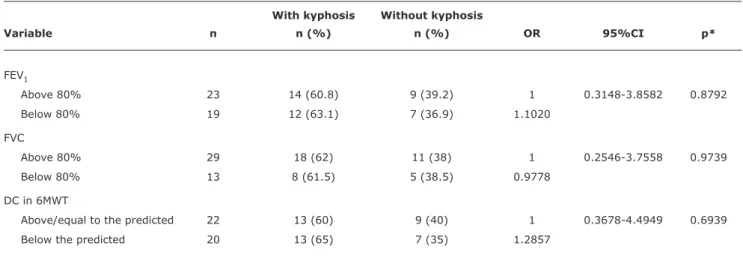
After conducting univariate logistic regression, it was found that there was a harmful association between DC
in 6MWT and children with kyphosis, as well as with FEV1 (odds ratio > 1.00). Patients with abnormal chest
showed DC below predicted and FEV1 below 80% (Table
4). In this study, a patient with CF and kyphosis showed a 0.972 chance having FEV1 below the predicted and
With kyphosis Without kyphosis
Variable n n (%) n (%) OR 95%CI p*
FEV1
Above 80% 23 14 (60.8) 9 (39.2) 1 0.3148-3.8582 0.8792
Below 80% 19 12 (63.1) 7 (36.9) 1.1020
FVC
Above 80% 29 18 (62) 11 (38) 1 0.2546-3.7558 0.9739
Below 80% 13 8 (61.5) 5 (38.5) 0.9778
DC in 6MWT
Above/equal to the predicted 22 13 (60) 9 (40) 1 0.3678-4.4949 0.6939
Below the predicted 20 13 (65) 7 (35) 1.2857
Table 4 - Distribution of frequencies and raw odds ratio in relation to spirometric variables (forced vital capacity and forced expiratory
volume in the irst second) and distance covered in the 6-minute walking test
% = relative frequency; 95%CI = 95% confidence interval; DC in 6MWT= distance covered in the 6-minute walk test; FEV1 = forced expiratory volume in the first second; FVC = forced vital capacity; n = absolute frequency; OR = odds ratio.
* Univariate logistic regression.
Discussion
The close relationship between breathing and posture is a consensus in the management of CF. Postural disorders are considered secondary to pulmonary disease. In this context, this study sought to assess the impact of increased thoracic kyphosis in pulmonary function parameters (spirometry) and functional capacity (6MWT) of children and adolescents with CF.
The identiication of the high prevalence of thoracic kyphosis in the studied population of children and adolescents with CF corroborates indings and conclusions that the literature presents. The pathophysiological process and the progression of pulmonary disease are pointed as having the major responsibility for the development of alterations in body posture. This is because the disease progression with pulmonary hypersecretion leads to hyperinlation and frequent coughing episodes. These events have repercussions on the musculoskeletal system through important muscle shortenings. The shortenings are able to cause postural changes that can alter the respiratory mechanics.2
This relationship between posture and respiratory mechanics has been the focus of discussion by various authors.2,4,16-20 According to them, air trapping is caused by recurrent inlammation, bronchial hypersecretion and decreased elastic recoil of the airways. These factors result in a remodeling of this structure, followed by chronic infection, with a consequent reduction of airway diameter and loss of lung elasticity. The hyperinlated thorax elevates the upper thorax and lattens the diaphragmatic domes. Such compensations raise the thoracic volume and the negativity of the pleural pressure, increasing transpulmonary pressure and airway diameter. The inspiratory muscles gradually
adapt to the new position of the bone structure, which will lead to a shortening of the ibers and a reduction of these muscles’ ability to generate strength.2,16 In individuals with
poor nutritional status, the development of spinal deformities seems to be enhanced.
Given this pathophysiological process and its intimate connection with the rib cage, the posture alteration most commonly seen in CF is increased thoracic kyphosis.4,17-21
This alteration was found in the present study, with a prevalence (62%) similar to that found in the study by Parasa & Maffulli.18 A lower incidence of around 15.1% had
been published in the 1980s.22
The high incidence of musculoskeletal complications in CF has been associated with increasing age and decreasing pulmonary function.4,18,23 Speciically on the posture, it is suggested that the presence of postural alterations has a correlation with reductions in lung function and in the capacity for physical exercise.5,21,24 However, this was not observed in
the current result, in which increased thoracic kyphosis was not related to spirometric data nor to 6MWT performance. The indings of Logvinoff & Erkilla4,17 corroborate those
presented here, because the kyphosis was also prevalent and had no correlation with the spirometric variables.
References
1. Dodge JA, Lewis PA, Stanton M, Wilsher J. Cystic ibrosis mortality and survival in the UK: 1947-2003. Eur Respir J. 2007;29:522-6.
2. Kraemer R, Baldwin DN, Ammann RA, Frey U, Gallati S. Progression of pulmonary hyperinlation and trapped gas associated with genetic and environmental factors in children with cystic ibrosis.
Respir Res. 2006;7:138. increased with age.4
The association between the severity of cystic ibrosis and the impairment of posture does not follow a standard in the literature, just as the relationship between a worse posture and a worse pulmonary function. Unlike the indings presented here, as well as the study of Erkilla et al.,4 Walshaw & Tattersall21 found a correlation between
posture and the severity of CF in adults. Denton et al.22
had found the same in 1981 with a sample of 91 children and adolescents, whose increased thoracic kyphosis was related to the severity of lung disease. The same behavior was published by Henderson & Specter,20 which also found
a correlation between the presence of kyphosis and age. In the results obtained here, although, statistically, the inluence of the severity of cystic ibrosis on pulmonary function of patients with increased thoracic kyphosis was not found, the second logistic regression analysis, even in the absence of this thoracic deformity, children with lesser severity showed higher values of FEV1 and FVC compared
to the more severe patients (95.6±12.2 versus 74.1±21.9, p = 0.027, and 97.6±13.2 versus 79.8±18,7, p = 0.027). Thus, irrespective of the posture, the worsening in disease severity is related to worsening pulmonary function. This probably is associated to the fact that clinical deterioration determines the progression of lung disease.
In contrast, the study by Tejero Garcia et al.19 found, in
a sample of 50 CF patients aged over 16 years, a correlation of disease severity with FEV1, FVC and increased thoracic
kyphosis, and the latter was also related to FEV1. However,
this study was a sample of spirometry values lower than those presented here, as well as with an older population. Moreover, disease severity was classiied by the spirometry data only and not by the broad SS, as done in the current investigation. All these factors may have inluenced the difference in results between studies. Another divergent inding in the study by this author is due to the correlation found between the severity of the disease and the DC in 6MWT, an event that did not happen here, since it was observed that the older the subject, the higher is the DC in 6MWT. There was no statistically signiicant difference in DC in 6MWT between groups of greater and lesser severity, in patients with and without thoracic kyphosis (p = 0.616 and p = 0.068, respectively).
According to literature, the development of thoracic kyphosis has many causes. Among the factors that contribute to this deformity are: reduction of muscle mass,25
of bone mineral density,26 excessive respiratory effort,22 osteopenia24 and chronic pain.26
Its presence is often reported in publications on adult cystic ibrosis populations with a high percentage identiied already in the pubertal phase,27 as is the proile of this study’s
sample. And, according to Elkin,27 even if those individuals
do not have repercussions on lung function yet, there is a need for an early multiprofessional intervention. Lannefors
et al.28 emphasize this preventive idea, considering that there is a high prevalence of increased thoracic kyphosis that can be reversed if treated properly. According to the author, the maintenance of postural alignment reduces the risk of low back pain and spinal complications, besides contributing to the preservation of physical function.28
Longitudinal studies show that the treatment of postural disorders can be successful through assessment methods and appropriate programs.16,21 Exercises for thoracic mobility,
muscle stretching and coordination activities to promote improved posture and chest wall compliance, resulting in the maintenance and optimization of pulmonary function. Patients with more severe pulmonary disease, lower aerobic capacity and more sedentary lifestyles deserve special attention because they are predisposed to lower bone mineral density, higher prevalence of vertebral fracture and development of major thoracic kyphosis increases.19
Although the logistic regression analysis was not statistically signiicant for any of the variables, the odds ratio value of the variables DC in 6MWT and FEV1 show a
tendency for the presence of kyphosis to be a risk factor for lower performance on the 6MWT and for lower spirometric value. A larger sample could sensitize statistical tests and contribute to the identiication of new results.
The absence of a larger sample, characterized by patients more severely impaired and older, may have restricted the identiication of results, as well as the application of a more sensitive instrument of postural assessment. Studies with longitudinal design to assess the evolution of thoracic deformity and the effect of therapeutic interventions, both in the musculoskeletal and respiratory systems, are suggestions for future supplementary investigations.
Conclusion
3. Hodges PW, Heijnen I, Gandevia SC. Postural activity of the diaphragm is reduced in humans when respiratory demand
increases. J Physiol. 2001 (Pt 3);537:999-1008.
4. Lannefors L. Inluences on posture. Eighteenth Annual North American Cystic Fibrosis Conference, St Louis, MO. Pediatr Pulmonol. 2004;27:155-6.
5. Erkkila JC, Warwick WJ, Bradford DS. Spine deformities and cystic ibrosis. Clin Orthop Relat Res. 1978;131:146-50.
6. Massery M. Musculoskeletal and neuromuscular interventions: a physical approach to cystic ibrosis.J R Soc Med. 2005;98 Suppl 45:55-66.
7. Kanga J, Kuhn R, Craigmyle L, Haverstock D, Church D. Cystic ibrosis clinical score: a new scoring system to evaluate acute pulmonary exacerbation. Clin Ther. 1999;21:1343-56.
8. Hafen GM, Ranganathan SC, Robertson CF, Robinson PJ.
Clinical scoring systems in cystic ibrosis. Pediatr Pulmonol. 2006;41:602-17.
9. Santos CI, Ribeiro JD, Ribeiro AF, Hessel G. Análise crítica dos escores de avaliação de gravidade da ibrose cística: estado da arte. J Bras Pneumol. 2004;30:286-98.
10. Althoff SA, Heyden SM, Robertson LD. Posture screening: a program that works. J Phys Educ Recreat. 1988;59:26-32. 11. Santos JB, Moro AR, Cezar MR, Reis PF, Luz JD, Reis DC. Descrição
do método de avaliação postural de Portland State University. Fisioter Brasil. 2005;6:392-5.
12. ATS Committee on Proiciency Standards for Clinical Pulmonary Function Laboratories. ATS statement: guidelines for the
six-minute walk test. Am J Respir Crit Care Med. 2002;166:111-7.
13. Miller MR, Hankinson J, Brusasco V, Burgos F, Casaburi R, Coates A, et al. Standardisation of spirometry. Eur Respir J.
2005;26:319-38.
14. Priesnitz CV, Rodrigues GH, Stumpf C da S, Viapiana G, Cabral CP, Stein RT, et al. Reference values for the 6-min walk test in healthy children aged 6-12 years. Pediatr Pulmonol. 2009;44:1174-9.
15. Doershuk CF, Matthews LW, Tucker AS, Nudleman H, Eddy G, Wise M, et al. A 5 year clinical evaluation of a therapeutic program for patients with cystic ibrosis. J Pediatr. 1964;65:677-93. 16. Sandsund CA, Roughton M, Hodson ME, Pryor JA. Musculoskeletal
techniques for clinically stable adults with cystic ibrosis:
a preliminary randomised controlled trial. Physiotherapy. 2011;97:209-17.
17. Logvinoff MM, Fon GT, Taussig LM, Pitt MJ. Kyphosis and pulmonary function in cystic ibrosis. Clin Pediatr (Phila). 1984;23:389-92.
18. Parasa RB, Maffulli N. Musculoskeletal involvement in cystic ibrosis. Bull Hosp Jt Dis. 1999;58:37-44.
19. Tejero García S, Giráldez Sánchez MA, Cejudo P, Quintana Gallego E, Dapena J, García Jiménez R, et al. Bone health, daily physical activity, and exercise tolerance in patients with cystic ibrosis.
Chest. 2011;140:475-81.
20. Henderson RC, Specter BB. Kyphosis and fractures in children and young adults with cystic ibrosis. J Pediatr. 1994;125:208-12. 21. Tattersall R, Walshaw MJ. Posture and cystic ibrosis. J R Soc Med.
2003;96 Suppl 43:18-22.
22. Denton JR, Tietjen R, Gaerlan PF. Thoracic kyphosis in cystic
ibrosis.Clin Orthop Relat Res. 1981;155:71-4.
23. Massie RJ, Towns SJ, Bernard E, Chaitow J, Howman-Giles R, Van Asperen PP. The musculoskeletal complications of cystic ibrosis.
J Paediatr Child Health. 1998;34:467-70.
24. Conway SP. Impact of lung inlammation on bone metabolism in adolescents with cystic fibrosis. Paediatr Respir Rev.
2001;2:324-31.
25. Elkin SL, Williams L, Moore M, Hodson ME, Rutherford OM.
Relationship of skeletal muscle mass, muscle strength and bone mineral density in adults with cystic ibrosis. Clin Sci (Lond).
2000;99:309-14.
26. Botton E, Saraux A, Laselve H, Jousse S, Le Goff P. Musculoskeletal manifestations in cystic ibrosis. Joint Bone Spine. 2003;70:327-35.
27. Elkin SL, Fairney A, Burnett S, Kemp M, Kyd P, Burgess J, et al.
Vertebral deformities and low bone mineral density in adults with cystic ibrosis: a cross-sectional study. Osteoporos Int.
2001;12:366-72.
28. Lannefors L, Button BM, McIlwaine M. Physiotherapy in infants
and young children with cystic ibrosis: current practice and future developments. J R Soc Med. 2004;97 Suppl 44:8-25.
Correspondence: Renata Tiemi Okuro