http://doi.org/10.1590/2317-6431-2017-1885 ISSN 2317-6431
Cervical vestibular evoked myogenic potentials in
children and adolescents without vestibular complaints
Potencial evocado miogênico vestibular cervical em crianças e
adolescentes sem queixas vestibulares
Bárbara Melissa Pereira da Silva1, Dayane Domeneghini Didoné2, Pricila Sleifer3
ABSTRACT
Introduction: The Cervical Vestibular Myogenic Evoked Potential (cVEMP) has become a reliable and complementary measure of vestibular assessment. The investigation of vestibular disorders in the pediatric population is of great importance because they can have a series of repercussions throughout development. Purpose: To evaluate and analyze cVEMP responses in children and adolescents and compare them among gender, age, and ears. Methods: A cross-sectional study with 35 children and adolescents, 18 females and 17 males, aged 7 to 18 years, with normal hearing thresholds and no otoneurological complaints. All of them underwent a peripheral auditory evaluation and cVEMP. Results: In the analysis, it was observed that the mean latencies of P1 and N1 waves were, respectively, 15.92 ms and 24.32 ms, the amplitude P1/N1 of 36.91 µv and the symmetry ratio presented an average of 22.95%. No statistically significant differences were found comparing gender and ears. The same happened in the asymmetry index between genders. There were also no statistically significant differences in latencies and amplitudes. Conclusion: It was possible to measure values for latency and the amplitude of P1 and N1 waves in children and adolescents. In the present study, there was no statistically significant difference in the comparisons among ears, gender, and age.
Keywords: Child; Adolescent; Vestibular evoked myogenic potentials; Evoked potentials; Vestibular function tests
RESUMO
Introdução: O Potencial Evocado Miogênico Vestibular Cervical (cVEMP) tem se tornado uma medida fidedigna e complementar da avaliação vestibular. A investigação das alterações de ordem vestibular na população pediátrica é de grande importância, pois essas alterações podem acarretar uma série de repercussões ao longo do desenvolvimento.
Objetivo: Avaliar e analisar as respostas do cVEMP em crianças e adolescentes e comparar esses achados entre gênero, idade e orelhas.
Métodos: Estudo transversal, constituído por 35 crianças e adolescentes, 18 do gênero feminino e 17 do masculino, de 7 a 18 anos de idade, possuindo limiares auditivos normais e sem queixas otoneurológicas. Todos realizaram avaliação auditiva periférica e cVEMP. Resultados:
Na análise, observou-se que a média das latências das ondas P1 e N1 foi, respectivamente, 15,92 ms e 24,32 ms, da amplitude de P1/N1 foi de 36.91 µv e a razão de simetria apresentou média de 22,95%. Não foram encontradas diferenças estatisticamente significativas na comparação de gênero e orelhas. O mesmo ocorreu no índice de assimetria que foi comparado entre os gêneros. Na comparação entre grupos de idade, também não foram evidenciadas diferenças estatisticamente significativas nas latências e amplitudes. Conclusão: Foi possível mensurar valores para latência e amplitude das ondas P1 e N1, em crianças e adolescentes. Não houve diferença nas comparações entre as orelhas, gênero e faixa etária.
Palavras-chave: Criança;Adolescente; Potenciais evocados miogênicos vestibulares; Potenciais evocados; Testes de função vestibular
Research conducted at the Department of Health and Human Communication, Universidade Federal do Rio Grande do Sul – UFRGS – Porto Alegre (RS), Brazil. (1) Universidade Federal do Rio Grande do Sul – UFRGS – Porto Alegre (RS), Brazil.
(2) Post Graduate Program in Child and Adolescent Health, Universidade Federal do Rio Grande do Sul – UFRGS – Porto Alegre (RS), Brazil. (3) Department of Health and Human Communication, Universidade Federal do Rio Grande do Sul – UFRGS – Porto Alegre (RS), Brazil.
Conflict of interests: No
Authors’ contribution:BMPS collected the data, analyzed and interpreted the results, and carried out the scientific writing of the study; DDD assisted in the analysis and interpretation of the results and aided in the scientific writing of the study; PS guided the data collection, analysis, and interpretation of results and carried out the scientific writing of the study.
Corresponding author: Bárbara Melissa Pereira da Silva. E-mail: [email protected]
INTRODUCTION
The first reports about evoked vestibular potentials date from 1964, when researchers observed auditory middle latency responses. They found that these responses were affected by the level of contraction of cervical musculature and, therefore, was classified as myogenic responses. Years later, it was observed that these responses were not present in patients with changes in the vestibular system, but were in those with auditory alterations and thereby pointing to the possibility of the responses being of vestibular origin, evidencing a vestibular myogenic potential(1).
The Cervical Vestibular Evoked Myogenic Potentials (cVEMP) evaluates the body balance from myogenic responses. This response is activated by a sound stimulation in high intensity, which activates the saccular macula, the inferior vestibular nerve, and the vestibular-spinal tract descendants. The responses are recorded by the surface electrodes of the cervical muscles in contraction force and head rotation(2).
The assessment of diseases in the vestibular system in pediatric populations is important because changes in this system may involve several effects in the future, such as delayed in motor and learning development, interfering in language, writing, and reading skills. Some studies show that childhood vertigo corresponds to approx. 1% of the consultations in pediatric neurology and approx. 13% of children in audiological evaluations. These numbers may be even higher, due mainly the difficulties in diagnosing and obtaining child anamnesis with dizziness due to their difficulty in naming the discomfort(3).
Differently than adults, children do not know how to describe what they feel, making the diagnosis difficult. For this reason, it is believed that the prevalence is underestimated(4).
Symptoms may vary, such as: undefined malaise, motion sickness, nausea, vomiting, visual disturbance, sudden change in behavior, agitation, sleep disorders, headache, inability to perform coordinated movements, difficulty in play and relating with friends, unfitness for some physical exercises, falls, motor development delay and language, both in writing and speech. These symptoms can lead to psychic impairment, school delay, anxiety, and panic(4).
The analysis and interpretation of vestibular tests are challenging, due to difficulties with cooperation, maintenance of alertness, and nauseating reactions. Therefore, the implementation of a test protocol for children with normative data at appropriate ages is of great importance in vestibular assessment(5).
There is no standard defined in the graphical findings of the current vestibular exams considered normal for the different pediatric age groups, making it more difficult to characterize normal and pathological results. Seventy-four percent of children with hearing loss present some type of vestibular abnormality when assessment with a combination of cervical rotation tests and electrophysiological procedures as vestibular myogenic evoked potentials, in contrast to a 60% rate of change
in myogenic evoked potentials and a 49% rate with isolated cervical rotation test(4).
The vestibular evoked myogenic potential (VEMP) is a complementary test in otoneurological assessment and has several favorable characteristics in many various population, being an objective, non-invasive, fast, and easy test. Besides that, it is inexpensive and does not bring discomfort to the patient(3,6,7,8).
There was a lack of studies in the scientific literature describing the results of cVEMP in children. In Brazil, only one study was found in this population. Researchers describe the P1 with a mean latency of 17.26ms and amplitude of 49.34µV, and the N1 with a mean latency of 24.78ms and amplitude of 66.23µV. The authors also point out that there are no statistical differences between genders and ears(3). In this study, the
individual amplitude of P1 and N1 was analyzed and not the value between the peaks. The values described in the study were similar to those in the international literature, with similar ages, ranging from 4 to 19 years, where the mean P1 latency ranged from 11.3 to 15.4 ms, and the mean N1 latency ranged from 18.2 to 23.7 ms. The mean total amplitude was 126.7 to 160.5, with asymmetry indexes between 16% and 20%(9,10).
This research is justified due to the great importance and applicability in clinical practice of otoneurological research and in order to contribute to the scarce national and international studies on cVEMP in children and adolescents.
Based on the clinical relevance of the subject and the demands of national literature, this study purposes to evaluate cVEMP responses in children and adolescents. In addition, it will analyze the latencies and amplitudes obtained from cVEMP and compare these findings with gender, age, and ears.
METHODS
This research is characterized as an observational and cross-sectional, study. It was approved by the Research Ethics Committee of Universidade Federal do Rio Grande do Sul by protocol 55.975.816. Resolution number 466/12, which deals with research with humans. Therefore, only the children and adolescents who signed the Free and Informed Consent Term (TCLE) participated in this study, in which the methodology was clarified, as well as the risks, discomfort, and confidentiality.
This study includes children and adolescents aged between 7 and 18 years old, without any history of complaints of body balance or otologic disorders and with bilateral normal hearing, according to Davis e Silverman(11). The subjects with genetic
and neurologic complaints, intellectual disabilities, changing in the external or middle ear, with neck rotations unable or who cannot perform the evaluation were excluded.
The information about age, gender, level of education, illness, use of medication, vestibular and learning complaints, among others were obtained through anamnesis. After that, the otoscopy and acoustic immitance measurements (MIA) were performed with an Interacoustics® Impedance Audiometer AT235h. The tympanometric curves were obtained by means of a probe that was inserted in the entrance of the external auditory canal of the patient. The ipsilateral and contralateral acoustic reflexes were investigated in the frequencies of 500, 1000, 2000, and 4000 Hz in both ears. The normal results were considered when the tympanometric curves were type A and acoustic reflexes were present in both ears. The Pure Tone Audiometry was obtained by screening the frequencies of 500, 1000, 2000, and 4000 Hz by air conduction in both ears. In sequence, the vocal audiometry was fulfilled starting with the speech recognition index and after the speech recognition threshold was met. For the percentage of the speech recognition index, 25 words, monosyllabic, were presented in a fixed intensity, 40 dBHL above the tritonal average of 500, 1000, and 2000 Hz by air conduction. For each ear, subjects had to repeat the words correctly(12). For the speech recognition threshold, the
initial intensity was 40 dBHL above the tritonal average air conduction. It was reduced until reaching the level of intensity in which the patient could adequately understand and repeat 50% of the trisyllabic words presented(12). The audiometer used
for tonal and vocal audiometry was the Inventis brand, model Harp Inventis, previously calibrated.
After the audiological assessment, the children and adolescents who fit the inclusion criteria were submitted to cVEMP, which was performed in an acoustically and electrically treated room with the equipment MASBE ATC Plus, brand Contronic®.
The individual was positioned in a comfortable chair. The skin was cleaned with a Nuprep®exfoliating cleanser and gauze. The electrodes were fixed with electrolytic paste (Ten20® conductive) and adhesive tape. The ground electrode was fixed on the forehead (Fz), the positive and negative were fixed on the right and left side of the sternocleidomastoid muscles (ECM), with the negative in the medial part of the muscle and the positive below. The impedance was less than 5
Ω in each derivation and the difference between the electrodes did not exceed 2 Ω. The electroencephalogram (EEG) was performed aimed to capture the spontaneous brain electrical activity in order to verify artifacts that may interfere in the test. The patient was instructed to not cross their legs and/or arms.
The stimuli were presented through earphones (Earphone
TONE™ GOLD) inserted in both ears. The type of auditory stimuli used was the tone burst at a frequency of 500 Hz, intensity of 118 dBHL, with alternating polarity, a band-pass filter of 5-1000 Hz, 200 stimuli per second, and a noise limit of 90 to 100%.
To perform the cVEMP, the patient was instructed to take the maximum lateral rotation of the head to the opposite side to the ear stimulated in order to capture the response of contraction muscle(2,3,9).
The database was made in Excel and analyzed by SPSS (Statistical Package for Social Sciences), version 20.0. The level of statistical significance was 5%. The continuous variables were described as mean, standard deviation, minimum, and maximum. The Student’s t-test for the independent groups (symmetric data distribution) was used to compare the variables and the Mann-Whitney test for the asymmetric data distribution.
RESULTS
The results refer to a sample of 35 children and adolescents, 18 females, and 17 males. In this sample, the overall mean latency was 15.92 in P1 and 24.32 in N1. The mean amplitude was 36.91 (Table 1).
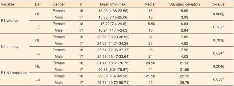
The comparison of variables by gender did not show a statistically significant difference between the ears in each group (Table 2). In the comparison of the variables between the ears, there was also no statistically significant difference between genders in each group (Table 3). The asymmetry index was compared between the genders and there was no statistically significant difference (Table 4).
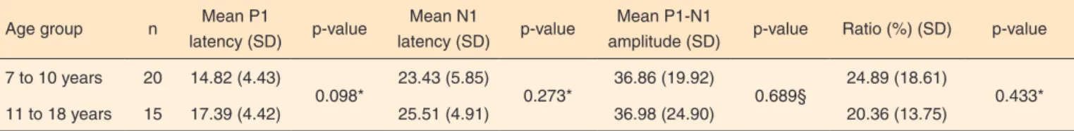
In order to verify the possible associations between age groups and cVEMP results, the subjects were divided into two groups, but there was no statistically significant difference in this comparison (Table 5).
DISCUSSION
There are several studies with different methodologies for obtaining cVEMP, but there is still no consensus on this. In this study, the protocol that was used to perform cVEMP was based on other related studies that objectified standardize this test(2,3,9).
The positioning of the electrodes is one of the most frequently found methodological differences in the literature. The positioning in the medial part of sternocleidomastoid muscle(13,14) used in this study was the most commonly used
technique in the literature, with more consistent results, besides being comfortable for the patient(15,16,17).
Despite the various methods described in the literature for the activation of the sternocleidomastoid muscle(2,18,19,20,21), the
Table 1. Descriptive data of sample
Mean Median Minimum Maximum Standard deviation
Age (years) 10.82 10 7 18 2.73
P1 latency (ms) 15.92 15.73 4.88 30.61 4.96
N1 latency (ms) 24.32 24.43 10.93 37.17 5.92
P1-N1 Amplitude (µV) 36.91 32.29 2.87 99.71 23.77
Ratio (%) 22.95 20.83 0.44 61.94 0.16
Subtitle: ms = milliseconds ; µV= microvolts
Table 2. Comparison of latencies and amplitudes between ears and by sex
Variable Gender Ear n Mean [min-max] Median Standard deviation p-value
P1 latency
Female RE 18 15.28 [4.88-23.55] 16 5.69 0.496*
LE 18 16.72 [7.4-29.6] 15.50 6.84
Male RE 17 15.35 [7.15-25.06] 16 3.93 0.337§
LE 17 16.24 [11.44-24.3] 16 2.84
N1 latency
Female RE 18 22.89 [10.23-36.92] 24 7.05 0.272*
LE 18 25.61 [12.95-37.17] 26 7.56
Male RE 17 24.35 [14.21-34.39] 25 4.62 0.972§
LE 17 24.59 [15.47-33.64] 24 4.03
P1-N1 Amplitude
Female RE 18 31.11 [10.51-70.73] 24.50 21.52 0.669§
LE 18 29.89 [2.87-69.93] 21.50 22.53
Male RE 17 40.88 [6.64-75.87] 44 21.66 0.490*
LE 17 46.71 [10.72-99.71] 42 26.75
§ Mann-Whitney test for non-parametric data; *T Student test for parametric data; p≤0.05
Subtitle: RE = right ear; LE = left ear; min = minimum; max = maximum
Table 3. Comparison of latencies and amplitudes between sexes and by ear
Variable Ear Gender n Mean [min-max] Median Standard deviation p-value
P1 latency
RE Female 18 15.28 [4.88-23.55] 16 5.69 0.868§
Male 17 15.35 [7.15-25.06] 16 3.93
LE Female 18 16.72 [7.4-29.6] 15.50 6.84 0.787*
Male 17 16.24 [11.44-24.3] 16 2.84
N1 latency
RE Female 18 22.89 [10.23-36.92] 24 7.05 0.703§
Male 17 24.35 [14.21-34.39] 25 4.62
LE Female 18 25.61 [12.95-37.17] 26 7.56 0.624*
Male 17 24.59 [15.47-33.64] 24 4.03
P1-N1 amplitude
RE Female 18 31.11 [10.51-70.73] 24.50 21.52 0.204§
Male 17 40.88 [6.64-75.87] 44 21.66
LE Female 18 29.89 [2.87-69.93] 21.50 22.53 0.058*
Male 17 46.71 [10.72-99.71] 42 26.75
§ Mann-Whitney test for non-parametric data; *T Student test for parametric data; p≤0.05
Subtitle: RE = right ear; LE = left ear; min = minimum; max = maximum;
Table 4. Comparison of the asymmetry index by sex
Asymmetry index n Mean [min-max] (%) Median (%) Standard deviation (%) p-value
Female 18 26.83 [1.32-61.94] 27.98 16.94
0.129§
Male 17 18.81 [0.44-60.64] 13.58 15.72
index obtained in this research (22.95%) corroborates with other studies(9,22) indicating that the method used was adequate
for cVEMP recording.
A variety of stimuli used to generate cVEMP was also described in the literature. In this study, a tone burst of 500 Hz was used due to the more effective responses than click stimuli(16,18,23). Frequencies equal or lower than 500 Hz are
more commonly used and evoked homogeneous and reliable responses(2,14,23).
The morphology and characteristics of responses depend on the type of stimuli. To evoke a response, the tone burst needs a lower threshold than click stimuli(24,25).
In this study, all of the subjects had responses in both ears, indicating the integrity of the saccular macula, inferior vestibular nerve, vestibular nuclei, vestibular-spinal and effector muscles(6,26).
In this sample, the P1 mean latency was 15.92 ms and N1 was 23.32 ms. These values corroborate with other studies carried out with the same population and similar methodology, in which P1 mean latency ranged from 11.3 to 15.4 ms and the N1 mean latency ranged from 18.2 to 23.7 ms(3,9,21).
In relation to the analysis of the amplitudes, a mean of 36.91 µV was verified. Different findings were found in the literature, both in studies with adult populations(18) and in
studies with children, in which the amplitude values ranged from 126.7 µV to 160.5 µV(3,9,21). This difference in values can
be justified depending on the equipment used, as there is no published standardization for the MASBE ATC PLUS device of the brand Contronic® for children. Therefore, it is believed that standardization studies are essential for each of the available equipment. In addition, it is assumed that the positioning of the electrodes, even in the mid-part of the ECM muscle, as most studies have reported, may suffer minor changes in the placement site, mainly due to the number of channels of the equipment. The positioning of the reference electrode and the ground electrode can also modify, thereby generating a different capture of muscle contraction at the moment of lateral rotation of the head. Another hypothesis is that the child population presents greater difficulty in maintaining muscle contraction, tiring faster and, therefore, generating a smaller amplitude.
Researchers report that older children with more developed musculature present larger amplitudes. These findings could be related to the variation of sternocleidomastoid muscle thickness(27).
Another important parameter of the analysis is the asymmetry index, which compares the interference of the muscular tonus of one side with respect to the other. It is calculated by the interaural difference of the amplitude, weighted by the mean of the response for each patient(2). In this
study, the average was 22.95%, a result that revalidates those of other studies in the child population(3,9,21).
In this research, there was no statistically significant difference between ears and gender compared to the latencies and amplitudes of waves. These findings agree with another national study that also described no differences between ears(3).
The same fact occurred in other populations that performed cVEMP(1,2,18).
Some studies reported that an increase in amplitude in the male gender, in relation to the female, could occur as a function of muscle strength(1,2), which was not observed in the
present study.
Studies(3,27) have shown that, from infancy to adulthood,
increasing age is accompanied by an increase in P1 and N1 amplitudes and an increase in N1 latency, but this does not occur in the P1 wave. As previously explained, the effect of age on the amplitude of the cVEMP waves is probably related to the variation of the ECM muscle thickness(2,3,27). Therefore,
older children with more developed musculature have larger amplitudes. As no age effects on P1 latency were observed, the increase in N1 latency is supposed to be associated with longer P1 wave duration. Therefore, it is also possibly dependent on muscular factors and not on the conduction velocity of the nerve pathway(3).
In this study, subjects were divided into two groups, one from seven to 10 years and other from 11 to 18 years old in order to verify the possible influence of age on cVEMP results, but no statistically significant difference was observed between groups. It may have occurred due to the size of the sample that is smaller in the older age group than the other.
From this sample, it was possible to characterize the cervical vestibular myogenic evoked potential in children and adolescents. The results of this study, when combined with other studies can be used as a reference for future research and contribute to a more accurate diagnosis of vestibular disorders in this studied population.
It is believed that this research may contribute to the scientific literature of vestibular myogenic evoked potentials, in addition to allowing professionals working in the field of
Table 5. Comparison of variables by age group
Age group n Mean P1
latency (SD) p-value
Mean N1
latency (SD) p-value
Mean P1-N1
amplitude (SD) p-value Ratio (%) (SD) p-value
7 to 10 years 20 14.82 (4.43)
0.098*
23.43 (5.85)
0.273*
36.86 (19.92)
0.689§
24.89 (18.61)
0.433*
11 to 18 years 15 17.39 (4.42) 25.51 (4.91) 36.98 (24.90) 20.36 (13.75)
§ Mann-Whitney test for non-parametric data; *T Student test for parametric data; p≤0.05
otoneurology and vestibular evaluation to obtain reference values in the child population.
CONCLUSION
It was possible to obtain values for the latency and amplitude of P1 and N1 waves in children and adolescents. There was no difference in the comparisons between the ears, gender, and age group.
REFERENCES
1. Oliveira AC. Potenciais Evocados na avaliação vestibular. In: Boéchat EM, Menezes PL, Couto CM, Frizzo ACF, Scharlach RC, Anastásio ART. Tratado de Audiologia. São Paulo: Guanabara Koogan; 2015. p. 331-42.
2. Felipe L, Santos MA, Gonçalves, DU. Potencial evocado miogênico vestibular (VEMP): avaliação das respostas em indivíduos normais. Pro Fono. 2008;20(4):249-54. https://doi.org/10.1590/S0104-56872008000400008
3. Pereira AB, Silva GSM, Assunção ARM, Atherino CCT, Volpe FE, Felipe L. Potencial evocado miogênico vestibular cervical em crianças. Braz J Otorhinolaryngol. 2015;81(4):358-62. https://doi. org/10.1016/j.bjorl.2014.08.019
4. Meirelles RC. Vertigem na infância. Rev Hospital Universitário Pedro Ernesto. 2015;14(1):60-5.
5. Maes L, De Kegel A, Van Waelvede H, Dhooge I. Rotatory and collic vestibular evoked myogenic potential testing in normal-hearing and hearing-impaired children. Ear Hear. 2014;35(2):e21-32. https://doi. org/10.1097/AUD.0b013e3182a6ca91
6. Halmagyi GM, Colebatch JG, Curthoys IS. New tests of vestibular function. Baillieres Clin Neurol. 1994;3(3):485-500.
7. Colebatch JG. Vestibular evoked potentials. Curr Opin Neurol. 2001;14(1):21-6.
8. David R, Colafémina JF. Potenciais miogênicos evocados vestibulares (VEMP): uma revisão bibliográfica. Rev Bras Otorrinolaringol. 2002;68(1):113-17. https://doi.org/10.1590/S0034-7299200200010002
9. Lee SK, Cha CI, Jung TS, Park DC, Yeo SG. Age-related differences in parameters of vestibular evoked myogenic potentials. Acta Otolaryngol. 2008;128(1):66-72. https://doi. org/10.1080/00016480701387108
10. Singh S, Gupta RK, Kumar RP. Vestibular evoked myogenic potentials in children with sensorineural hearing loss. J Pediatr Otorhinolaryngol. 2012;76(9): 1308-11. https://doi.org/10.1016/j. ijporl.2012.05.025
11. Davis H, Silverman SR. Hearing and deafness. 3rd ed. New York: Holt, Rinehart and Winston; 1970.
12. Russo ICP, Lopes LQ, Brunetto-Borgianni LM, Brasil LA. Logoaudiometria. In: Momensohn- Santos TM, Russo ICP. Prática da audiologia clínica. 8a ed. São Paulo: Cortez, 2011. p. 115-54. 13. Lim CL, Clouston P, Sheean G, Yiannikos C. The influence of
voluntary EMG activity and click intensity on the vestibular click
evoked myogenic potential. Muscle Nerve.1995;18(10):1210-3. https://doi.org/10.1002/mus.880181021
14. Sheykholeslami K, Kaga K, Murofushi T, Hughes DW. Vestibular function in auditory neuropathy. Acta Otolaryngol. 2000;120(7):849-54. https://doi.org/10.1080/000164800750061714
15. Ferber-Viart C, Duclaux R, Colleaux B, Dubreuil C. Myogenic vestibular evoked potentials in normal subjects: comparison between responses obtained on sternomastoid and trapezius muscles. Acta Otolaryngol.1997;117(4):472-81. https://doi. org/10.3109/00016489709113424
16. Murofushi T, Matsuzaki M, Wu CH. Short tone burst-evoked myogenic potentials on the sternocleidomastoid muscle: are these potentials also of vestibular origin? Arch Otolaryngol Head Neck Surg. 1999;125(6):660-4. https://doi.org/10.1001/archotol.125.6.660 17. Guillén VP, García EG, Piñero AG, Rey APD, Pérez CM. Potencial
vestibular miogénico evocado: un aporte al conocimiento de la fisiología y patología vestibular. Patrones cuantitavos en la población normal. Acta Otorrinolaringol Esp. 2005;56(8):349-53. https://doi. org/10.1016/S0001-6519(05)78628-3
18. Cal R, Maia FCZ, Araújo MS, Brusco TR. Potenciais evocados miogênicos vestibulares (VEMP). In: Zuma e Maia FC, Mangabeira-Albernaz PL., Carmona S. Otoneurologia atual. Rio de Janeiro: Revinter; 2014. p. 105-19.
19. Versino M, Colnaghi S, Callieco R, Bergamaschi R, Romani A, Cosi V. Vestibular evoked myogenic potentials in multiple sclerosis patients. Clin Neurophysiol. 2002;113(9):1464-69.
20. Ito K, Karino S, Murofushi T. Effect of head position on vestibular evoked myogenic potentials with toneburst stimuli. Acta Otolaryngol. 2007;127(1):57-61. https://doi. org/10.1080/00016480600740597
21. Ozdek A, Tulgar M, Saylam G, Tatar E, Korkmaz H. Comparison of head rotation versus head elevation methods for vestibular evoked myogenic potentials by using logon stimulus. Int J Pediatr Otorhinolaryngol. 2009;73(5):645-9. https://doi.org/10.1016/j. ijporl.2008.12.023
22. Mudduwa R, Kara N, Whelan D, Banerjee A. Vestibular evoked myogenic potentials: review. J Laryngol Otol. 2010;124(10):1043-50. https://doi.org/10.1017/S0022215110001234
23. Akin FW, Murnane OD, Panus PC, Caruthers SK, Wilkinson AE, Proffitt TM. The influence of voluntary tonic EMG level on the vestibular-evoked myogenic potential. J Rehabil Res Dev. 2004;41(3B):473-80.
24. Akin FW, Murnane OD, Proffitt TM. The effects of click and tone-burst stimulus parameters on the vestibular evoked myogenic potential (VEMP). Acad Audiol. 2003;14(9):500-9. https://doi. org/10.3766/jaaa.14.9.5
25. Welgampola MS, Colebatch, JG. Characteristics and clinical applications of vestibular-evoked myogenic potentials. Neurology. 2005;64(10):1682-8. https://doi.org/10.1212/01. WNL.0000161876.20552.AA