Psidium cattleianum (PC) has been displaying inhibitory effect against a variety of microorganisms, but this effect has not yet been tested against endodontic pathogens. The aim of this study was to evaluate the antimicrobial activity and biocompatibility of the aqueous (PCAE) and hydroethanolic (PCHE) extracts from Psidium cattleianum
(PC) leaves. Minimum inhibitory concentration (MIC) and minimum lethal concentration (MLC) were determined using the microdilution broth method in order to analyze the antimicrobial effect against Enterococcus faecalis, Pseudomonas aeruginosa, Actinomyces israelii and Candida albicans in planktonic conditions. Biofilm assays were conducted only with the extracts that were able to determine the MLC for microorganisms in planktonic conditions. Immediate and late tissue reactions against PC extracts were evaluated using edemogenic test and histological analysis of subcutaneous implants in Wistar rats. The results showed that the MIC and MLC values ranged between 0.25 and 4 mg/mL. The MLC obtained for PCHE inhibited 100% growth of all the tested strains, except for C. albicans. PCAE had the same effect for E. faecalis and P. aeruginosa. Both PC extracts were able to eliminate E. faecalis biofilms and only the PCHE eliminated P. aeruginosa biofilms. The positive controls inhibited the growth of all tested strains in MIC and MLC essays, but no CHX tested concentrations were able to eliminate A. israelii biofilm. PCAE caused a discrete increase in the edema over time, while PCHE caused a higher initial edema, which decreased progressively. Both PCAE and PCHE extracts were biocompatible, but PCHE showed better results with slight levels of inflammation at 28 days. In conclusion, PCHE was biocompatible and presented better antimicrobial effect against important pathogens associated with persistent endodontic infections
A n t i m i c r o b i a l A c t i v i t y a n d
B i o c o m p a t i b i l i t y o f t h e
P s i d i u m c a t t l e i a n u m
E x t r a c t s
f o r E n d o d o n t i c P u r p o s e s
Loiane Massunari, Renata Zoccal Novais, Márcio Teixeira Oliveira, Diego Valentim, Eloi Dezan-Junior, Cristiane Duque
Department of Pediatric Dentistry and Public Health, Department of Endodontics, UNESP - Universidade Estadual Paulista, School of Dentistry, Araçatuba, SP, Brazil
Correspondence: Cristiane Duque, Rua José Bonifácio, 1193, 16015-050 Araçatuba, SP, Brasil. Tel: +55-18-3636-3315. e-mail: [email protected]; [email protected]
Key Words: dental pulp diseases, phytotherapeutic drugs, antimicrobial activity.
Introduction
The microbiota remaining after conventional endodontic treatment has been related to the maintenance or development of periapical diseases. The persistence of some microorganisms, like Enterococcus faecalis and Candida albicans in cases of endodontic treatment failures and Actinomyces israelii and Pseudomonas aeruginosa in refractory periapical lesions, associated with the complex anatomy of the root canal system require irrigants and dressings with a large spectrum of antimicrobial action for better disinfection (1-3). The most commonly used antimicrobial agents in endodontics are calcium hydroxide (CH) and chlorhexidine (CHX) (4,5). Although most endodontic pathogens are effectively eliminated by CH, E. faecalis and C. albicans can be resistant, depending on the CH vehicles (6). CHX has a reasonably wide range of activity against aerobic and anaerobic bacteria, as well as Candida species. Several studies have reported a better antimicrobial activity for CHX when used as an endodontic dressing, compared with CH and the association of CHX and CH (7-9). Although CHX is also a potent antimicrobial
irrigant, its capacity of tissue dissolution is reduced and when in contact with a common endodontic irrigant, sodium hypochlorite, it results in a “potentially toxic precipitate” (10).
Biological evaluation of Psidium cattleianum extracts obtained from PC leaves against persistent endodontic
pathogens.
Materials and Methods
Plants Extracts
The leaves were collected in the Araçatuba School of Dentistry, UNESP, Araçatuba, SP, Brazil. The specimen, number HLF2006/7, was deposited at the Pharmacology and Research Herbarium, Phytotherapy Lab, São José do Rio Preto, SP, Brazil. The leaves were washed and dried at 37 °C for one week, when they become brittle and ground to a fine powder. Aqueous extract was obtained by decoction in deionized water (20 g/250 mL) for 5 min at 100 °C and at 55 °C for an additional 1 h. Hydroethanolic extract was obtained mixing 20 g of powdered leaves with 250 mL of 80% ethanol and shaking vigorously five times a day for 12 days (16). Both solutions were then filter sterilized with 0.22 µm mixed cellulose ester membranes (EMD Millipore, Billerica, MA, USA). The hydroethanolic extract was concentrated under reduced pressures by a rotary evaporator (MA120/ TH; Marconi Marconi Equipamentos para Laboratórios Ltda, Piracicaba, SP, Brazil) linked to a vacuum pump (TE-058; Tecnal Equipamentos Científicos; Piracicaba, SP, Brazil). The aqueous extract was incubated at 37 °C for 72 h. Both dried extracts were stored in dark bottles at -20 °C until use.
Antimicrobial Properties of the Plant Extracts
Microbial Conditions
The following microbial strains used in the present study were kindly provided by the Oswaldo Cruz Foundation (FIOCRUZ, Rio de Janeiro, RJ, Brazil): E. faecalis (ATCC 51299), Actinomyces israelii (ATCC 12102), P. aeruginosa (ATCC 15442)and C. albicans (ATCC 26790). The purity of the strains was confirmed by the Gram method. Microbial suspensions were prepared from overnight grown culture in Brain Heart Infusion broth (Ref.M210/ Lot.35340, Himedia, Mumbai, Maharashtra, India) for bacteria strains or Sabouraud Dextrose broth (Ref. 238230/ Lot. 2082199, Difco, Bordeaux, France) for C. albicans and incubated at 37 °C for 24 h in a 5% CO₂ (HF212-UV Ultra Safe Incubator; Progen Scientific Ltd, London, UK). All microorganisms were incubated in these atmospheric conditions to simulate low oxygen concentration inside root canals. Growth curve assays were performed for each microorganism in order to determine the optical density at the mid-log phase [approximately 0.5 (5-10×10⁸ CFU/mL) for bacteria and 0.3 (5-10×10⁶ CFU/mL) for C. albicans at 550 nm] to be used in the next experiments. The absorbance was measured using a microplate reader (Eon Microplate Spectrophotometer; BioTek Instruments,
Winooski, VT, USA) to assess the cell growth.
Determination of Minimum Inhibitory Concentration (MIC) and Minimum Lethal Concentration (MLC)
MIC and MLC were determined by microdilution broth method, in 96-well microtiter plates, following the criteria previously described by Clinical and Laboratory Standard Institute M7-A9 and M27-A3. The final concentration of bacterial suspension in the wells was 5-9x10⁵CFU/mL
and 2.5-5×10³ CFU/mL for C. albicans. Initially, the plant extracts were serially diluted in water and then the microbial suspensions (correctly adjusted for the concentrations), were inoculated in each well. The plates were incubated at 37 °C for 24 h in a 5% CO₂ atmosphere. Afterwards, 15 µL of 0.01% Resazurin (R7017; Sigma-Aldrich, St. Louis, MI, USA) was applied in each well and incubated for 4 h to promote oxidation-reduction and determine the cell viability by visually detected color change. Posteriorly, the wells corresponding to MIC (the least blue well) and at least three previous wells were homogenized, six times diluted and plated on Mueller-Hinton agar or Sabouraud Dextrose agar to determine the MLC. The plates were incubated at 37 °C for 24 h in a 5% CO₂ atmosphere. The colonies were counted and the number of viable bacteria was determined in CFU/mL. MLC was considered when the extracts/fractions killed over 50% to 100% of the tested microbial culture. Chlorhexidine digluconate (CHX) and Amphotericin B (AB) were used as positive controls. The negative controls were cultures without antimicrobial agents in their media. Assays were repeated three times for each microorganism, in three independent experiments.
Biofilm Formation in Polystyrene Microplates
L. Massunari et al.
5× or 10× MLC concentrations) and AB (at 5× or 10× MLC concentrations) were used as positive controls and biofilm in water without antimicrobial agents, as negative control. Assays were repeated three times for each microorganism, in three independent experiments.
Biologic Tissue Responses to PCAE and PCHE
Immediate Tissue Reaction (Edemogenic Test)
The animal experiment was approved by Animal Experimentation Ethics Committee of Araçatuba School of Dentistry (Process #2008-000166). Eighteen 4-6 month-old Wistar male rats weighting 250-280g were divided in 6 groups of 3 animals according to the experimental period (3 and 6 h) and treatment with hydroethanolic (PCHE) or aqueous (PCAE) extract, both with a 4.0 mg/mL concentration or saline (S) as control. Anesthetized animals received an intravenous injection of 2% Evan’s blue (0.1 mL/100 g of body weight) (Evan’s Blue; Difco Lab). After 30 min, 0.1 mL of the extracts or saline solution were injected at the dorsum region, near the tail and using the median line as a reference (16). After 3 and 6 h the animals were euthanized by anesthetic overdose and a fragment of the tissue with a 23 mm diameter containing a blue halo in the center was removed. The tissue was fixed in 4 mL of formamide for 72 h at 45 °C and filtered for posterior analysis in a spectrophotometer at 630 nm.
Late Tissue Reaction (Histological Analysis)
Thirty 4-6 month-old Wistar male albino rats weighting 250-280g were divided in groups of 5 for each experimental group (PCAE, PCHE and S) and period of time (7 and 28 days). Sixty polyethylene tubes with 1.0 mm inner diameter, 1.6 mm external diameter and 10 mm length with a paper cone, were prepared and filled with the extracts (at 40 mg/mL) and saline and subcutaneously implanted according to Machado et al. (16). After 7 and 28 days of implantation the animals were killed by anesthetic overdose and tubes/surrounding tissue removed and fixed in 10% formalin. Histological analysis was performed with a light microscope and original magnification of 40 x after hematoxylin-eosin staining. The quantitative analysis of inflammation was performed according to the number of inflammatory cells present and classified in the following scores (16,18): 0: absent (absence of
inflammatory cells), 1: low (<25 inflammatory cells), 2: moderate (25-125 inflammatory cells) and 3: severe (>125 inflammatory cells). For the thickness of fibrous capsule, the scores were: 0: thin (thickness <150 µm) and 1: thick (thickness >150 µm).
Statistical Analysis
Data from planktonic growth in MIC and MLC conditions were converted to logarithm scale (log10 (CFU+1) and the
percentage (%) of microbial reduction was compared to normal growth (control group). Box-whisker plots were performed to represent the distribution of non-parametric data obtained in the biofilm assays. Mann-Whitney tests were applied to compare experimental groups and the ones with positive control for microbiological assays. Results of edemogenic test were analyzed by two-way ANOVA complemented by Tukey test (p<0.05). Data from histological study were analyzed by Mann-Whitney and Wilcoxon tests (p<0.05).
Results
The MIC and MLC values in the present study ranged between 0.25 mg/mL and 4 mg/mL (Table 1). The results showed strong antibacterial activity of the Psidium cattleianum aqueous extract (PCAE) and Psidium cattleianum hydroethanolic extract (PCHE) against E. faecalis and A. israelii, and weak activity for P. aeruginosa in
Table 1. Minimum inhibitory concentration (MIC) and minimal lethal concentration (MLC) of Psidium cattleianum extracts
Antimicrobial tested Microorganism MIC (mg/mL)
MLC 50%-99.8%
(mg/mL)
MLC 99.9-100%
(mg/mL)
Psidium cattleianum aqueous extract (PCAE)
E. faecalis 0.5 - 0.5
P. aeruginosa 4 - 4
C. albicans >4 >4 >4
A. israelii 0.25 >4 >4
Psidium cattleianum hidroethanolic extract (PCHE)
E. faecalis 0.5 - 0.5
P. aeruginosa 4 - 4
C. albicans >4 >4 >4
A. israelii 0.25 - 0.25
Chlorhexidine digluconate (CHX)
E. faecalis 0.006 - 0.010
P. aeruginosa 0.04 - 0.08
A .israelii 0.001 - 0.001
Biological evaluation of Psidium cattleianum extracts planktonic conditions. Both extracts were unable to inhibit
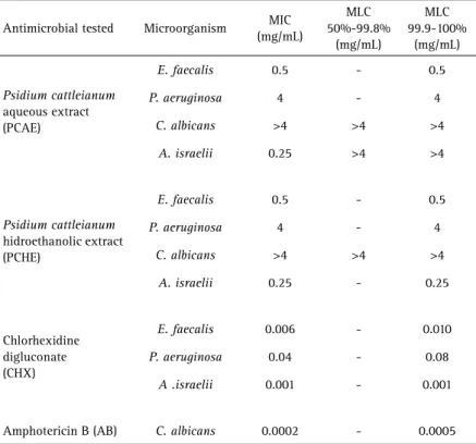
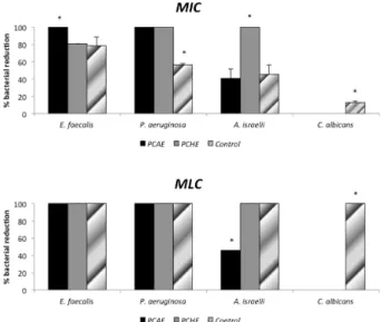
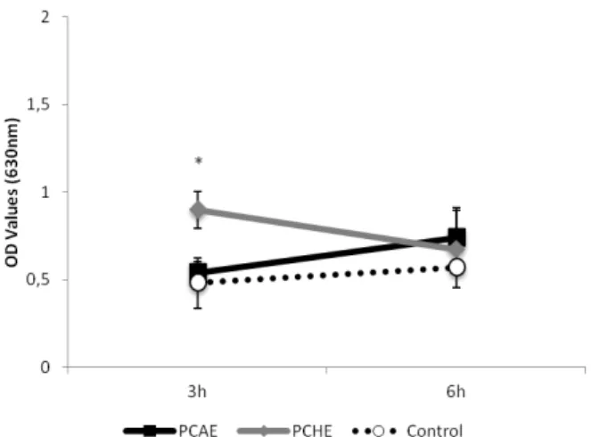
C. albicans growth by the microdilution broth method. The percentages of microbial reduction in MIC and MLC for Psidium cattleianum extracts are in Figure 1A and B, respectively. The values of MIC inhibited completely the growth of E. faecalis and P. aeruginosa for PCAE and P. aeruginosa and A. israelii for PCHE. P. aeruginosa was the most sensitive microorganism and presented 100% growth reduction in presence of MIC obtained for both extracts. C. albicans was the most resistant strain for both tested substances and no extract had effect on its growth. MLC obtained for PCHE inhibited 100% growth of all the tested strains, except for C. albicans. The MLC for PCAE had the same effect for E. faecalis and P. aeruginosa. The positive controls (Chlorhexidine and Amphotericin B) inhibited the growth of all tested strains. Figure 2 shows box-whisker plots of the activity of the plant extracts against biofilms. PCHE (5× and 10× MLC), CHX and PCAE (10× MLC) were able to completely eliminate the E. faecalis biofilm. The same was observed for PCHE (10×) and CHX (5× and 10× MLC) for P. aeruginosa. PCHE (10× MLC) reduced the growth of A. israelii after 24 h of exposition. Both CHX (5× and 10× MLC) tested were not able to eliminate A. israelii biofilm. Figure 3 shows the results of edemogenic test. Saline and PCAE caused a discrete increase of edema over time while PCHE caused a higher initial edema that decreased gradually. Statistically significant differences were observed for edema between saline and PCHE (p<0.05).
Figure 1. A: Percentage (%) of microbial reduction compared to normal growth under minimum inhibitory concentration (MIC) of Psidium cattleianum extracts. B: Percentage (%) of microbial reduction compared to normal growth under minimum lethal concentration (MLC) of Psidium cattleianum extracts. Data were expressed in mean of log (CFU+1). Control for bacteria - CHX, Control for Candida albicans - AB. * - Statistical difference among the groups according to Mann-Whitney tests).
Figure 2. Box-whisker plots of the activity of the Psidium cattleianum extracts against biofilm of pathogenic microorganisms. Different capital letters show statistical difference among the groups and control (H2O), according to Mann-Whitney tests. Bars
indicate minimum and maximum values. Black and white boxes indicate lower and upper quartiles, respectively. Line in the middle of boxes is median.
L. Massunari et al.
implant. A layer of macrophages can be observed near the implant, and some cells were dispersed in the repair tissue. There are lymphocytes and mononucleated cells, characterizing a chronic inflammation with the presence of angiogenesis (Figs. 4A and 4C). The saline group at 7 days presented more aggressive inflammatory response. The area of tissue reaction was higher and the fibrous capsule was thicker with macrophages, lymphocytes, mononucleated cells and new fibroblasts. Collagen fibers were distributed in a complex position (Fig. 4E). The PCAE and saline groups at 28 days presented a great reduction of inflammatory reaction and a higher tissue organization. The fibrous capsule was thinner with organized collagen fibers parallel to the implant. The number of inflammatory cells was reduced. There were few vessels with small diameter in the connective tissue (Figs. 4B and 4F). The hydroethanolic group (PCHE) at 28 days presented a more homogenous repair than the other groups. The fibrous capsule was thin and condensed and the collagen fibers were thicker and organized. The fibroblasts showed an elongated and thin nucleus. The macrophages around the implant presented a slight diameter indicating attenuation on the phagocytic activity. Occasional plasma cells may be observed between the implant and the macrophages, suggesting a humoral response to PCHE (Fig. 4D).
Discussion
The complexity of root canals, the limitations of the chemomechanical preparation and the vulnerability of each species involved in the infection are well recognized factors related to the failure of an endodontic treatment (8). In alternative to solve this problem, several authors have been studying medicinal plants to improve the endodontic treatment (19,21,22). In the present study, the objective was
to analyze the antimicrobial activity of Psidium cattleianum extracts against microorganisms involved in the persistent endodontic infection, apart from analyzing their immediate and late biologic responses.
The antimicrobial properties of PC have been recently studied (11,13-15), but the direct comparison of the obtained results with the literature is difficult, since different methodologies and microorganisms were used in these investigations, only a parallel analysis was done. Similar to this study results, de Souza et al. (9) did not observe an effect against C. albicans in any of the 18 tested extracts, including the PC extract (11). PCAE showed antibacterial effect at high concentration in a reduced exposure time (5 min) or at a low concentration in 60 min against the Streptococcus mutans biofilm (13). After treatment with PCAE, in an in situ biofilm assay, total anaerobic microorganism, total streptococci and S. mutans counts decreased when compared with water or antiseptic treatments (14). In the present study, the PCAE also presented antibacterial effect against the E. faecalis and P. aeruginosa biofilms and eliminated the E. faecalis biofilm at higher concentrations (10×MLC). In order to reduce the time for calcium hydroxide activity as an endodontic dressing, PCHE was used as a vehicle for calcium hydroxide in a biofilm assay. The tested formulation eliminated E. faecalis in 24 h (19). This study results also showed E. faecalis elimination when exposed to PCHE (5× and 10× MLC), suggesting that only the presence of the PC extract produced bactericidal effect.
The large range between the MIC and MLC values were observed in the results for PCAE extract - MIC (0.25 mg/mL) and MLC (>4 mg/mL) against A. israelii. The same was also reported in studies using antibiotics and propolis against the same bacterium (20,21). This suggested that the
Figure 3. Means (standard deviations) values of edema on both periods of time for the PCAE, PCHE and control groups. *Statistical difference between PCHE and the other groups, according to ANOVA/Tukey tests.
Table 2. Medians (minimum-maximum) of scores for inflammatory cell infiltrate and thickness of fibrous capsule after 7 and 28 days of extract exposure
Extract
Inflammatory cell infiltrate
Fibrous capsule
7 days 28 days 7 days 28 days
PCAE 2 (1-2) 2(1-2) 0(0-1) 0 (0-1)
PCHE 2 (1-2) 1(0-2)# 0(0-1) 0 (0-1)
Saline 3 (2-3)* 2 (0-2) 1 (0-1)* 0(0-1)
Biological evaluation of Psidium cattleianum extracts slow rate of A. israelii elimination is due to the genotypic
tolerance of the species (20).
P. aeruginosa was resistant to PC hexane, ethyl acetate and methanol fruit extracts by disc diffusion (15). In the present study, although others solvents and other tree parts were employed to obtain the PC extracts may observed the effect of PC extracts for both solvents against P. aeruginosa. Different results can be observed in other studies with plant material. This can occur due to the different methods of extraction employed and the different solvents used for
obtaining the crude extracts (23). Furthermore, the presence and concentration of the compounds may be different in each part of the tree (24).
The presence of the phenolic compounds - three flavonoids (kaempferol, quercetin and cyaniding) and tannin (ellagic acid) - have been related as the factor responsible for the PC antibacterial activity (13). Flavonoids are secondary metabolites naturally synthesized by plants, capable of penetrating into bacterial cells due to their lipophilic characteristic (25). Quercetin-5.3-dimethylether
L. Massunari et al.
from Combretum erythrophyllum displayed MICs values of 0.05 and 0.1 mg/mL for E. faecalis and P. aeruginosa, respectively (25). Tannins can be toxic to filamentous fungi, yeasts and bacteria due to their ability for inactivating microbial adhesins, enzymes and cell envelopes that transport proteins (26).
The present study demonstrated that the extracts are safe for use in biological tissues causing benefits to biologic response during repair. During the studied period, the wound healing was in an advanced stage presenting reduced levels of inflammation for PCAE and PCHE groups. These results were different from another study that followed the same methodology using Myracrodruon urundeuva aqueous and ethanolic extracts (16). The Myracrodruon urundeuva ethanolic extract demonstrated persistency of inflammation until the 28th day (16).
The edemogenic test showed a higher edema in the PCHE group at 3 h, which probably occurred due to the ethanol. However, this edema reduced progressively. The morphological analysis showed repair progress over time proving that the implants did not interfere with natural repair progression. Both extracts induced better tissue responses than control group (saline) suggesting a beneficial effect of these extracts for inflammation control. This was observed at 7 days when there were moderate inflammatory responses mainly due to the surgical trauma to tissues. Besides, the inflammatory tissue reactions caused by PCAE and PCHE extracts were lower than those caused by saline (control). At 28 days there was a regression of inflammatory process in all groups, but PCHE showed better results with slight levels of inflammation.
This present study has some limitations. First of all, the complete neutralization of antimicrobial agents was not confirmed before plating. Two common methods are used for inhibition of residual biocides: dilution and chemical neutralization. In this study was used the dilution of the antimicrobial agents, 6 times before plating. The choice was to not use chemical neutralization of biocides because of the toxicity displayed by several types of neutralizers. The use of neutralizers could not distinguish between neutralization of the biocide versus recovery of organisms injured by sub-lethal exposure to the biocide (27). In the present study, was used a population of organisms without exposure to antimicrobial agents as a growth control. However, studies have appointed for the importance of the complete neutralization of biocides, like chlorhexidine, because the accuracy of a biocidal assay and low levels of residuals would lead to exaggerated results of microbicidal activity (27). Another limitation of this study is absence of dentin as a substrate for bacterial growth. This is the first study that found antimicrobial activity for the Psidium cattleianum extracts against endodontic pathogens and
new studies are required to confirm this property simulating the canal root environment.
In conclusion, PCHE was biocompatible and presented good antimicrobial effect against important pathogens associated with persistent endodontic infections.
Resumo
Psidium cattleianum (PC) tem apresentado atividade inibitória frente diversos microrganismos, entretanto esse efeito ainda não foi testado contra microrganismos de interesse endodôntico. O objetivo desse estudo foi avaliar a atividade antimicrobiana e a biocompatibilidade dos extratos aquoso (EAPC) e hidroetanólico (EHPC) das folhas de Psidium cattleianum. As concentrações inibitória mínima (CIM) e letal mínima (CLM) foram determinadas pelo método de microdiluição em caldo, com o objetivo de analisar o efeito antimicrobiano frente Enterococcus faecalis, Pseudomonas aeruginosa, Actinomyces israelii e Candida albicans em condições planctônicas. Os ensaios de biofilme foram realizados somente com os extratos em que se determinou a CLM frente os microrganismos em condições planctônicas. Respostas teciduais imediata e tardia frente aos extratos de Psidium cattleianum foram avaliadas por teste edemogênico e análise histológica de implantes subcutâneos em ratos Wistar. Os resultados mostraram que CIM e CLM variaram entre 0,25 e 4 mg/mL. As CLMs determinadas pelo EHPC inibiram 100% do crescimento de todas as cepas testadas, exceto Candida albicans. EAPC apresentou o mesmo efeito para E. faecalis e P. aeruginosa. Ambos os extratos de PC conseguiram eliminar o biofilme de E. faecalis, e somente o EHPC eliminou o biofilme de P. aeruginosa. Os controles positivos inibiram o crescimento de todos os microrganismos testados nos ensaios de CIM e CLM, mas nenhuma das concentrações de clorexidina testadas foi capaz de eliminar o biofilme de A. israelii. O EAPC provocou um discreto aumento de edema com o tempo, enquanto EHPC provocou um edema inicial severo, que diminuiu progressivamente. Ambos os extratos EAPC e EHPC foram biocompatíveis, entretanto, EHPC apresentou melhores resultados com baixos níveis de inflamação em 28 dias. Pode-se concluir que EHPC foi biocompatível e apresentou melhor efeito antimicrobiano frente importantes patógenos associados a infecções endodônticas persistentes.
Acknowledgements
The microbial strains were provided by Oswaldo Cruz Foundation (FIOCRUZ). This study was funded by CAPES (Coordenação de Aperfeiçoamento de Pessoal de Nível Superior) from Brazil.
References
1. Sakamoto M, Siqueira JF Jr, Rôças IN, Benno Y. Molecular analysis of the root canal microbiota associated with endodontic treatment failures. Oral Microbiol Immunol 2008;23:275-281.
2. Siqueira JF Jr, Rôças IN. Clinical implications and microbiology of bacterial persistence after treatment procedures. Journal of Endodontics 2008;34:1291-1301.e3
3. Zhang S, Wang QQ, Zhang CF, Soo I. Identification of dominant pathogens in periapical lesions associated with persistent apical periodontitis. Chin J Dent Res 2010;13:115-121.
4. Mohammadi Z, Dummer PM. Properties and applications of calcium hydroxide in endodontics and dental traumatology. Int Endod J 2011;44:697-730.
5. Vaghela DJ, Kandaswamy D, Venkateshbabu N, Jamini N, Ganesh A. Disinfection of dentinal tubules with two different formulations of calcium hydroxide as compared to 2% chlorhexidine: As intracanal medicaments against Enterococcus faecalis and Candida albicans: An in vitro study. J Cons Dent 2011;14:182-186.
Biological evaluation of Psidium cattleianum extracts
7. Gomes BP, Souza SF, Ferraz CC, Teixeira FB, Zaia AA, Valdrighi L, et al.. Effectiveness of 2% chlorhexidine gel and calcium hydroxide against Enterococcus faecalis in bovine root dentine in vitro. Int Endod J 2003;36:267-275.
8. Gomes BP, Vianna ME, Sena NT, Zaia AA, Ferraz CC, de Souza Filho FJ. In vitro evaluation of the antimicrobial activity of calcium hydroxide combined with chlorhexidine gel used as intracanal medicament. Oral Surg Oral Med Oral Pathol Oral Radiol Endod 2006;102:544-550. 9. de Souza-Filho FJ, Soares A de J, Vianna ME, Zaia AA, Ferraz CC, Gomes
BP. Antimicrobial effect and pH of chlorhexidine gel and calcium hydroxide alone and associated with other materials. Braz Dent J 2008;19:28-33.
10. Cintra LT, Watanabe S, Samuel RO, da Silva Facundo AC, de Azevedo Queiroz IO, Dezan-Júnior E, et al.. The use of NaOCl in combination with CHX produces cytotoxic product. Clin Oral Invest 2014;18:935-940. 11. de Souza GC, Haas AP, von Poser GL, Schapoval EE, Elisabetsky E.
Ethnopharmacological studies of antimicrobial remedies in the south of Brazil. J Ethnopharmacol 2004;90:135-143.
12. Biegelmeyer R, Andrade JM, Aboy AL, Apel MA, Dresch RR, Marin R, et al.. Comparative analysis of the chemical composition and antioxidant activity of red (Psidium cattleianum) and yellow (Psidium cattleianum var. lucidum) strawberry guava fruit. J Food Sci 2011;76:C991-996. 13. Brighenti FL, Luppens SB, Delbem AC, Deng DM, Hoogenkamp MA,
Gaetti-Jardim E Jr, et al.. Effect of Psidium cattleianum leaf extract on Streptococcus mutans viability, protein expression and acid production. Caries Res 2008;42:148-154.
14. Brighenti FL, Gaetti-Jardim E Jr, Danelon M, Evangelista GV, Delbem AC. Effect of Psidium cattleianum leaf extract on enamel demineralisation and dental biofilm composition in situ. Arch Oral Biol 2012;57:1034-1040.
15. McCook-Russell KP, Nair MG, Facey PC, Bowen-Forbes CS. Nutritional and nutraceutical comparison of Jamaican Psidium cattleianum (strawberry guava) and Psidium guajava (common guava) fruits. Food Chem 2012;15;134:1069-1073.
16. Machado AC, Dezan Junior E, Gomes-Filho JE, Cintra LT, Ruviére DB, Zoccal R, et al.. Evaluation of tissue reaction to aroeira (Myracrodruon urundeuva) extracts: a histologic and edemogenic study. J Appl Oral Sci 2012; 20:414-8.
17. Budzynska A, Wieckowska-Szakiel M, Sadowska B, Kalemba D, Rózalska B. Antibiofilm activity of selected plant essential oils and their major components. Pol J Microbiol 2011;60:35-41.
18. Fédération Dentaire International, Commission of Dental Materials, Instruments, Equipment and Therapeutics. Recommended standard practices for biological evaluation of dental materials. Int Dent J 1980;30:140-188.
19. Dezan-Jr E, Sangalli J, Gomes-Filho JE, Gaetti-Jardim Jr E. Psidium cattleianum plus Ca(OH)2 antimicrobial efficacy against Enterococcus faecalis. IADR; 15-16,2010, J Dent Res; 2010;89, Special Issue B. 20. Barnard D, Davies J, Figdor D. Susceptibility of Actinomyces israelii to
antibiotics, sodium hypochlorite and calcium hydroxide. Int Endod J 1996;29:320-326.
21. Ferreira FB, Torres SA, Rosa OP, Ferreira CM, Garcia RB, Marcucci MC, et al.. Antimicrobial effect of propolis and other substances against selected endodontic pathogens. Oral Surg Oral Med Oral Pathol Oral Radiol Endod 2007;104:709-716.
22. Abbaszadegan A, Dadolahi S, Gholami A, Moein MR, Hamedani S, Ghasemi Y, et al.. Antimicrobial and cytotoxic activity of Cinnamomum zeylanicum, calcium hydroxide, and triple antibiotic paste as root canal dressing materials. J Contemp Dent Pract 2016;1;17:105-113. 23. Passari LM, Scarminio IS, Bruns RE. Experimental designs characterizing
seasonal variations and solvent effects on the quantities of coumarin and related metabolites from Mikania laevigata. Anal Chim Acta 2014;22;821:89-96.
24. Tortoriello J, Meckes-Fischer M, Villarreal ML, Berlin B, Berlin E. Spasmolytic activity of medicinal plants used to treat gastrointestinal and respiratory diseases in the Highland of Chiapas. Phytomedicine 1995;2:57-66.
25. Martini ND, Katerere DR, Eloff JN. Biological activity of five antibacterial flavonoids from Combretum erythrophyllum (Combretaceae). J Ethnopharmacol 2004;93:207-12.
26. Cowan MM: Plant products as antimicrobial agents. Clin Microbiol Rev 1999;12:564-582.
27. Sutton SV, Proud DW, Rachui S, Brannan DK. Validation of microbial recovery from disinfectants. PDA J Pharm Sci Technol 2002;5:255-266.