Volume 2012, Article ID 904963,6pages doi:10.5402/2012/904963
Research Article
Biocompatibility of Intracanal Medications Based on
Calcium Hydroxide
Carolina Andolfatto,
1Guilherme Ferreira da Silva,
1Ana Livia Gomes Corn´elio,
1Juliane Maria Guerreiro-Tanomaru,
1Mario Tanomaru-Filho,
1Gisele Faria,
1Idomeo Bonetti-Filho,
1and Paulo S´ergio Cerri
21Department of Restorative Dentistry, Araraquara Dental School, Universidade Estadual Paulista (UNESP),
14801-903 Araraquara, SP, Brazil
2Laboratory of Histology and Embryology, Department of Morphology, Araraquara Dental School,
Universidade Estadual Paulista (UNESP), 14801-903 Araraquara, SP, Brazil
Correspondence should be addressed to Paulo S´ergio Cerri,[email protected]
Received 6 November 2012; Accepted 27 November 2012
Academic Editors: H. S. Cardash, J. H. Jeng, and G. Mount
Copyright © 2012 Carolina Andolfatto et al. This is an open access article distributed under the Creative Commons Attribution License, which permits unrestricted use, distribution, and reproduction in any medium, provided the original work is properly cited.
Objective. The aim of this study was to evaluate the rat subcutaneous tissue reaction to calcium hydroxide-based intracanal
medicaments, UltraCal XS (calcium hydroxide, barium sulphate, aqueous matrix), Hydropast (calcium hydroxide, barium sulphate, and propyleneglycol), and Calen (Calcium hydroxide, zinc oxide, colophony, and polyethyleneglycol), used as a control.
Methods. Forty-eight rats (Rattus Norvegicus Holtzman) were distributed in three groups: Calen, UltraCal XS, and Hydropast.
Polyethylene tubes filled with one of the medicaments were implanted in the dorsal subcutaneous. After 7 and 30 days, the implants were removed and the specimens were fixed and embedded in paraffin. Morphological and quantitative analyses were carried out in the HE-stained sections. The numerical density of inflammatory cells in the capsule was evaluated and statistical analyses were performed (P≤0.05).Results. At 7 days, all materials induced an inflammatory reaction in the subcutaneous tissue adjacent to the implants. In all groups, a significant reduction in the number of inflammatory cells and giant cells was verified in the period of 30 days.Conclusion. These results indicate that the calcium hydroxide-based medicaments evaluated present biocompatibility similar to Calen.
1. Introduction
The success of endodontic treatment of teeth with periapical lesion depends on the reduction or elimination of the
intraradicular infection [1, 2]. The root canal mechanical
preparation is not enough to eliminate this infection because many microorganisms are not only in the main root canal, but also disseminated throughout the root canal system. Therefore, the use of an intracanal dressing to eliminate the microorganisms is indicated [3–6].
Antimicrobial activity and biocompatibility are charac-teristics that an ideal intracanal dressing has to show [7]. Calcium hydroxide [Ca(OH)2] has been widely used for its
biological and antimicrobial activity [4,8], ability to dissolve
organic tissue [9], and capacity to inactivate bacterial
endotoxin [10,11]. Despite these properties, the Ca(OH)2
has no satisfactory physical properties such as radiopacity to visualize on dental radiographs and flow capacity to facilitate
its insertion in the root canal [12, 13]. For this reason, it
needs the incorporation of a radiopacifying agent and a
vehicle to improve these characteristics [8,14].
Although the Ca(OH)2 shows an excellent
biocom-patibility, the addition of other substances can affect its
biological properties [8,12]. In the last years, it has been
demonstrated that UltraCal XS has a high pH value [15]
and an effective antimicrobial activity against common
endodontic bacteria of teeth with pulp necrosis [16]. On the other hand, Brazilian paste Hydropast, composed by 38% of calcium hydroxide, barium oxide, as a radiopacifying agent, and propilenoglycol as vehicle, is a recent material, and, therefore, until now, there are no studies of its biological properties.
Considering the recommendation of International
Orga-nization for Standardization [17], it is necessary in vitro
and/orin vivostudies for evaluation of the biocompatibility
of these new materials. Thus, the purpose of this study was to evaluate the tissue reaction of these calcium hydroxide-based medicaments in rat subcutaneous.
2. Materials and Methods
2.1. Animals and Experimental Proceedings. This study was
performed in accordance with the principles of animal care on animal experiments. The research protocol was autho-rized by the Ethical Committee for Animal Research of the S˜ao Paulo State University, Brazil (Dental School, UNESP, Araraquara).
Forty-eight male Holtzman rats (Rattus norvegicus
albi-nus), weighing 250±10 g, were kept in individual stainless
steel cages under 12 : 12 light-dark cycle at controlled
temperature (23±2◦C) and humidity (55 + 10%), with food
and water providedad libitum. The animals were randomly
distributed into three groups (n = 16) on the basis of the
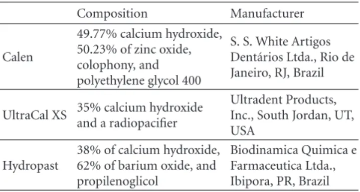
intracanal medicaments analysed, Calen group, Calen paste (S. S. White Artigos Dent´arios Ltda., Rio de Janeiro, RJ, Brazil), used as control group; UltraCal group, UltraCal XS paste (Ultradent Products, Inc., South Jordan, UT, USA), and Hydropast group, Hydropast (Biodinˆamica Qu´ımica e Farmacˆeutica Ltda., Ibipora, PR, Brazil). The composition of
these materials is described at Table1.
The polyethylene tubes (Embramed Ind. Com. Ltda., S˜ao Paulo, SP, Brazil) with 10.0 mm length and 1.5 mm diameter previously sterilized with ethylene oxide were filled with Calen, UltraCal XS paste, or Hydropast intracanal medicaments.
The animals were anaesthetized with an intraperitoneal injection containing 80 mg/Kg of body weight of ketamine (Uni˜ao Qu´ımica Farmacˆeutica Nacional S/A-Brazil) and 4 mg/Kg of body weight of xylazine (Virbac do Brasil Ind ´ustria e Com´ercio Ltda., Brazil). After shaved and disinfection with 5% iodine solution, a 20 mm-long incision in a head-to-tail orientation was made using a scalpel (no. 15, Fibra Cir ´urgica, Joinvile, SC, Brazil) in the dorsal skin. Subsequently, the polyethylene tube containing Calen paste, UltraCal XS, or Hydropast was immediately implanted into the dorsal subcutaneous connective tissue. After implan-tation, the skin was closed with 4.0 silk suture (Vicryl; Johnson & Johnson: Ethicon Inc., New Brunswick, NJ, USA). One polyethylene tube filled with an intracanal paste was implanted in each animal and left for periods of 7 and 30 days.
Table1: Tested materials and composition.
Composition Manufacturer
Calen
49.77% calcium hydroxide, 50.23% of zinc oxide, colophony, and polyethylene glycol 400
S. S. White Artigos Dent´arios Ltda., Rio de Janeiro, RJ, Brazil
UltraCal XS 35% calcium hydroxideand a radiopacifier
Ultradent Products, Inc., South Jordan, UT, USA
Hydropast
38% of calcium hydroxide, 62% of barium oxide, and propilenoglicol
Biodinamica Quimica e Farmaceutica Ltda., Ibipora, PR, Brazil
After experimental periods, the animals were killed by overdose of anesthetic solution, and the tubes were removed
with surrounding connective tissue and prepared for paraffin
embedding.
2.2. Histological Procedures and Analysis. The specimens
containing the implanted polyethylene tubes were fixed in 4% formaldehyde (prepared from paraformaldehyde)
buffered at pH 7.2 with 0.1 M sodium phosphate at room
temperature for 48 hours. Subsequently, the specimens were
dehydrated and embedded in paraffin. Serial 6µm-thick
sections were made parallel to the tube long axis and stained with hematoxylin and eosin (H&E) for morphological and morphometric analyses. The morphological analysis of the capsule in contact with the material on the opening of the tube was performed considering the following parameters: presence of inflammatory process, main cells (inflammatory cells or fibroblasts) present in the capsule, presence of multinucleated giant cells, and presence of collagen fibers.
The numerical density of inflammatory mononucleated cells and multinucleated giant cells was undertaken using a light microscope (BX51, Olympus, Tokyo, Japan) and an image analysis system (Image Pro-Express 6.0, Olympus). Three H&E-stained sections per animal were selected at
intervals of at least 100µm; in each section, a standardized
field of 0.09 mm2 of the connective tissue adjacent to the
opening of the tube implanted was analyzed, totaling
0.27 mm2 per animal. In each area, the total number of
inflammatory cells was counted using the image analysis
sys-tem at×40 magnification; in each animal, the total number
of inflammatory cells was divided by total area, and, then,
the number of inflammatory cells/mm2 was obtained. The
differences between the groups were statistically analyzed
using SigmaStat 2.0 software (Jandel Scientific, Sausalito, CA, USA); the data were submitted to ANOVA and Tukey test.
The significance level accepted wasP≤0.05.
3. Results
3.1. Morphological and Quantitative Analyses. After 7 days
I BV BV CG
CG
IC
IC
I
Fb CF
Fb
CF
BV BV
(a)
(b)
(c)
Figure1: Light micrographs of sections showing portions of capsules adjacent to the opening of the tubes (I) filled with Calen paste after 7
(a) and 30 days (b and c) of implantation in the subcutaneous. In (a), numerous inflammatory cells (ICs) are observed in the inner portion of the capsule adjacent to the tube opening (I). The inset of the outlined area shows multinucleated giant cells (GCs) in close juxtaposition to the material implanted. BV, blood vessels.×110; inset:×250 (b) shows the capsule exhibits several fibroblasts (Fb) dispersed among
the collagen fiber bundles (CF)×130. In (c), outlined area in (b), shows some inflammatory cells (arrows), mainly lymphocytes, situated adjacent to the blood vessels (BV)×250.
I BV
BV
I IC
IC CG
BV
IC
(a)
I BV
BV
FC Fb
Fb
Fb
(b)
Figure2: Light micrographs of sections showing portions of capsules adjacent to the opening of the tubes (I) filled with UltraCal XS paste
after 7 (a) and 30 days (b) of implantation in the subcutaneous. In (a), The capsule exhibits several inflammatory cells (IC) and blood vessels (BV). Note that dense masses of inflammatory cells (ICs) are observed in the inner portion of the capsule, adjacent to the tube opening (I). The inset, outlined area, shows multinucleated giant cells (GCs).×120; inset:×230. (b) The capsule contains several fibroblasts (Fbs) and numerous blood vessels (BV). Inflammatory cells (arrows), lymphocytes and macrophages are situated mainly in the inner portion of the capsule and next to the blood vessels×150.
evident in the innermost portion of the capsule, that is, in
close juxtaposition to the materials (Figures1(a),2(a)and
3(a)). According to Table 2, no significant difference was
verified in the numerical density of inflammatory cells among the groups. Multinucleated giant cells were also observed in the capsules formed in all groups (Figures
1(a),2(a), and3(a)). However, in the Hydropast group the
number of multinucleated giant cells was significantly higher
in comparison to other groups; on the other hand, the capsule of the Calen group exhibited the lower number of
multinucleated giant cells (Table2).
A significant decrease in the number of inflammatory cells and multinucleated giant cells was verified from 7 to
30 days, in all groups (Figures1(b),2(b), and3(b); Table2).
I N
IC
IC
IC BV
CG CG
(a)
I CF
BV
CF
BV
FC BV
(b)
Figure3: Light micrographs of sections showing portions of capsules adjacent to the opening of the tubes (I) filled with Hydropast after 7
(a) and 30 days (b) of implantation in the subcutaneous. In (a), numerous inflammatory cells (ICs) and multinucleated giant cells (GCs) are present by thorough capsule. BVs, blood vessels×120. (b) The capsule contains predominantly inflammatory cells (arrows); scarce collagen
fibers (FC) distributed irregularly are observed in the capsule. BVs, blood vessels×120.
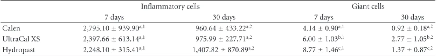
Table2: Number of inflammatory cells and giant cells/mm2of the capsule adjacent to the implants in the subcutaneous.
Inflammatory cells Giant cells
7 days 30 days 7 days 30 days
Calen 2,795.10±939.90a,1 960.64±433.22a,2 4.14±0.90a,1 0.92±0.18a,2
UltraCal XS 2,397.66±613.14a,1 975.99±227.71a,2 6.00±1.03b,1 2.77±1.05b,2
Hydropast 2,248.10±315.41a,1 1,407.82±870.89a,2 8.77±1.46c,1 1.37±0.87c,2
Values are expressed as mean±standard deviation.
Equal letters indicate no statistically significant difference (P >0.05) between the materials in the same experimental period. Different numbers indicate difference statistically significant (P≤0.05) of each material in the different experimental periods.
bundles of collagen fibers between fibroblasts; lymphocytes and plasma cells were mainly present next to blood vessels
(Figure1(b)). In the UltraCal group, the capsule contained
several fibroblasts among the inflammatory cells; usually, bundles of collagen fibers were only present in the outermost
portion of the capsule (Figure2(b)). The connective tissue
of the capsule of the Hydropast exhibited several cells and
scarce collagen fibers (Figure3(b)). Although no significant
difference was found in the number of inflammatory cells
between the groups, the mean of the numerical density of inflammatory cells in the Hydropast group was around
1,400 cells/mm2, whereas in the other groups was around
965 cells/mm2. Moreover, in the Calen group the number
of multinucleated giant cells was significantly lower in
comparison to UltraCal and Hydropast groups (Table2).
4. Discussion
Implantation in the subcutaneous connective tissues of experimental animals has been extensively used to evaluate
the biocompatibility of endodontic materials [18,19]. Our
findings indicate that UltraCal XS and Hydropast exhibit biological behavior similar to Calen (control group). At 7 days, an intense inflammatory reaction and foci of coag-ulative necrosis were seen in the adjacent capsule to the implanted materials. After 30 days, significant reduction
in the inflammatory process was verified in all analyzed groups; usually, the capsule formed juxtaposed to the Calen paste exhibited inflammatory cells among typical bundles of collagen fibers and fibroblasts. The inflammatory reaction observed in the period of the 7 days may be attributed to the superficial necrosis promoted by calcium hydroxide-based materials [20]. Calcium hydroxide has an alkaline pH [21] and, when in contact with the connective tissue, induces the formation of a coagulative necrosis zone [22]. Coagulative necrosis refers to a spectrum of morphological changes in living tissue resulting from the action of enzymes on lethally injured cells. The mass of necrotic cells is characterized by preservation of the basic outline of the coagulated cells for a span of at least some days. As necrotic cells are unable to maintain membrane integrity, their contents leak out and elicit an inflammatory response that removes the cellular debris by phagocytosis, followed by healing [20].
The vehicles mixed with calcium hydroxide powder play an important role in the ionic dissociation process and so in the disinfection of the root canal and biocompatibility
[8,23]. There are three main types of vehicle: water-soluble
substances, viscous, and oil-based [8]. In the present study,
the vehicles of the different medicaments did not interfere
to Hydropast showed a significantly lower number of giant cells than the UltraCal XS. The giant cells are derived from the fusion of 20 or more monocytes/macrophages and are formed for removing exogenous agents [24]. In the period of 30 days, the high number of giant cells verified in the UltraCal XS group suggests that this material may release more irritant substances than the Hydropast and Calen.
In Calen paste, the calcium hydroxide is mixed to a viscous vehicle, polyethylene glycol 400, one of the most commonly used vehicles in root canal medicaments with low toxicity, high solubility in aqueous solutions, low immuno-genicity and antiimmuno-genicity [25], and antibacterial activity [5]. This vehicle releases calcium and hydroxyl ions more slowly and for longer periods than water-soluble and oil-based
materials [26]. Because the releasing of H+, the polyethylene
glycol 400 neutralizes the OH−released by calcium hydroxide
and, thereby, reduces the superficial necrosis area [27]. The propylene glycol is used as vehicle in the Hydropast; the vehicle used in the Hydropast is classified as a viscous vehicle with high molecular weight and, as well as polyethy-lene glycol 400, prolong the action of calcium hydroxide in the root canal system [26]. Moreover, propylene glycol shows
low toxicity and antimicrobial properties [28, 29]. It was
demonstrated that the addition of propylene glycol may not interfere in the biocompatibility of MTA in rat subcutaneous tissue [30].
Our results also suggest that the different radiopacifying
agents of the pastes did not interfere in the tissue reaction.
The endodontic materials should present sufficient
radiopac-ity to be distinguished from adjacent anatomical structures, such as bone and teeth [31]. Zinc oxide, barium sulfate, bismuth oxide, and other components with iodine and
bromine are some examples of radiopacifiers [12,14,27,32].
The zinc oxide of Calen paste does not affect the
biological properties of calcium hydroxide [27,33]. Barium
sulphate, radiopacifying agent of Hydropast, is also
bio-compatible because its cause no detrimental effect in rat
subcutaneous tissue [34] or in periapical tissue in association with calcium hydroxide [14]; barium oxide in association with the Norian, an skeletal repair system (SRS), in tibiae defects of rats, maintains the properties of biocompatibility and osteoconductive materials of the SRS [35]. Although the manufacturer does not inform the radiopacifying agent of the UltraCal XS, our results demonstrated that this material has a good biological behavior.
Considering the methodology used in the present study, our findings indicate that UltraCal XS and Hydropast are biocompatible in subcutaneous tissue of rats.
Conflict of Interests
The authors declared that they have no any conflict of inte-rests related to this study.
Acknowledgments
The authors thank Mr. Pedro S´ergio Sim˜oes for technical support. This paper was supported by Coordenac¸˜ao de
Aperfeic¸oamento de Pessoal de N´ıvel Superior (CAPES) and Conselho Nacional de Desenvolvimento Cient´ıfico e Tecnol ´ogico (CNPq), Brazil.
References
[1] L. Tronstad, “Recent development in endodontic research,”
Scandinavian Journal of Dental Research, vol. 100, no. 1, pp.
52–59, 1992.
[2] J. F. Siqueira Jr., “Aetiology of root canal treatment failure: why well-treated teeth can fail,”International Endodontic Journal, vol. 34, no. 1, pp. 1–10, 2001.
[3] G. Faria, P. Nelson-Filho, A. C. Freitas, S. Assed, and I. Y. Ito, “Antibacterial effect of root canal preparation and calcium hydroxide paste (Calen) intracanal dressing in primary teeth with apical periodontitis,”Journal of Applied Oral Science, vol. 13, pp. 351–355, 2005.
[4] M. R. Leonardo, M. E. F. T. Hernandez, L. A. B. Silva, and M. Tanomaru-Filho, “Effect of a calcium hydroxide-based root canal dressing on periapical repair in dogs: a histological study,” Oral Surgery, Oral Medicine, Oral Pathology, Oral
Radiology and Endodontology, vol. 102, no. 5, pp. 680–685,
2006.
[5] R. K. P. Lima, J. M. Guerreiro-Tanomaru, N. B. Faria-J ´unior, and M. Tanomaru-Filho, “Effectiveness of calcium hydroxide-based intracanal medicaments against Enterococcus faecalis,”
International Endodontic Journal, vol. 45, no. 4, pp. 311–316,
2012.
[6] J. F. Siqueira Jr., K. M. Magalh˜aes, and I. N. R ˆoc¸as, “Bacterial reduction in infected root canals treated with 2.5% NaOCl as an irrigant and calcium hydroxide/camphorated para-monochlorophenol paste as an intracanal dressing,”Journal of
Endodontics, vol. 33, no. 6, pp. 667–672, 2007.
[7] P. Carrotte, “Endodontics: part 9 Calcium hydroxide, root resorption, endo-perio lesions,”British Dental Journal, vol. 197, no. 12, pp. 735–743, 2004.
[8] Z. Mohammadi and P. M. H. Dummer, “Properties and appli-cations of calcium hydroxide in endodontics and dental traumatology,”International Endodontic Journal, vol. 44, no. 8, pp. 697–730, 2011.
[9] G. Hasselgren, B. Olsson, and M. Cvek, “Effects of calcium hydroxide and sodium hypochlorite on the dissolution of necrotic porcine muscle tissue,”Journal of Endodontics, vol. 14, no. 3, pp. 125–127, 1988.
[10] K. E. Safavi and F. C. Nichols, “Effect of calcium hydroxide on bacterial lipopolysaccharide,”Journal of Endodontics, vol. 19, no. 2, pp. 76–78, 1993.
[11] J. M. G. Tanomaru, M. R. Leonardo, M. Tanomaru Filho, I. Bonetti Filho, and L. A. B. Silva, “Effect of different irrigation solutions and calcium hydroxide on bacterial LPS,”
Interna-tional Endodontic Journal, vol. 36, no. 11, pp. 733–739, 2003.
[12] T. Alac¸am, G. G¨org¨ul, and H. ¨Om¨url¨u, “Evaluation of diagnos-tic radiopaque contrast materials used with calcium hydrox-ide,”Journal of Endodontics, vol. 16, no. 8, pp. 365–368, 1990. [13] L. R. G. Fava and W. P. Saunders, “Calcium hydroxide pastes:
classification and clinical indications,”International
Endodon-tic Journal, vol. 32, no. 4, pp. 257–282, 1999.
[15] S. Heward and C. M. Sedgley, “Effects of intracanal mineral trioxide aggregate and calcium hydroxide during four weeks on ph changes in simulated root surface resorption defects: an in vitro study using matched pairs of human teeth,”Journal of
Endodontics, vol. 37, no. 1, pp. 40–44, 2011.
[16] M. L. Blanscet, P. A. Tordik, and G. G. Goodell, “An agar dif-fusion comparison of the antimicrobial effect of calcium hydroxide at five different concentrations with three different vehicles,” Journal of Endodontics, vol. 34, no. 10, pp. 1246– 1248, 2008.
[17] International Organization for Standardization, ISO 7405: dentistry—preclinical evaluation of biocompatibility of medi-cal devices used in dentistry—test methods for dental materi-als, 2008.
[18] M. S. S. Pereira, G. Faria, L. A. B. da Silva, M. Tanomaru-Filho, M. C. Kuga, and M. A. Rossi, “Response of mice connec-tive tissue to intracanal dressings containing chlorhexidine,”
Microscopy Research and Technique, vol. 75, no. 12, pp. 1653–
1658, 2012.
[19] N. N. Viola, J. M. Guerreiro-Tanomaru, G. F. da Silva, E. Sasso-Cerri, M. Tanomaru-Filho, and P. S. Sasso-Cerri, “Biocompatibility of an experimental MTA sealer implanted in the rat subcu-taneous: quantitative and immunohistochemical evaluation,”
Journal of Biomedical Materials Research B, vol. 100, pp. 1773–
1781, 2012.
[20] G. Faria, M. R. N. Celes, A. de Rossi, L. A. B. Silva, J. S. Silva, and M. A. Rossi, “Evaluation of chlorhexidine toxicity injected in the paw of mice and added to cultured L929 fibroblasts,”
Journal of Endodontics, vol. 33, no. 6, pp. 715–722, 2007.
[21] J. M. Guerreiro-Tanomaru, D. G. Chula, R. K. de Pontes Lima, F. L. V. C. Berbert, and M. Tanomaru-Filho, “Release and diffusion of hydroxyl ion from calcium hydroxide-based medicaments,”Dental Traumatology, vol. 28, no. 4, pp. 320– 323, 2012.
[22] R. Holland, C. E. Pinheiro, W. de Mello, M. J. Nery, and V. de Souza, “Histochemical analysis of the dogs’ dental pulp after pulp capping with calcium, barium, and strontium hydroxides,”Journal of Endodontics, vol. 8, no. 10, pp. 444–447, 1982.
[23] N. V. Ballal, G. V. Shavi, R. Kumar, M. Kundabala, and K. S. Bhat, “In vitro sustained release of calcium ions and pH main-tenance from different vehicles containing calcium hydroxide,”
Journal of Endodontics, vol. 36, no. 5, pp. 862–866, 2010.
[24] P. S. Cerri, E. Freym¨uller, and E. Katchburian, “Light and electron microscopic study of autologous implants of dental roots in the subcutaneous tissue of rats,”The Bulletin of Tokyo
Dental College, vol. 38, no. 2, pp. 113–122, 1997.
[25] B. Athanassiadis, P. V. Abbott, and L. J. Walsh, “The use of calcium hydroxide, antibiotics and biocides as antimicrobial medicaments in endodontics,”Australian Dental Journal, vol. 52, no. 1, pp. S64–S82, 2007.
[26] B. P. Gomes, C. C. Ferraz, M. E. Vianna et al., “In vitro antimi-crobial activity of calcium hydroxide pastes and their vehicles against selected microorganisms,”Brazilian dental journal, vol. 13, no. 3, pp. 155–161, 2002.
[27] P. Nelson Filho, L. A. Silva, M. R. Leonardo, L. S. Utrilla, and F. Figueiredo, “Connective tissue responses to cal-cium hydroxide-based root canal medicaments,”International
Endodontic Journal, vol. 32, no. 4, pp. 303–311, 1999.
[28] K. S. Bhat and S. Walkevar, “Evaluation of bactericidal prop-erty of propylene glycol for its possible use in endodontics,”
Arogya Journal of Health Science, vol. 1, pp. 54–59, 1975.
[29] P. A. Thomas, K. S. Bhat, K. M. Kotian, and S. K. Manipal, “Antibacterial properties of dilute formocresol and eugenol
and propylene glycol,”Oral Surgery Oral Medicine and Oral
Pathology, vol. 49, no. 2, pp. 166–170, 1980.
[30] R. Holland, L. Mazuqueli, V. de Souza, S. S. Murata, E. Dezan J ´unior, and P. Suzuki, “Influence of the type of vehicle and limit of obturation on apical and periapical tissue response in dogs’ teeth after root canal filling with mineral trioxide aggregate,”Journal of Endodontics, vol. 33, no. 6, pp. 693–697, 2007.
[31] F. C. S. Pires, L. C. Pardini, D. R. Cruvinel, H. M. Hamida, and L. F. Garcia, “In vitro comparison of the radiopacity of cavity lining materials with human dental structures,” Journal of
Conservative Dentistry, vol. 13, pp. 65–70, 2012.
[32] A. L. G. Corn´elio, L. P. Salles, M. Campos da Paz, J. A. Cirelli, J. M. Guerreiro-Tanomaru, and M. Tanomaru Filho, “Cytotoxicity of Portland cement with different radiopacifying agents: a cell death study,”Journal of Endodontics, vol. 37, no. 2, pp. 203–210, 2011.
[33] A. M. de Queiroz, S. Assed, A. Consolaro et al., “Subcutaneous connective tissue response to primary root canal filling materials,”Brazilian Dental Journal, vol. 22, no. 3, pp. 203– 211, 2011.
[34] D. O. Adams, “The granulomatous inflammatory response: a review,”American Journal of Pathology, vol. 84, no. 1, pp. 164– 191, 1976.
Submit your manuscripts at
http://www.hindawi.com
Hindawi Publishing Corporation
http://www.hindawi.com Volume 2014
Oral Oncology
Journal ofDentistry
International Journal ofHindawi Publishing Corporation
http://www.hindawi.com Volume 2014
Hindawi Publishing Corporation
http://www.hindawi.com Volume 2014 International Journal of
Biomaterials
Hindawi Publishing Corporation
http://www.hindawi.com Volume 2014
BioMed
Research International Hindawi Publishing Corporation
http://www.hindawi.com Volume 2014
Case Reports in
Dentistry
Hindawi Publishing Corporation
http://www.hindawi.com Volume 2014
Oral Implants
Journal ofHindawi Publishing Corporation
http://www.hindawi.com Volume 2014
Anesthesiology Research and Practice
Hindawi Publishing Corporation
http://www.hindawi.com Volume 2014 Radiology
Research and Practice Environmental and
Public Health Journal of
Hindawi Publishing Corporation
http://www.hindawi.com Volume 2014
The Scientiic
World Journal
Hindawi Publishing Corporationhttp://www.hindawi.com Volume 2014
Hindawi Publishing Corporation
http://www.hindawi.com Volume 2014
Dental Surgery
Journal ofDrug Delivery
Journal of Hindawi Publishing Corporationhttp://www.hindawi.com Volume 2014
Hindawi Publishing Corporation
http://www.hindawi.com Volume 2014
Oral Diseases
Journal ofHindawi Publishing Corporation
http://www.hindawi.com Volume 2014
Computational and Mathematical Methods in Medicine
Scientifica
Hindawi Publishing Corporationhttp://www.hindawi.com Volume 2014
Pain
Research and Treatment Hindawi Publishing Corporation
http://www.hindawi.com Volume 2014
Hindawi Publishing Corporation
http://www.hindawi.com Volume 2014
Endocrinology
International Journal ofHindawi Publishing Corporation
http://www.hindawi.com Volume 2014
Hindawi Publishing Corporation