Submitted: Dec 13, 2016
Accepted for publication: May 03, 2017 Last revision: May 21, 2017
Immunohistochemical expression of
TGF-
β
1 and MMP-9 in periapical lesions
Abstract: The objective of this study was to evaluate the expression of matrix metalloproteinase 9 (MMP-9) and transforming growth factor beta (TGF-β1) in periapical lesion samples correlated with the intensity of the inlammatory iniltrate and thickness of the epithelial lining. Forty-ive cases of periapical lesions (23 periapical granulomas and 22 radicular cysts) were subjected to morphological and immunohistochemical analyses using anti-MMP-9 and anti-TGF-β1 antibodies. The data were analyzed using the following tests: non-parametric Mann-Whitney, chi-square, Fisher’s exact test and Spearman’s correlation test (P<0.05). Analysis of inlammatory iniltrate revealed that 78% of periapical granulomas presented iniltrate grade III, in contrast with 32% of radicular cysts (P<0.001). Morphological evaluation of the epithelial thickness in radicular cysts revealed the presence of atrophic epithelium in 86% of the cysts. The immunostaining of MMP-9 was score 2 in 67% of the granulomas and 77% of the cysts. Both lesions were predominantly score 1 for TGF-β1. Signiicant differences were conirmed between the expression scores of TGF-β1 and MMP-9 in periapical granulomas (p = 0.004) and in radicular cysts (p < 0.001). Expression of TGF-β1 was different for periapical granulomas and radicular cysts. This immunoregulatory cytokine seems more representative in asymptomatic lesions. The extracellular matrix remodeling process dependent on MMP-9 seems to be similar for both periapical granulomas and radicular cysts. TGF-β1 and MMP-9 may play an important role in the maintenance of periapical lesions.
Keywords: Inlammation; Immunohistochemistry; Periapical Granuloma; Radicular Cyst; Biopsy.
Introduction
Periapical granulomas and radicular cysts are osteolytic inlammatory lesions affecting the jaws, and representing up to two-thirds of all radiolucent periapical lesions.1 They develop in periapical tissues as a direct immunological response mainly to the progression and maintenance of endodontic bacterial infection. In addition, mechanical and/or thermic injuries that may result in pulp necrosis, as well as infection by human cytomegalovirus and Epstein-Barr virus, may also be implicated as etiological factors in a small number of cases.2,3
When an immune/inlammatory response is maintained in periapical tissues, it can lead to the accumulation of lymphocytes, plasma cells,
Pâmella Recco ÁLVARES(a)
José Alcides Almeida de ARRUDA(a)
Leorik Pereira da SILVA(b)
George João Ferreira do NASCIMENTO(c)
Marcia Maria Fonseca da SILVEIRA(d)
Ana Paula Veras SOBRAL(a)
(a) Universidade de Pernambuco – UPE, School
of Dentistry, Department of Oral Pathology, Camaragibe, PB, Brazil.
(b) Universidade Federal do Rio Grande do
Norte – UFRN, School of Dentistry, Department of Oral Pathology, Natal, RN, Brazil.
(c) Universidade Federal de Campina
Grande – UFCG, Department of
Stomatology, School of Dentistry, Campina Grande, PB, Brazil.
(d) Universidade de Pernambuco – UPE, School
of Dentistry, Department of Stomatology, Camaragibe, PB, Brazil.
Declaration of Interest: The authors certify that they have no commercial or associative interest that represents a conflict of interest in connection with the manuscript.
Corresponding Author:
José Alcides Almeida de Arruda E-mail: [email protected]
macrophages —mediating the scarce neutrophils — and eosinophils, resulting in a well-circumscribed lesion called periapical granuloma.4,5 On the other hand, continuous and chronic inlammation, resulting from the host immune system acting to fully eradicate infection, may reactivate the epithelial cell rests of Malassez and other epithelial sources that proliferate to form radicular cysts.6,7
Several immune/inlammatory mediators and proteases may be involved in the formation of periapical granulomas and radicular cysts, such as transforming growth factor-β1 (TGF-β1) and matrix metalloproteinase-9 (MMP-9).8,9 TGF-β1 is a polypeptide member of the transforming growth factor-β superfamily of cytokines, which is secreted by platelets and inlammatory cells. It performs many cellular functions, including the control of cell growth, cell proliferation, cell differentiation, apoptosis and inflammation, as well as tissue repair.10 Additionally, TGF-β1 immune/inlammatory functions include (a) chemotaxis for monocytes, neutrophils and ibroblasts; (b) initiation, growth and differentiation of inlammatory cells; and (c) formation of bone structures.6
Several cells, such as neutrophils, macrophages, T cells, mast cells and odontoblasts, may secrete MMP-9, a metalloenzyme belonging the gelatinase group of matrix metalloproteinases (MMPs) that degrade a vast number of extracellular matrix (ECM) components, including denatured collagen, basement membrane and bone matrix. These MMPs are essential for the initiation of bone resorption, and also participate as modulators of acute and chronic inlammatory responses, since they can activate both proinlammatory and immunoregulatory (such as TGF-β1) mediators.11,15,16,17,18
Despite controversial evidence implicating proinlammatory modulators in the pathogenesis of periapical granulomas and radicular cysts, little is known about their immunoregulatory and modulatory mechanisms. This study descriptively analyzed and veriied possible correlations between the immunohistochemical expression of TGF-β1 and MMP-9 in periapical lesions and clinical/morphological variables in an endeavor to gain a better understanding
of the pathogenesis of periapical lesions, and conirm protein expression related to progression.
Methodology
Study design and tissue sample
The study included a descriptive analysis of the immunohistochemical expression pattern of TGF-β1 and MMP-9 in forty-ive randomly selected cases of periapical lesions, including 23 periapical granulomas and 22 radicular cysts. Data related to the patients (age and gender) and their clinical signs (symptomatology, radiographic findings and anatomical site) were collected through biopsy records, and information on the immunoexpression of TGF-β1 and MMP-9 was obtained after performing the experimental study. The selection and retrieval of all cases, their biopsy records and laboratorial experiments were performed at the Surgical Pathology Laboratory of the University of Pernambuco, Brazil. The study was approved by the Research Ethics Committee of the University of Pernambuco (0082.0.097.000-10).
Morphological analysis
Five-micrometer-thick tissue sections obtained from paraffin-embedded blocks of periapical granulomas and radicular cysts were placed on a glass slide, stained with hematoxylin-eosin and visualized under light microscopy. The intensity of the inlammatory iniltrate was evaluated according to the method adopted.19,20 Each specimen was graded according to its inlammatory condition, in three consecutive microscopic ields, starting with the inner portion of the specimen and proceeding deeper into the connective tissue. In brief, each specimen was graded at 400X magniication as follows:
1. Grade I, inlammatory cells restricted to the
irst microscopic ield;
2. Grade II, inlammatory cells extending to the
second microscopic ield;
3. Grade III, inlammatory cells in all three
microscopic ields.
arranged into proliferating arcades), based on the predominant pattern found in each case.20
Immunohistochemical method
The immunohistochemical study was performed by obtaining 3-mm-thick sections from parafin-embedded tissue blocks and placing them on a silanized glass slide. The sections were deparafinized, then washed in phosphate-buffered saline (PBS) and submitted to antigen retrieval (Table 1). Afterwards, the samples were immersed in 3% hydrogen peroxide to block endogenous peroxidase activity. After treatment with normal serum, the sections were incubated with the anti-TGF-β1 and anti-MMP-9 primary antibodies (Table 1) in a moist chamber at room temperature, according to the streptavidin-biotin peroxidase method optimized by LSAB ampliication system (Streptavidin biotin-labeled primary mouse antibodies, DAKO, Carpinteria, CA, USA). Peroxidase activity was visualized by immersing the tissue sections in diaminobenzidine (Liquid DAB Substrate, DAKO), which resulted in a brown reaction product. Finally, the sections were counterstained with Harris hematoxylin and coverslipped. Sections of human breast carcinoma were used as positive controls for the anti-TGF-β1 antibody, and sections of spleen tissue were used for the anti-MMP-9 antibody. As negative controls, the samples were treated as described previously, except that the primary antibody was omitted and replaced by non-immune murine IgG1 (X-0931, DAKO) or 1% BSA-PBS for both antibodies studied.
Immunostaining assessment and statistical analysis
Immunohistochemical analysis was performed by two oral pathologists with a Nikon E200 light microscope (Nikon, Tokyo, Japan). Tissue sections were examined under light microscopy (100X magnification) to identify areas containing the largest number of immunoreactive cells, and the
selected microscopic ields were then analyzed at 400X magniication. The following aspects were considered in evaluating the TGF-β1 and MMP-9 immunostaining: presence (+) or absence (-) of immunostaining, type of immunopositive cells and their tissue distribution (focal or diffuse). Immunoexpression scores were attributed semi-quantitatively to the percentage of positive cells in each case: score 0 - no positive cells; score 1 - 1–50% positive cells; and score 2 - > 50% positive cells.
A descriptive statistical analysis was conducted using Windows Ofice Excel 2015®. WinPepi software for Windows, version 11.32, 2013, was used to test the possible differences and/or inferential statistical associations between the immunoexpression scores in periapical granulomas and radicular cysts, using the non-parametric Mann-Whitney, Pearson´s chi-square, Fisher’s exact tests and Spearman’s correlation test. The conidence interval was deined as 95% and a value of p < 0.05 was considered statistically signiicant.
Results
Analysis of clinical and radiographic aspects
An analysis of the 23 cases of periapical granuloma showed a higher incidence found in female patients (n = 17; 72%), aged between 30 and 40 years (n = 14; 60%), asymptomatic (n = 15; 64%), showing radiolucencies (n = 22; 96%), and anatomically sited mostly in the anterior maxilla (n = 16; 68%). The 22 radicular cysts were more prevalent in male patients (n = 12; 56%), aged between 20 and 30 years (n = 12; 53%), with no symptoms (n = 18; 82%). As for the radiographic appearance and anatomical location, a radiolucent image was observed in 21 cases (96%), mostly in the anterior region of the maxilla (n = 13; 60%). In relation to the clinical characteristics and radiographic indings of periapical granulomas and radicular cysts, the only signiicant differences were found in relation to gender (p < 0.001, 95%CI.: 1.81 to 5.92; Pearson’s
Table 1. Manufacturer, clone, antigen retrieval, dilution, and incubation period of the monoclonal primary antibodies.
Antibody Manufacturer Clone Antigen retrieval Dilution Incubation
TGF-β1 Abcam Ab79781 Pepsin 1%, pH 1.8, Pascal, 37°C, 3 min 01:50 60 min
chi-square) and symptoms (p = 0.004, 95%CI.: 0.20 to 0.75; Pearson’s chi-square).
Morphological analysis
Analysis of inlammatory iniltrate in periapical granuloma revealed 18 cases (78%) with inlammatory iniltrate grade III, 3 cases (13%) with grade II and 2 cases (9%) with grade I. In radicular cysts, 5 cases (23%) had inlammatory iniltrate grade III, 7 cases (32%) had grade II, and 10 cases (45%) had grade I. The non-parametric Mann-Whitney test revealed a significant difference between the degree of inlammatory iniltrate in periapical granulomas versus radicular cysts (p < 0.001). Morphological evaluation of the epithelial thickness in radicular cysts revealed the presence of an atrophic epithelium in 19 cases (86%) and a hyperplastic epithelium in 3 cases (14%). The degree of inlammatory iniltrate and
epithelial thickness was not signiicantly correlated to any clinical or radiographic inding for the periapical lesions examined.
Immunohistochemical assessment
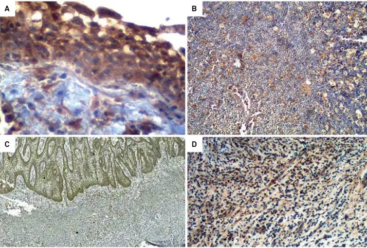
The immunohistochemical expressions of TGF-β1 and MMP-9 were distributed diffusely in all the cases of both periapical granuloma and radicular cysts (Figure 1) (Table 2). In the periapical granulomas, the inlammatory cells and the stromal connective tissue exhibited positivity to both proteins studied, whereas the epithelial cystic lining (atrophic or hyperplastic), as well as the inlammatory cells and connective tissue of the cystic capsule in radicular cysts, showed cellular positivity. Nevertheless, there were no signiicant associations and/or differences in the immunoexpression of TGF-β1 and MMP-9, in relation to clinical signs, radiographic indings,
Figure 1. Representative photomicrographs of immunohistochemical staining of the studied markers in periapical lesions. (a) MMP-9 expression in radicular cysts (IHC, 200X), (b) MMP-9 expression in periapical granuloma (IHC, 100X), (c) TGF-β1 expression in radicular cysts (IHC, 100X), (d) TGF-β1 expression in periapical granuloma (IHC, 100X).
A B
and level of inlammatory iniltrate visualized in the periapical lesions evaluated.
The majority of periapical granulomas had score 1 for TGF-β1 immunoexpression (n = 12; 54%), and score 2 for MMP-9 (n = 16; 67%). Similarly, in radicular cysts, score 1 was more prevalent for TGF-β1 (n = 15; 68%), whereas score 2 was higher for MMP-9 (n = 17; 77%) (Table 2). Signiicant differences could be seen between the expression scores of TGF-β1 and MMP-9 in periapical granulomas (p = 0.004; 95%CI: 1.34 to 4.24) and radicular cysts (p < 0.001; 95%CI: 0.31 to 0.58). Notwithstanding, when the immunohistochemical expression of each protein was evaluated in the periapical lesions, only TGF-β1 revealed relevant differential expression between these lesions (Pearson´s chi-square: p = 0.042; 95%CI: 0.31 to 0.99) (Table 2).
In periapical cysts, no significant correlation (Spearman’s correlation test) was observed between TGF-β1 and MMP-9 (r = 0.370; p = 0.09). As for periapical granulomas, a signiicant moderate positive correlation (Spearman’s correlation test) was veriied between these proteins (r = 0.633; p = 0.001).
Discussion
The development of periapical lesions is a complex and dynamic process in which multiple types of
inlammatory cells and their derivatives are involved, but the details are not well understood. Substantial evidence suggests that these lesions are the result of a long-term endodontic infection or pulp necrosis leading to a progressive immune/inflammatory response, whose persistence in periapical tissues can result in the resorption of the surrounding bone and reactivation of epithelial cell rests of Malassez10,21,22; which are probably the main source of the epithelial lining of radicular cysts.9 In fact, it seems that this persistent, chronic inlammation and destruction of bone depends on the inability of a host defense to eradicate the infection.10
The participants in this study were predominantly females, aged 20-40 years, and exhibited a well-demarcated radiolucency image, and asymptomatic lesions affecting the anterior maxilla. This patient proile was similar to that of several other recent epidemiological reports.23,24 However, only gender and symptoms revealed differences between periapical granulomas and radicular cysts; however, these differences could not be correlated with other clinical variables or with the TGF-β1 and MMP-9 immunohistochemical assessments. The results suggest that there is no speciic relationship between the clinical, demographic and immunohistochemical features of the periapical lesions examined.
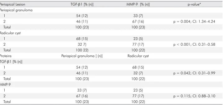
Table 2. Distribution of immunohistochemical expression scores of TGF-β1 and MMP-9 in periapical granulomas and radicular cysts
Periapical Lesion TGF-β1 [% (n)] MMP-9 [% (n)] p-value*
Periapical granuloma
1 54 (12) 33 (7)
2 46 (11) 67 (16) p = 0.004; CI: 1.34–4.24
Total 100 (23) 100 (23)
Radicular cyst
1 68 (15) 23 (5)
2 32 7) 77 (17) p < 0.001; CI: 0.31–0.58
Total 100 22) 100 (22)
Proteins Periapical granuloma [ (n)] Radicular cyst
TGF-β1 [% (n)]
1 54 (12) 68 (15)
2 46 (11) 32 (7) p = 0.042; CI: 0.31–0.99
Total 100 (23) 100 (22)
MMP-9
1 33 (7) 23 (5)
2 67 (16) 77 (17) p = 0.115; CI: 0.88–3.10
Total 100 (23) 100 (22)
The morphological analysis showed that periapical granulomas exhibited higher grades of inlammatory iniltrate than the cysts, but this signiicant difference was not associated with the scores attributed to the immunohistochemical expression of TGF-β1 and MMP-9. In contrast, Gazivoda et al.10 used a luorescent bead immunoassay and/or ELISA to prove that immunoregulatory cytokines such as TGF-β are more important for suppression of inflammation in asymptomatic lesions. Based on this inding, the effect of TGF-β was stronger than that of IL-10. In addition, no differences were established in the present study between the epithelial thickness of radicular cyst lining and the immunoexpression of TGF-β1 and MMP-9, as corroborated by other authors.25,26
In this research, the TGF-β1 expression revealed differences between periapical granulomas and radicular cysts, thus corroborating the observation by Marçal et al.27 and Teixeira-Salum et al.28. Although the data could not conirm any signiicant association between symptoms and TGF-β1 expression, their descriptive analysis showed that the majority of granulomas were asymptomatic and presented a greater number of cases with higher TGF-β1 scores (score 2). Divergently, there were more cases of cysts with symptoms, but fewer cases with higher scores for this protein (Table 2). Thus, these results corroborate those of other authors,demonstrating that asymptomatic periapical lesions exhibited higher levels of TGF-β1 and other immunoregulatory cytokines.10,21 Furthermore, substantial evidence suggests that asymptomatic periapical lesions expressing high levels of immunoregulatory cytokines are less immunologically active.3,10
The MMP-9 immunohistochemical expression was not different among the periapical lesions evaluated in this study. Notwithstanding, an immunohistochemistry assay by Carneiro et al.29 found that the non-epithelialized periapical lesions exhibited higher MMP-9-positive cells, but no signiicant differences were observed among these lesions. Andrade et al.26 also evidenced a higher expression of MMP-9 in granulomas than in radicular and residual radicular cysts. These data suggest that MMP-9 presents a similar remodeling activity of extracellular matrix in periapical lesions.
Additionally, the expression of MMP-9 was not relevant concerning the symptoms and grade of inlammatory iniltrate of the lesions. These results are corroborated by Faustino et al.30 and Andrade et al.26, who showed that the MMP-9 immunoexpression was no different between symptomatology and grade of inlammation among periapical granulomas, radicular cysts and residual radicular cysts.
Nevertheless, MMP-9 showed significantly higher expression scores than TGF-β1, both in periapical granulomas and radicular cysts. MMP-9 is a gelatinase that plays an important role in bone resorption,because of its ability to degrade the denatured collagen of the extracellular matrix, and also activates uncleaved TGF-β in tissues.16,17,29,30,31 A long-term inflammation maintained in periapical tissues could increase the levels of MMP-9, inducing the remodeling of bone and activation of TGF-β, which is important to avert osteoclast formation and stimulate tissue repair mechanisms.2,31,33 Particularly noteworthy is that expressions of TGF-β1 and MMP-9 may depend on the secretion of other proteins, such as cytokines, growth factors, transcriptional factors, other matrix metalloproteinases and tissue inhibitors of matrix metalloproteinases (TIMPs).2,3,4,5,6,31,32,33,34,35,36
Experiments using MMP-9 knockout mice14 evidenced that these animals exhibited larger periapical lesions with greater inflammation, indicating a central participation of MMP-9 in the development of periapical lesions. The presence of TGF-β1 and MMP-9 in periapical lesions denotes their important role in the maintenance, development and exacerbation of chronic processes,5,14,34,35 although further studies are needed to evaluate the possible molecular mechanisms involved in the progression of these lesions.
Conclusions
1. Koivisto T, Bowles WR, Roherer M. Frequency and distribution of radiolucent jaw lesions: a retrospective analysis of 9,723 cases. J Endod. 2012;38(6):729-32. https://doi.org/10.1016/j.joen.2012.02.028
2. Hernádi K, Gyöngyösi E, Mészáros B, Szakács L, Szalmás A, Csoma E et al. Elevated tumor necrosis factor-alpha expression in periapical lesions infected by Epstein-Barr virus. J Endod. 2013;39(4):456-60. https://doi.org/10.1016/j.joen.2012.12.028 3. Jakovljevic A, Andric M, Knezevic A, Soldatovic I, Nikolic N,
Karalic D et al. Human Cytomegalovirus and Epstein-Barr Virus Genotypes in Apical Periodontitis Lesions. J Endod. 2015;41(11):1847-51. https://doi.org/10.1016/j.joen.2015.08.027 4. Piattelli A, Rubini C, Fioroni M, Favero L. Strocchi
R. Expression of transforming growth factor-beta 1
(TGF-beta 1) in odontogenic cysts. Int Endod J. 2004;37(1):7-11. https://doi.org/10.1111/j.1365-2591.2004.00739.x
5. Márton IJ, Kiss C. Overlapping protective and destructive regulatory pathways in apical periodontitis. J Endod. 2014;40(2):155-63. https://doi.org/10.1016/j.joen.2013.10.036 6. Muglali M, Komerik N, Bulut E, Yarim GF, Celebi N,
Sumer M. Cytokine and chemokine levels in radicular and residual cyst fluids. J Oral Pathol Med. 2008;37(3):185-9. https://doi.org/10.1111/j.1600-0714.2007.00595.x 7. Nonaka CF, Maia AP, Nascimento GJ, Freitas RA, Souza LB,
Galvão HC. Immunoexpression of vascular endothelial growth factor in periapical granulomas, radicular cysts, and residual radicular cysts. Oral Surg Oral Med Oral Pathol Oral Radiol Endod. 2008;106(6):896-902. https://doi.org/10.1016/j.tripleo.2008.06.028 8. Alcantara BA, Carli ML, Beijo LA, Pereira AA,
Hanemann JA. Correlation between inflammatory infiltrate and epithelial lining in 214 cases of periapical cysts. Braz Oral Res. 2013;27(6):490-5. https://doi.org/10.1590/S1806-83242013005000023 9. Bernardi L, Visioli F, Nör C, Rados PV. Radicular
Cyst: An Update of the Biological Factors Related to Lining Epithelium. J Endod. 2015;41(12):1951-61. https://doi.org/10.1016/j.joen.2015.08.036 10. Gazivoda D, Dzopalic T, Bozic B, Tatomirovic Z,
Brkic Z, Colic M. Production of proinflammatory and immunoregulatory cytokines by inflammatory
cells from periapical lesions in culture. J Oral Pathol Med. 2009;38(7):605-11.
https://doi.org/10.1111/j.1600-0714.2009.00788.x 11. Tsuji M, Yamasaki M, Amano K, Matsui H, Morimoto
T, Nakamura H. Histochemical localization of neutral proteases released during development of rat periradicular lesion. Arch Oral Biol. 2009;54(12):1128-35.
https://doi.org/10.1016/j.archoralbio.2009.10.003 12. Bradley LM, Douglass MF, Chatterjee D, Akira S, Baaten BJ.
Matrix metalloprotease 9 mediates neutrophil migration into the airways in response to influenza virus-induced toll-like receptor signaling. PLoS Pathog. 2012;8(4):e1002641. https://doi.org/10.1371/journal.ppat.1002641
13. Ahmed GM, El-Baz AA, Hashem AA, Shalaan AK. Expression levels of matrix metalloproteinase-9 and gram-negative bacteria in symptomatic and asymptomatic periapical lesions. J Endod. 2013;39(4):444-8. https://doi.org/10.1016/j.joen.2012.11.009 14. Wan C, Yuan G, Yang J, Sun Q, Zhang L, Zhang J, et al.
MMP9 deficiency increased the size of experimentally induced apical periodontitis. J Endod. 2014;40(5):658-64. https://doi.org/10.1016/j.joen.2014.01.003
15. D’addazio G, Artese L, Piccirilli M, Perfetti G. Role of matrix metalloproteinases in radicular cysts and periapical granulomas. Minerva Stomatol. 2014;63(11-12):411-20. 16. Sternlicht MD, Werb Z. How matrix metalloproteinases regulate
cell behavior. Annu Rev Cell Dev Biol. 2001;17(1):463-516. https://doi.org/10.1146/annurev.cellbio.17.1.463
17. Folgueras AR, Pendás AM, Sánchez LM, López-Otín C. Matrix metalloproteinases in cancer: from new functions to improved inhibition strategies. Int J Dev Biol. 2004;48(5-6):411-24. https://doi.org/10.1387/ijdb.041811af
18. Henriques ÁC, Vasconcelos MG, Galvão HC,
de Souza LB, de Almeida Freitas R. Comparative analysis of the immunohistochemical expression of collagen IV, MMP-9, and TIMP-2 in odontogenic cysts and tumors. Oral Surg Oral Med Oral Pathol Oral Radiol Endod. 2011;112(4):468-75. https://doi.org/10.1016/j.tripleo.2011.05.033
19. Tsai CH, Weng SF, Yang LC, Huang FM, Chen YJ, Chang YC. Immunohistochemical localization of tissue-type plasminogen activator and type I plasminogen activator inhibitor in radicular cysts. J Oral Pathol Med. 2004;33(3):156-61. https://doi.org/10.1111/j.0904-2512.2004.00133.x
References
may be inluenced by TGF-β1. There was no difference and/or association between TGF-β1 and MMP-9 immunoexpression in respect to clinical or radiographic results, grade of inlammatory iniltrate and type of cystic epithelial lining. TGF-β1 and MMP-9 may be factors involved in the maintenance of periapical lesions.
Acknowledgements
20. Costa Neto H, Andrade AL, Gordón-Núñez MA, Freitas RA, Galvão HC. Immunoexpression of tryptase-positive mast cells in periapical granulomas and radicular cysts. Int Endod J. 2015;48(8):729-35. https://doi.org/10.1111/iej.12366 21. Colić M, Gazivoda D, Vucević D, Vasilijić S, Rudolf R,
Lukić A. Proinflammatory and immunoregulatory mechanisms
in periapical lesions. Mol Immunol. 2009;47(1):101-13. https://doi.org/10.1016/j.molimm.2009.01.011 22. Aminoshariae A, Kulild JC. Association of
Functional Gene Polymorphism with Apical Periodontitis. J Endod. 2015;41(7):999-1007. https://doi.org/10.1016/j.joen.2015.03.007
23. Akinyamoju AO, Gbadebo SO, Adeyemi BF. Periapical lesions of the jaws: a review of 104 cases in Ibadan. Ann Ib Postgrad Med. 2014;12(2):115-9.
24. Jamshidi S, Shojaei S, Roshanaei G, Modabbernia S, Bakhtiary E. Jaw intraosseous lesions biopsied extracted from 1998 to 2010 in an Iranian population.
Iran Red Crescent Med J. 2015;17(6):e20374. https://doi.org/10.5812/ircmj.17(5)2015.20374 25. Andrade AL, Nonaka CF, Gordón-Núñez MA,
Freitas Rde A, Galvão HC. Immunoexpression of
interleukin 17, transforming growth factor β1, and forkhead box P3 in periapical granulomas, radicular cysts, and residual radicular cysts. J Endod. 2013;39(8):990-4. https://doi.org/10.1016/j.joen.2013.04.028
26. Andrade A, Santos EM, Carmo AF, Freitas RA, Galvão HC. Analysis of tryptase-positive mast cells and immunoexpression of MMP-9 and MMP-13 in periapical lesions. Int Endod J. 2017;50(5):446-54. https://doi.org/10.1111/iej.12638 27. Marçal JR, Samuel RO, Fernandes D,
Araujo MS, Napimoga MH, Pereira SA et al. T-helper cell type 17/regulatory T-cell immunoregulatory balance in human radicular cysts and periapical granulomas. J Endod. 2010;36(6):995-9. https://doi.org/10.1016/j.joen.2010.03.020 28. Teixeira-Salum TB, Rodrigues DB, Gervásio AM, Souza CJ,
Rodrigues Junior V, Loyola AM. Distinct Th1, Th2 and Treg cytokines balance in chronic periapical granulomas and
radicular cysts. J Oral Pathol Med. 2010;39(3):250-6. https://doi.org/10.1111/j.1600-0714.2009.00863.x 29. Carneiro E, Menezes R, Garlet GP, Garcia RB,
Bramante CM, Figueira R et al. Expression analysis of matrix metalloproteinase-9 in epithelialized and nonepithelialized apical periodontitis lesions. Oral Surg Oral Med
Oral Pathol Oral Radiol Endod. 2009;107(1):127-32. https://doi.org/10.1016/j.tripleo.2008.07.030
30. Faustino ISP, Azevedo RS, Takahama Junior A. Metalloproteinases 2 and 9 immunoexpression in periapical lesions from
primary endodontic infection: possible relationship with the histopathological diagnosis and the presence of pain. J Endod. 2016;42(4):547-51. https://doi.org/10.1016/j.joen.2015.12.020 31. Engsig MT, Chen QJ, Vu TH, Pedersen AC, Therkidsen B,
Lund LR et al. Matrix metalloproteinase 9 and vascular endothelial growth factor are essential for osteoclast recruitment into developing long bones. J Cell Biol. 2000;151(4):879-89. https://doi.org/10.1083/jcb.151.4.879 32. Corotti MV, Zambuzzi WF, Paiva KB, Menezes R,
Pinto LC, Lara VS et al. Immunolocalization of matrix metalloproteinases-2 and -9 during apical periodontitis development. Arch Oral Biol. 2009;54(8):764-71. https://doi.org/10.1016/j.archoralbio.2009.04.013 33. Danin J, Linder L, Lundqvist G, Wretlind B.
Cytokines in periradicular lesions: the effect of linezolid treatment. Oral Surg Oral Med Oral Pathol Oral Radiol Endod. 2003;96(4):492-8. https://doi.org/10.1016/S1079-2104(03)00059-3 34. Zehnder M, Wegehaupt FJ, Attin T. A first study on the
usefulness of matrix metalloproteinase 9 from dentinal fluid to indicate pulp inflammation. J Endod. 2011;37(1):17-20. https://doi.org/10.1016/j.joen.2010.10.003