Cynthia Soares de Azevedo(a) Luciana Cardoso Espejo Trung(a) Maria Regina Lorenzetti Simionato(b)
Anderson Zanardi de Freitas(c) Adriana Bona Matos(a)
(a) Department of Operative Dentistry, Dental School, University of São Paulo (USP), São Paulo, SP, Brazil.
(b) Department of Microbiology, Institute of Biomedical Sciences (ICB), University of São Paulo (USP), São Paulo, SP, Brazil.
(c) Nuclear and Energy Research Institute (IPEN-CNEN/SP), São Paulo, SP, Brazil.
Corresponding Author:
Adriana Bona Matos E-mail: [email protected]
Received for publication on May 20, 2011 Accepted for publication on Aug 26, 2011
Evaluation of caries-affected dentin
with optical coherence tomography
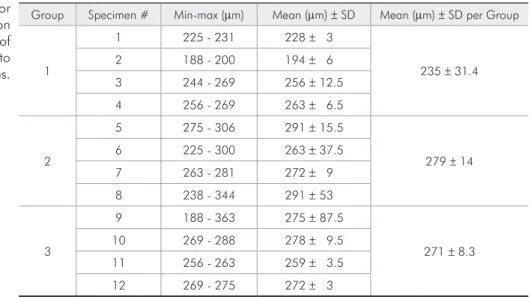
Abstract: The purpose of this study was to evaluate the degree of demin-eralization of artiicially induced caries-affected human dentin by an in vitro microbiological method. The occlusal surfaces of 6 human molar teeth were abraded until a lat surface was obtained, and the enamel was removed to expose the occlusal dentin surface. These teeth were sectioned in 12 halves in the vestibular-lingual direction and divided into 3 groups according to the period length of the microbiological essay (n = 4): G1, 7 days; G2, 14 days; and G3, 21 days. The surfaces of all specimens were protected by an acid-resistant nail varnish, except for a window where the caries lesion was induced by a Streptoccocus mutans bioilm in a batch-culture model supplemented with 5% sucrose. The specimens were then analyzed by optical coherence tomography (OCT) with a super-lu-minescent light diode (λ = 930 nm) with 6.0-µm lateral and longitudinal resolution (in the air). Qualitative and quantitative results (images and average dentin demineralization, respectively) were obtained. The mean demineralization depths were (µm) 235 ± 31.4, 279 ± 14, and 271 ± 8.3 in groups 1, 2, and 3, respectively. In addition, no signiicant change was observed in the lesion mean depth from 7 days of cariogenic challenge on. In conclusion, OCT was shown to be an eficient and non-invasive method to detect the depths of lesions caused by demineralization. Fur-ther, a seven-day demineralization time was considered suficient for car-ies-affected dentin to be obtained.
Descriptors: Tomography; Dental Caries; Microbiology.
Introduction
Dental caries is one of the most prevalent chronic diseases. Its treat-ment has changed signiicantly in recent years, since the current man-agement of caries involves non-invasive techniques and partial removal of carious dentin.1 Therefore, adhesive restorations are widely
indicat-ed, and adhesives must adequately interact with caries-affected dentin, which is demineralized but prone to remineralization.
It is important to point out that most bond strength tests are per-formed on healthy dental tissue, which is not considered a clinically rel-evant substrate. Since the characteristics of caries-affected dentin are different from those of healthy dentin, the hybrid layer associated with it will also be different.2 Also, contradictory results have been obtained
from bond strength tests performed on natural caries-affected dentin.3,4
Therefore, studies in which standardized caries-affected dentin is used Declaration of Interests: The authors
contribute signiicantly to this area of knowledge. Numerous in vitro caries models have been re-ported.5 In the chemical method, the acidiied gel
technique and pH-cycling method are included; in contrast, microorganism strains with known cario-genicity are used in the microbiological method in a way whereby the acid product of their metabolism demineralizes the dental structure.5 More elaborate
systems involving chemostats, lowcells, artiicial mouths, and constant-depth ilm fermentors have been developed in an attempt to better mimic the environment of the oral cavity.5 However, their high
cost and complex apparatus requirements are often limiting factors.5 Some authors6 believe that the
bac-terial model is the closest to the conditions found
in vivo. In the present study, the microbiological method modiied by Gama-Teixeira et al.7 was used
to obtain caries-affected dentin, but the amount of time necessary to produce it has not yet been estab-lished.
Since the artiicially obtained substrate will be used in future laboratory tests, demineraliza-tion depth must be measured by a non-destructive method. Image diagnosis modalities such as ultraso-nography, computerized tomography, and magnetic resonance are technologies that had a signiicant im-pact on medical research and clinical practice, since new tools are needed for the non-destructive assess-ment of the depth and severity of caries lesions.8 In
optical coherence tomography (OCT), spatial reso-lution can be as high as 10 times that obtained by the ultrasound technique.9 OCT is based on
low-coherence laser interference principles, and is simi-lar to the ultrasound technique in various respects, although electromagnetic radiation is used instead of sound waves.10
OCT is a non-invasive technique for caries detec-tion that allows for both following the process of dental tissue demineralization11 and premature
de-tection of the presence of caries. In addition, it has the potential to determine the locations and depths of lesions, as well as to detect hidden and second-ary caries that occurs at the tooth-restoration inter-face.11 The use of electromagnetic radiation in the
near-infrared region, instead of ionizing radiation (x-rays), which is potentially mutagenic, is among its
advantages.
The aim of this study was to evaluate the degree of demineralization of artiicially induced caries-af-fected human dentin by an in vitro microbiological method.
Methodology
Artificial occlusal caries
Healthy human molar teeth were cleaned with periodontal curettes and a rubber cup with pumice paste and water, and were stored in distilled water (4 °C) until use. Approval was obtained from the Ethics Committee (University of São Paulo, Dental School, FOUSP, protocol #115/2010).
The occlusal surfaces of 6 human molar teeth were abraded with a sandpaper disc until a lat sur-face was obtained and the occlusal dentin sursur-face was exposed. These teeth were sectioned in half in the buccal-lingual direction, and the 12 resulting halves were randomized and distributed among 3 experimental groups (n = 4).
Half the area of the prepared teeth was covered with acid-resistant nail varnish (Colorama, CEIL Coml Exp Ind Ltda., São Paulo, Brazil). The den-tal halves were sterilized with gamma ray irradia-tion (25 kGy). Caries lesions were then induced in vitro with the use of a bacterial system, following the method modiied by Gama-Teixeira et al.7
of bacterial contaminants, and after the speciied time periods, the dental halves were removed from the bacterial system.
At the end of the cariogenic challenge, the bio-ilm adhering to the surface was carefully removed, and the specimens were washed with deionized wa-ter. Immediately before the demineralization depth was read, the acid-resistant nail varnish was me-chanically removed from the surfaces of all speci-mens.
OCT imaging
System configuration
In the most common OCT systems, a Michelson interferometer is used to provide a cross-section im-age of the scattering specimens, with non-invasive evaluation and without contact with their surfaces. In this system, the light is conducted by an optical iber as far as a 2 × 2 coupler, where the light is split into 2 beams, one of which is sent to a reference mirror and the other to the sample.
In this OCT system (SR930 Thorlabs Inc., New-ton, USA), a SLED (super-luminescent light-emitting diode; λ = 930 nm) provided A and B scans with 4.0- and 6.0-µm resolution, respectively. The system was able to produce up to 4 frames per sec, thus provid-ing real-time images (2000 × 512 pixels; equivalent to a width × height area of 6000 × 1581 µm2).
The images were acquired from the central re-gions of the exposed windows, generating images
from both exposed and varnish-protected (control) surfaces. The images of the caries-affected dentin were obtained by OCT for different exposure peri-ods (7, 14, and 21 days) and were compared accord-ing to demineralization depth (described above). If any tissue loss occurred due to the demineralization process, the amount of lost tissue was included in the calculation, with the adjacent sound dentin as the initial reference.
Results
Quantitative analysis
Depth values (in µm) of minimum, maximum, and mean demineralization are shown in Table 1. Regarding the mean demineralization depth, the values calculated for all groups were similar, when demineralization depths for 7, 14, and 21 days were compared.
Qualitative analysis
OCT renders 3 types of image: traditional (simi-lar to that generated by ultrasound equipment), col-ored, and inverted (white, with the demineralized region/line in gray or black). In the present study, traditional and inverted images were obtained for each of the 12 specimens. Representative images of each experimental group were selected to illustrate the OCT characteristics of the specimens subjected to cariogenic challenge (Figures 1A, 1B, 2A, 2B, 3A, and 3B). All Figures present a vertical bar that
di-Group Specimen # Min-max (µm) Mean (µm) ± SD Mean (µm) ± SD per Group
1
1 225 - 231 228 ± 3
235 ± 31.4 2 188 - 200 194 ± 6
3 244 - 269 256 ± 12.5
4 256 - 269 263 ± 6.5
2
5 275 - 306 291 ± 15.5
279 ± 14 6 225 - 300 263 ± 37.5
7 263 - 281 272 ± 9
8 238 - 344 291 ± 53
3
9 188 - 363 275 ± 87.5
271 ± 8.3 10 269 - 288 278 ± 9.5
11 256 - 263 259 ± 3.5
12 269 - 275 272 ± 3 Table 1 - Values for
vides the dentin surface into demineralized (right) and non-demineralized (left) sides. Demineraliza-tion depth is indicated by the presence of arrows.
The severity of demineralization could be detect-ed by OCT and is indicatdetect-ed by the amount of light gray in the traditional image (Figures 1A, 2A, 3A) and dark gray (Figures 1B, 2B, 3B) in the inverted image mode. Much severe demineralization could be detected at 14 and 21 days, when compared with that at 7 days of cariogenic challenge.
Discussion
Caries-affected substrate obtained in vitro by the microbiological method contributes signiicantly to the ield of dentin adhesion, because it will allow laboratory tests to be performed on standard caries-affected dentin, which is a clinically relevant
sub-strate.12-14 The microbiological method allowed us to
artiicially produce caries-affected dentin effectively induced from 7 days on.
The achievement of artiicial caries lesions that are morphologically similar to natural caries le-sions14 was a relevant factor in the choice of the
mi-crobiological method, although caries lesions thus obtained show low hardness levels compared with those obtained by chemical or pH-cycling meth-ods.15 However, the same authors state that the
mi-crobiological method is more adequate for obtaining lesions with an evident infected dentin layer, which can be removed.
OCT has already been shown to be effective in detecting premature and secondary caries,16,17 and
was chosen as the diagnostic method for the evalu-ation of demineralizevalu-ation depth. The images ob-Figures 1A and 1B - OCT image after 7 days of cariogenic challenge. A slight loss of structure is observed on the surface. Higher demineralization and loss of structure are observed at the tooth-varnish interface, due to a higher accumulation of bac-terial plaque at both the center and the right of the image. In the images, demineralization is observed in white (1A) and dark gray/black (1B) (arrows). Bar = 500 µm.
Figures 2A and 2B - OCT image after 14 days of cariogenic challenge. The loss of structure is more pronounced, and a step can be observed between the exposed surface and that protected by the acid-resistant varnish. In the images, demineralization is observed in white (2A) and dark gray/black (2B) (arrows). Bar = 500 µm.
Figures 3A and 3B - OCT image after 21 days of cariogenic challenge. Significant variation was not observed in dentin de-mineralization in the period between 14 and 21 days. Loss of structure (step between the exposed surface and that protected by acid-resistant varnish) is observed. In the images, demineralization is observed in white (3A) and dark gray/black (3B) (ar-rows). The regions where higher loss of structure is observed are directly related to the higher accumulation of bacterial plaque. Bar = 500 µm.
B
B
B
A
tained by OCT conirm that this method is effective in evaluating the depth of demineralization caused by induction of caries in vitro from 7 days on. The images clearly show that the demineralization pro-cess is occurring in the specimens after different pe-riods of exposure to the bacterial challenge. Associ-ating qualitative and quantitative results, we could observe that the demineralization depth was almost the same when the 3 periods of challenge were com-pared. However, a close examination of the images allows for the identiication of a more severe and uniform demineralization at 14 and 21 days, when compared with samples demineralized for only 7 days.
Freitas et al.18 evaluated the demineralization
process up to 11 days, and observed exponential bacterial growth until about 9 days. These authors relate this fact to the exhaustion of nutrients avail-able in the culture, accumulation of inhibitory me-tabolites and metabolic end-products, or lack of biological space.18 This result was conirmed in the
present study, because an increase in the deminer-alization depth was not observed from 7 days on. We believe that the nutrient exhaustion hypothesis does not apply to our conditions, since the nutrients were supplied every 24 h. However, accumulation of metabolites and end-products as well as lack of biological space could be the causes for the non-pro-gression in lesion depth from 7 days on.
Such indings can be explained: The bioilm that naturally accumulates on tooth surfaces is a complex community comprised of more than 500 bacterial species.19,20 The early colonizers are
pre-dominantly streptococci. To initiate heterogeneous bacterial interactions, diffusible signals may play important roles, and processes of co-adhesion and co-aggregation of distinct microorganisms contrib-ute to bioilm maturation. In the presence of sucrose, glucosyltransferase enzymes produce water-insol-uble glucan polysaccharides and may contribute to
S. mutans colonization.21 It is likely that the initial
colonizing bacteria create a favorable environment for new invaders, due to the presence of metabolites inhibitory to those bacteria. This would favor the survival of other species more able to adapt to the new habitat and replace the initial species. In this
study, the bioilm consisted of only S. mutans, and, consequently, there was no competitive or inhibitory substance produced by other species. Moreover, the bacterial deposition that occurred during the cario-genic challenge days rendered the bioilm thicker. Nevertheless, the development of a bioilm having a high bacterial cell density increases the concentra-tion of signaling molecules, increasing the possibili-ties of expression of mechanisms for bioilm control, such as quorum sensing or production of surface proteins.22,23 Although no biomass measurements
were made in this study, the results show a non-deepening of demineralization in the period up to 21 days, which would be explained because a too-thick layer of bacterial deposit was formed in the caries induction process.
S. mutans, used in the present study, is an initial colonizer, and has a recognition system that allows the bacterial surface to connect with the acquired dental bioilm. S. mutans has characteristics, espe-cially in the presence of sucrose (the source of nutri-ents used in this study), which trigger its adhesion to the tooth and allow other bacterial cells to co-aggregate therein. In the accumulation phase, mul-tiplication of the initial colonizers and their volume increase occur, and adhesion of different colonizers is likely to occur. Thus, microbial succession is the next phase in the development of dental caries. This last process did not occur because our study used a mono-species bioilm. Perhaps additional studies using different strains of S. mutans with known car-iogenicity would be important for comparing results obtained with ATCC 25175.
with false (inverted) colors of the same image were generated, which can improve tenuous color varia-tions present in the traditional image to be differen-tiated. In all images, the demineralization depth is the same, although it differs in severity.
OCT is a method that allows qualitative and quantitative information,24 such as demineralization
depth and size, to be obtained through 3D images,25
and shows great potential for future use in vivo. Also, as a non-invasive method, it provides a signii-cant contribution to the achievement of standard, artiicially produced caries-affected dentin that can be used in laboratory tests performed over a more clinically relevant substrate. Additional studies are required to validate this laboratory technique as a
protocol.
Conclusions
OCT has been shown to be an eficient and non-invasive method for the detection of the deminer-alization depth of caries-affected dentin produced through cariogenic challenge. It was observed that a 7-day period of cariogenic challenge can be consid-ered adequate to obtain caries-affected dentin.
Acknowledgements
The authors acknowledge the inancial support from FAPESP (grant # 2009/07709-0), and Dr. Pau-lo Boschcov for his revision of the inal version of the manuscript.
References
1. Pugach MK, Strother J, Darling CL, Fried D, Gansky SA, Marshall SJ, et al. Dentin caries zones: mineral, structure, and properties. J Dent Res. 2009 Jan;88(1):71-6.
2. Haj-Ali R, Walker M, Williams K, Wang Y, Spencer P. His-tomorphologic characterization of noncarious and caries-affected dentin/adhesive interfaces. J Prosthodont. 2006 Mar-Apr;15(2):82-8.
3. Mobarak EH, El-Korashy DI, Pashley DH. Effect of chlorhexi-dine concentrations on micro-shear bond strength of self-etch adhesive to normal and caries affected dentin. Am J Dent. 2010 Aug;23(4):217-22.
4. Kunawarote S, Nakajima M, Foxton RM, Tagami J. Effect of pretreatment with mildly acidic hypochlorous acid on adhe-sion to caries-affected dentin using a self-etch adhesive. Eur J Oral Sci. 2011 Feb;119(1):86-92.
5. Steiner-Oliveira C, Rodrigues LKA, Zanin ICJ, de Carvalho CL, Kamiya RU, Hara AT, et al. An in vitro microbial model associated with sucrose to produce dentin caries lesions. Cent Eur J Biol. 2011 Jan;6(3):414-21.
6. Gilmour ASM, Edmunds DH, Dummer PMH. The produc-tion of secondary caries-like lesions on cavity walls and the assessment of microleakage using an in vitro microbial caries system. J Oral Rehabil. 1990 Nov;17(6):573-8.
7. Gama-Teixeira A, Simionato MRL, Elian SN, Sobral MAP, Luz MAACL. Streptococcus mutans-induced secondary car-ies adjacent to glass ionomer cement, composite resin and amalgam restorations in vitro. Braz Oral Res. 2007 Oct-Dec;21(4):368-74.
8. Le HM, Darling CL, Fried D. Automated analysis of lesion depth and integrated reflectivity in PS-OCT scans of tooth demineralization. Lasers Surg Med. 2010 Jan;42:62-8.
9. Fujimoto JG, Brezinski ME, Tearney GJ, Boppart SA, Bouma B, Hee MR, et al. Optical biopsy and imaging using optical coherence tomography. Nat Med. 1995 Sep;1(9):970-2. 10. Otis LL, Colston BW Jr, Everett MJ, Nathel H. Dental optical
coherence tomography: a comparison of two in vitro systems. Dentomaxillofac Radiol. 2000 Mar;29(2):85-9.
11. Bakhsh TA, Sadr A, Shimada Y, Tagami J, Sumi Y. Non-invasive quantification of resin-dentin interfacial gaps using optical coherence tomography: validation against confocal microscopy. Dent Mater. 2011 Sep;27(9):915-25.
12. Perdigão J. Dentin bonding - Variables related to the clini-cal situation and the substrate treatment. Dent Mater. 2010 Feb(2);26:e24-e37.
13. Pereira PNR, Nunes MF, Miguez PA, Swift EJ Jr. Bond strengths of a 1-step self-etching system to caries-affected and normal dentin. Oper Dent. 2006 Nov-Dec;31(6):677-81. 14. Carvalho FG, Gonçalves LS, Carlo HL, Soares CJ, Correr-Sobrinho L, Puppin-Rontani RM. Influence of sterilization method on the bond strength of caries-affected dentine. Braz Oral Res. 2009 Jan-Mar;23(1):11-6.
15. Marquezan M, Corrêa FNP, Sanabe ME, Rodrigues Filho LE, Hebling J, Guedes-Pinto AC, et al. Artificial methods of dentine caries induction: a hardness and morphological comparative study. Arch Oral Biol. 2009 Dec;54(12):1111-7. 16. Jones RS, Fried D. Remineralization of enamel caries can de-crease optical reflectivity. J Dent Res. 2006 Sep;85(9):804-8. 17. Na J, Baek JH, Ryu SY, Lee C, Lee BH. Tomographic imaging
18. Freitas AZ, Zezell DM, Mayer MPA, Ribeiro AC, Gomes ASL, Vieira Jr ND. Determination of dental decay rates with optical coherence tomography. Laser Phys. 2009 Dec;6(12):896-900. 19. Kolenbrander PE, Andersen RN, Blehert DS, Egland PG, Fos-ter JS, Palmer RJ Jr. Communication among oral bacFos-teria. Microbiol Mol Biol Rev. 2002 Sep;66(3):486-505.
20. Paster BJ, Boches SK, Galvin JL, Ericson RE, Lau CN, Leva-nos VA, et al. Bacterial diversity in human subgingival plaque. J Bacteriol. 2001 Jun;183(12):3770-83.
21. Banas JA, Vickerman M. Glucan-binding proteins of the oral streptococci. Crit Rev Oral Biol Med. 2003;14(2):89-99. 22. Lee SF, Li YH, Bowden GH. Detachment of Streptococcus
mutans biofilm cells by an endogenous enzymatic activity. Infect Immun. 1996 Mar;64(3):1035-8.
23. Vats N, Lee SF. Active detachment of Streptococcus mutans cells adhered to epon-hydroxyapatite surfaces coated with salivary proteins in vitro. Arch Oral Biol. 2000 Apr;45(4):305-14.
24. Gimbel C. Optical coherence tomography diagnostic imaging. Gen Dent. 2008 Nov-Dec;56(7):750-7.