w w w . j c o l . o r g . b r
Journal
of
Coloproctology
Original
Article
Microscopic
analysis
of
patients
with
chronic
diarrhea
without
macroscopic
disease
夽
Ary
Santos
Silva
a,∗,
Doryane
Maria
dos
Reis
Lima
a,baHospitalSãoLucas,FaculdadeAssisGurgacz(FAG),Cascavel,PR,Brazil
bSectorofAnorectalPhysiology,GastroclínicaCascavel,Cascavel,PR,Brazil
a
r
t
i
c
l
e
i
n
f
o
Articlehistory:
Received18June2015 Accepted28August2015
Availableonline21December2015
Keywords:
Chronicdiarrhea Microscopiccolitis Colonoscopy
a
b
s
t
r
a
c
t
Background:Colonoscopyis partofthecurrentdiagnosticarmamentarium.However, in
somepatientswithchronicdiarrhea,acolonoscopymayshownormalmucosa;inthese cases,serialbiopsiescanprovideimportantinformationforthediagnosisandtreatmentof patients.
Aim: To analyze patients with chronic diarrhea having a macroscopically normal colonoscopy,byevaluatinghistologicalchanges.
Methods:30patientswithchronicdiarrheaandnormalcolonoscopywereprospectively
eval-uatedandsubmittedtoserialbiopsiesoftheterminalileum,ascendingcolonandrectum.
Results:Thesampleof30patientsshowedaratioof18men(60%)and12women(40%).
On histologicaltypes, itwasfoundthat13patients (43.3%)hadlymphoid hyperplasia, eosinophilicinflammationin4(13.3%),nonspecificinflammationin4(13.3%), regenera-tivechangesin3(10%),lymphocyticcolitisin2(6.6%)andchangesconsistentwithCrohn’s diseasein1(3.3%).
Conclusions: Onecanobservethatevenchronicdiarrheapatients,withoutotherassociated
factors,benefitedfromcolonoscopywithbiopsy,becauseitheldtheetiologicdiagnosisin somecasesasalsoexcludedbyhistopathology.Itwasnoticedthatthefrequencyofpatients withalteredbiopsyandlessdraggeddiarrhealepisodes(84.2%)waslarge,shouldconsider theirachievement.
©2015SociedadeBrasileiradeColoproctologia.PublishedbyElsevierEditoraLtda.All rightsreserved.
夽
StudyconductedattheGeneralSurgeryService,HospitalSãoLucas,FacultyAssisiGurgacz(FAG);ConcurrentwithGastricCascavel, Cascavel,PR,Brazil.
∗ Correspondingauthor.
E-mail:[email protected](A.S.Silva).
http://dx.doi.org/10.1016/j.jcol.2015.08.009
Análise
microscópica
de
pacientes
com
diarreia
crônica
sem
doenc¸a
macroscópica
Palavras-chave:
Diarreiacrônica Colitemicroscópica Colonoscopia
r
e
s
u
m
o
Introduc¸ão: Acolonoscopiafazpartedoarsenaldediagnósticoatual.Porém,emalguns
pacientescomdiarreiacrônica,a colonoscopiapodeevidenciarmucosanormal;nesses casosbiópsiasseriadaspodemtrazerinformac¸õesimportantesparaodiagnósticoe trata-mentodospacientes.
Objetivo: Analisarpacientescomdiarreiacrônicasubmetidosàcolonoscopia
macroscopi-camentenormal,avaliandoassimhistologicamenteasalterac¸ões.
Métodos:Análiseprospectivadahistologia30pacientescomdiarreiacrônicaecolonoscopias
normais,submetidosabiópsiasseriadasdeíleoterminal,cólonascendenteereto.
Resultados: Aamostrade30pacientesmostrouumaproporc¸ãode18homens(60%)e12
mulheres(40%).Sobreostiposdealterac¸õeshistológicas,foiverificadoque13pacientes (43,3%)apresentaramhiperplasialinfóide,inflamac¸ãoeosinofílicaem4(13,3%),inflamac¸ão inespecíficaem4(13,3%),alterac¸õesregenerativasem3(10%),colitelinfocíticaem2(6,6%) ealterac¸õescompatíveiscomDoenc¸adeCrohnem1(3,3%).
Conclusões: Observou-sequemesmopacientescomdiarreiacrônica,semoutrosfatores
associados,beneficiaram-sedacolonoscopiacombiópsia,poisamesmarealizouo diag-nóstico etiológico em algunscasos comotambém oexcluiu atravésda histopatologia. Verificou-sequeafrequênciadepacientescombiópsiaalteradaequadrosdiarreicosmenos arrastados(84,2%)foigrande,devendo-seconsiderararealizac¸ãodoexame.
©2015SociedadeBrasileiradeColoproctologia.PublicadoporElsevierEditoraLtda. Todososdireitosreservados.
Introduction
Chronicdiarrheais acommondisorder, characterizedbya courselongerthan30days;ontheotherhand,itbrings dis-comfort–notonlyphysicalbutalsosocial–tothepatient.1
Consideringthatyoungadults,constitutedbyapopulationof individualswho,intheirmajority,arehealthy,whenexposed to symptoms such as diarrhea tend to belittle the event, postponing this diagnosis. But often this diagnosis can be verycomplexandcomprehensive,asitmayreflectnumerous infectious, endocrine-metabolic, neoplastic, functional and drug-derivedethiologies.1,2
Currently,colonoscopyisoneofthemostcomprehensive researchmethodsofcolorectaldiseases.2,3However,insome
patients with chronic diarrhea, this method may show a normal mucosa. In such cases, obtaining serial biopsies can provide important information for the diagnosis and treatment.4,5 Thismicroscopicanalysisisvery useful,both
forthe diagnosis ofinflammatorybowel diseases and irri-table bowel syndrome, and for the differential diagnosis for lymphocytic, collagenous, actinic, and ischemic colitis, besides cases ofinfectious colitis (tuberculosis, amebiasis, histoplasmosis,and pseudomembranous colitis)and infec-tionsassociatedwithacquiredimmunedeficiencysyndrome, by enabling the visualization of mucosa and allowing the collectionofmaterialforhistopathologicalanalysis.3,6
Thus,thesearchformicroscopicchangesinyoungpatients is very useful, sincethe conditions (including those most severe ones, such as inflammatory bowel disease) can be diagnosed only by histopathology, considering that these
conditions are in sucha nascent stage, atthe point ofan absenceofmacroscopiclesions.
Objectives
To analyze patients with chronic diarrhea with a macro-scopicallynormalcolonoscopy,withtheaimtoidentifythe variablesofage,gender,durationofdiarrheaand histopatho-logicalchanges,anddrawaparallelbetweensuchvariables.
Methodology
ThisstudywasapprovedbytheResearchEthicsCommittee, FaculdadeAssisGurgacz(FAG),opinionnumber1026986/2015 – CEP/FAG. Thisisa prospectivestudy inwhich data were stored in the database ofthe Clinic (in the form of medi-calcharts)andcollectedinachecklist.Thirtypatientswith chronicdiarrhea,undergoingcolonoscopiesandserial biop-siesfromterminalileum,ascendingcolonandrectumwere selected atGastroclínica Cascavel – PR from October 2014 to March 2015. The inclusion criteria were: young adults (18–50 years old) with diarrhealastingmore than 30 days, without endoscopic mucosal changes and considered as colonoscopically normal subjects. The samples were sub-jected topathologists’assessmentaccordingtotheAtlasof Nontumor Pathology –GastrointestinalDiseases(2007)5: (1)
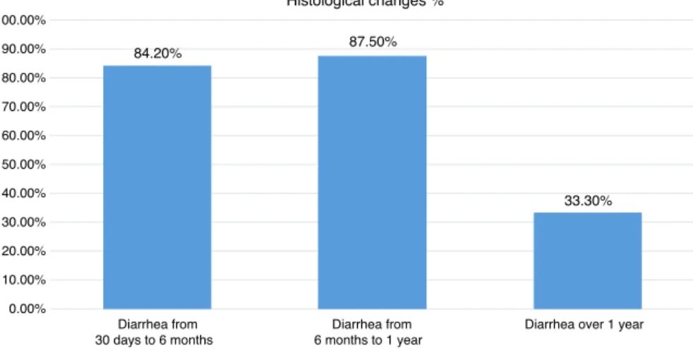
84.20% 87.50%
33.30%
0.00% 10.00% 20.00% 30.00% 40.00% 50.00% 60.00% 70.00% 80.00% 90.00% 100.00%
Histological changes %
Diarrhea from 30 days to 6 months
Diarrhea from 6 months to 1 year
Diarrhea over 1 year
Fig.1–Incidenceofhistologicalchangeswithrespecttodiarrheaduration.
(2) unspecificinflammation: findings that donotmeet the criteriaofanspecificinflammation,howeverexceedingthe limitsofanormalorreactionalmucosa;(3)lymphoid hyper-plasia:activationofmucosaproperlymphoidfollicles,with enlargementofthegerminalcenter;(4)eosinophiliccolitis: anincreaseof20eosinophilsperfield,compromisingthe sub-mucosaandthepropermusclelayer.
HIV-positivesubjects,individualswithlactoseintolerance or celiac disease,or withinflammatorybowel disease pre-viouslydiagnosed,patientswithdiverticulardiseaseandits complications,aswellaspatientswithincompletedata,were excludedfromthisstudy.Theparametersevaluatedwere:age, gender,durationofdiarrhea,medicationsinuse,andresults ofmicroscopy.Datawerestatisticallyanalyzedforsignificance andcomparedtotheavailableliteraturefordiscussion.
Results
Regardinggender,thesamplewascomposedof18men(60%) and12women(40%).Withregardtoage,11patients(36.6%) wereaged18–35yearsand19patients(63.3%)wereaged35–50 years,withamean ageof30.5years.Itwas foundthat20 patients(66.6%)didnotuseanychronicmedication,and10 patients(33.3%)wereusingcontinuousmedication.Ofthese latterpatients,3were usingoralcontraceptives,3wereon hypertensives(losartanandpropranolol),2wereon omepra-zole,1wasinuseoflevothyroxine,and1wasamultivitamin user.Nopatientrelatedanyofthesepharmacologicalproducts withtheonsetofdiarrhealsymptoms.
Regardingtimelineofsymptoms,19patients(63.3%)were sufferingdiarrhealastingfrom30daysto6months,8patients (26.6%) from 6 monthsto 1 year, and 3 patients (10%) for morethan 1year. Themean duration ofsymptoms was 3 months.Withregardtohistology,therewerechangesin84.2% ofpatientswithdiarrhealastingfrom30daysto6months,in 87.5%ofthosewithdiarrheafrom6monthsto1year,andin 33.3%ofpatientswithdiarrhealastingover1year(Fig.1).
Astohistopathology,itwasfoundthat4patients(13.3%) exhibitednochangeand26patients(86.6%)exhibitedsome histologicalchange.Amongthose withchanges,5patients (19.2%)exhibitedchangesinmorethanonesegmentbiopsied:
1patient(3.84%)withchanges inall threesegments (distal ileum,ascendingcolon,andrectum)and4patients(15,38%) presentedterminalileumandrectumchanges.
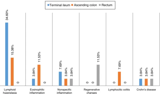
Astothetypesofhistologicchanges,itwasfoundthat13 patients(50%)exhibitedlymphoid hyperplasia:eosinophilic inflammation in 4 (15.38%) patients, unspecific inflamma-tionin4(1538%)patients,regenerativechangesin3(1153%) patients,lymphocyticcolitisin2(7.69%)patients,andchanges consistentwithCrohn’sdisease in1(3.84%)patient(Fig.2). Terminalileumwasthemostaffectedsegment,withchanges observed in12 patients(46.1%); lymphoid hyperplasia was themostcommonlyobservedchange,followedbychangesin theascendingcolonin9(34.6%)patients,witheosinophilic inflammationasthemostcommonchange(withrectum)in5 (19.2%)patients,withpredominanceofregenerativechanges (Table1).
Discussion
Considering that this is a population composed mostly of healthy,youngadults,whenexposed tosymptomssuchas diarrhea, tendtoneglect itsimportance,delayingthe diag-nosis.However,oftenthediagnosiscanbeverycomplexand wide-ranging, thankstothe potential fornumerous (infec-tious,endocrine-metabolic,neoplastic,functionalanddrug) etiologies.4,6Colonoscopyhasbeenincreasinglyusedinthese
patients, especiallyinthose withahistoryofchronic diar-rhea,andforwhomonecannotgettoadiagnosiswithother methods.6Withtheuseofcolonoscopy,itispossibleto
ana-lyzethepresenceofmacroscopiclesions;butinsomepatients withchronic diarrhea,colonoscopymay showonlynormal mucosa.Insuchcases,performingserialbiopsiescanresult inimportantinformationforthediagnosisandtreatmentof thesepatients.4,6
Although macroscopically the colonoscopies were nor-mal in our study, 86.6% of patients had some histological change. Evenconsideringsuchansmallsample,thisfigure demonstratestheimportanceofaninvestigationintocases ofchronicdiarrhea.Anhistologicalstudy7ofcolonicmucosa
34.
60%
3.84
% 7.69
%
0 0
3.84
%
1
5.
38%
1
1.
53%
3.84
%
0
7.69
%
3.84
%
0 0
3.84
%
11.
53%
0
3.84
%
Lymphoid hyperplasia
Eosinophilic inflammation
Nonspecific inflammation
Regenerative changes
Crohn’s disease Lymphocitic colitis
Terminal ileum Ascending colon Rectum
Fig.2–Pathologicalchanges(inpercentages)foundinbiopsiesoftheterminalileum,ascendingcolonandrectumbiopsies.
(10.5%)withhistopathologicalfindings withpotential clini-calsignificance,suggestiveofcollagenouscolitis,lymphocytic colitis and melanosiscoli; and 35/119 (21.6%)with defined histopathological findings: collagenous colitis, lymphocytic colitis, minimum microscopic colitis, eosinophilic colitis, pericrypteosinophilicenterocolitis,intestinalspirochaetosis, schistosomiasis, and Crohn’s disease – findings similar to thosediagnosesfoundinourstudy,showingthatmicroscopic colitisandincipientinflammatoryboweldiseasecanbemore oftendiagnosed.
In2002,onecasereportofcollagenouscolitisinapatient withchronicdiarrheaofunknowncauseassociatedwiththe use of lansoprazole was published.8 The histopathological
changesresolvedwiththediscontinuationofthemedication andrelapsedwithitsreintroduction.Weobservedno associ-ationwithantidiarrhealmedications;butinsomecases,drug interactionisanimportantfactor.
Autoimmunity is a condition common to several dis-eases, being also observed in lymphocytic colitis. A study was conducted9 with colonoscopies of 50 patients with
Hashimoto’s thyroiditis, 5ofthem withdiarrhea. Of these 50patients,20(40%)hadintestinalhistologicalchanges con-sistentwith lymphocytic colitis. Theauthors of this study concludedinfavorofahighincidenceoflymphocyticcolitis
in patients with Hashimoto’s thyroiditis,although mostof themwereasymptomatic,whichleadsusagaintodwellon theimportanceofusingthehistoryofotherpreviousdiseases –speciallyautoimmunediseases–asastartingpointinthe investigation.
Paralleltothediscussionontheneed,ornot,forroutine biopsiesinpatientswithdiarrheawithnormalcolonoscopy, liesthe debateontheneed, ornot,toexamineand biopsy theterminalileum.10Otherauthors11retrospectivelystudied
683 ileocolonoscopies, finding that 499 (73.06%) had nor-malresults,123 (18%)exhibitedlymphoid hyperplasia,and 25 (3.66%) suffered from Crohn’s disease,demonstrating a high percentage of coincidence between endoscopic and histopathological diagnoses (96.87% in normal ileocolono-scopies, 77.77% in lymphoid hyperplasia, and 77.27% in Crohn’sdisease), which revealedlargenumbers ofpatients withlymphoidhyperplasia,asoccurredinourstudy,and espe-ciallyinthecaseoftheterminalileumthat,inourpatients, wasthemostaffectedsegment.
What can be said is that the study and treatment of patients with chronicdiarrhea are not aneasy task,often depending onmonitoringofseveral steps.Several diseases are includedinthe checklistofdifferentialdiagnoses,and, among them, microscopic colitis (collagenous colitis and
Table1–Numberofalterationsfound,accordingtotheaffectedsegmentandthemostfrequentinjury.
Affectedsegment Patients Mostfrequentinjuries Numberofcases
Terminalileum,ascendingcolonandrectum 1 InflammationsuggestiveofChron’sdisease 1
Terminalileumandrectum 4 Lymphoidhyperplasia 4
Terminalileum 7 Lymphoidhyperplasia 7
Ascendingcolon 8 Eosinophilicinflammation 4
Rectuma 0 0
lymphocytic colitis) in patients with normal macroscopic appearance on their colonoscopy.12–14 In cases of
collage-nouscolitis,athickening ofthebasementmembrane with increasedcollagentissueatthislevelcanbeobserved,andin casesoflymphocyticcolitisthereisanincreased inflamma-toryinfiltrateintothelaminapropria,withapredominance ofcytotoxicCD8lymphocytes.13–15Thesetwoconditionscan
causechronicorintermittentdiarrhea,beingdiagnosedonly byhistopathology.Insuspectedcasesofmicroscopiccolitis, theentirecolonshouldbebiopsiedforhistopathology.15,16
Inanalyzingthehistopathologyofthecases,wefoundthat thereisnouniformpatternforreportingbothpositivedata and the presence of inflammatorycell infiltrateor cryptal damage, nor for negative data, for instance, the presence ofgranulomas,fibrosis,micro-organisms, abscesses, malig-nancy,specificity,parasites,etc.14–17
Onegetstheimpressionthatpathologistsdonotgivedue importancetothe necessary measurementsand countings forcharacterization,especiallyincasesofcollagenous coli-tisandlymphocytic colitis,conditionsthat, fromwhatwas discussed,canjustifythepresenceofdiarrhea,evenwitha normalcolonoscopy.18–20Similartowhatwasdonefor
gastri-tiswiththeuseoftheSydneyclassification,21afutureideato
bediscussedamongclinicians,pathologistsandendoscopists isthesearch forsomehistopathologicaluniformityforthe diagnosisofmicroscopiccolitis.22–24
Conclusion
Microscopicanalysisinpatientswithchronicdiarrheawith normalmacroscopicresultsrevealedthefrequentoccurrence oflymphoidhyperplasia,andthatthemostaffected intesti-nalsegmentwastheterminalileum.Themostaffectedsites wereterminalileumandascendingcolon(21/26patients), sug-gesting that these areas deserve tobe remembered inthe investigation,regardlessoftheirmacroscopicappearance.
Conflicts
of
interest
Theauthorsdeclarenoconflictsofinterest.
Acknowledgements
Our sincere thanks to Dr. Carlos Floriano de Morais from LaboratórioAPC,andtoDr.AlexandreGalvãoBuenofrom Lab-oratórioAnatom,fortheirreviewofslidesandcellularrecount withreviewofpathology reports; and alsoto Mrs. Claudia Duarte,headofthescientificsectorofGastroclínicaCascavel, forherhelpintheselectionandorganizationofpatientdata.
r
e
f
e
r
e
n
c
e
s
1. AmericanGastroenterologicalAssociationMedicalPosition Statement.Guidelinesfortheevaluationandmanagementof chronicdiarrhea.Gastroenterology.1999;6:146–63.
2. FineKD,SchillerLR.AGAtechnicalreviewontheevaluation andmanagementofchronicdiarrhea.Gastroenterology. 1999;116:1464.
3.FerreiraS,MagalhãesM,CotrimI,PereiraA,SaraivaR. Diarreiacrónica.JPortGastrenterol.2012;19:140–2.
4.RafiUD,ManzarZ,Mujeeb-Ur-RehmanAB.Chronicdiarrhea: largegutcauses.ProfMedJ.2008;15:479–85.
5.NoffsingerA,Fenoglio-PreiserCM,MaruD,GiliniskyN. Gastrointestinaldiseases,AFIPatlasofnontumorpathology. Firstseries,vol.5;2007.p.656–8.
6.NogueiradaSilvaJG.Colonoscopianospacientescom diarreiacrônica.RevGastroenterolFugesp.2000.
7.daSilvaJG,DeBritoT,CintraDamiaoAO,LaudannaAA, SipahiAM.Histologicstudyofcolonicmucosainpatients withchronicdiarrheaandnormalcolonoscopicfindings.J ClinGastroenterol.2006;40:44–8.
8.WilcoxGM,MattiaA.Collagenouscolitisassociatedwith lansoprazole.JClinGastroenterol.2002;34:164–6.
9.CindorukM,TuncerC,DursunA,YetkinI,KarakanT,CakirN, etal.Increasedcolonicintraepitheliallymphocytesin patientswithHashimoto’sthyroiditis.JClinGastroenterol. 2002;34:237–9.
10.CuvelierC,DemetterP,MielantsH,VeysEM,DeVosM. Interpretationofilealbiopsies:morphologicalfeaturesin normalanddiseasedmucosa.Histopathology.2001;38:1–12.
11.GonzálezAH,GarciaOMH,JimenezG.Estudioendoscópico delíleonterminal.RevCubaMed.2002;41:141–5.
12.ElliotVJ,BatemanAC,GreenB.Theendoscopicallynormal colonwhenismappingbiopsyhistopathologicallyjustifiable? FrontlineGastroenterol.2012;3:104–8.
13.HowatA,BoydK,DouceG.Histopathologyandcytopathology oflimitedornoclinicalvalue.TheRoyalCollegeof
Pathologists.2ed.London:RCPath;2005.
14.KoksalAR,BogaS,AlkimH,ErgunM,BayramM,SakizD.How doesabiopsyofendoscopicallynormalterminalileum contributetothediagnosis?Whichpatientsshouldundergo biopsy?[sérieonline].LibyanJMed.2014;9:23441.
15.MeloMMC,CuryPM,RonchLS,Gonc¸alves-FilhoFA,Cunrath GS,NetinhoJG.Íleoterminaldepacientessubmetidosà colonoscopia:aspectosendoscópicos,histológicoseclínicos. ArqGastroenterolSãoPaulo.2009;46:102–6.
16.MeloMMC,NetinhoJG.Aspectosendoscópicosno diagnósticodedoenc¸asqueacometemoíleoterminal.Rev ColBrasCir.2010;37:234–9.
17.MünchA,LangnerC.Microscopiccolitis:clinicaland pathologicperspectives.Linköping,Sweden/Graz,Austria: DivisionofGastroenterologyandHepatology,Departmentof ClinicalandExperimentalMedicine,FacultyofHealth Science,LinköpingsUniversity/InstituteofPathology,Medical UniversityofGraz;2014.
18.NahasSC,AlvesPA,NahasOS,Habr-GamaA,PinottiHW. Colonoscopiacomométododiagnósticoeterapêuticona doenc¸adointestinogrossonoHospitaldasClínicasda FaculdadedeMedicinadaUniversidadedeSãoPaulo.Rev BrasColoproctol.1989;9:20.
19.NahasSC,AlvesPRA,AraújoSEA,SousaJAHS,SobradoJCW, NahasCSR,etal.Empregodacolonoscopiacomométodo diagnósticoeterapêuticodasdoenc¸asdointestinogrosso. Resultadosobservadosem1.715exames.RevHospClínFac MedSãoPaulo.1998;53:117–21.
20.NahasSC,OliveiraFDES,AraújoSE,Lourenc¸ãoJL,Sobrado JCW,NahasCSR,etal.Colonoscopia:indicac¸ões,
contraindicac¸õesecomplicac¸ões.RevHospClínFacMedSão Paulo.1998;53:91–9.
21.MainguetP,JouretA,HaotJ.TheSidneySystem,anew classificationofgastritis.GastroenterolClinBiol. 1993;17:T13–7.
23.NossaFLC,PaulaNBLBB,TodinovLR,BarretoNPF,SilvaJH, FormigaGJS.Colonoscopiadiagnósticaeterapêutica. Avaliac¸ãodasindicac¸õeseresultados.RevBrasColoproctol. 1999;19:168–71.
24.SilvaJG,BritoT,CintraDAO,LaudannaAA,SipahiAM. Histologicstudyofcolonicmucosainpatientswithchronic diarrheaandnormalcolonoscopicfindings.JClin