Nutr Hosp. 2015;31(5):1947-1956 ISSN 0212-1611 • CODEN NUHOEQ S.V.R. 318
Revisión
Hepatic inflammatory biomarkers and its link with obesity and chronic
diseases
Ana Carolina Pinheiro Volp1, Fernanda Cacilda Santos Silva2 and Josefina Bressan3
1Department of Clinical and Social Nutrition, Federal University of Ouro Preto, Ouro Preto. 2Department of Biological Sciences,
Federal University of Ouro Preto, Ouro Preto. 3Department of Nutrition and Health, Federal University of Viçosa, Viçosa, Brazil.
Abstract
Introduction: The low-grade inflammation and insulin resistance are two events that could be present in varying degrees, on obesity and chronic diseases. The degree of subclinical inflammation can be gauged by measuring the concentrations of some inflammatory biomarkers, including the hepatic origin ones. Some of those biomar-kers are sialic acid, α1-antitrypsin and the C-terminal fragment of alpha1-antitrypsin, ceruloplasmin, fibrino-gen, haptoglobin, homocystein and plasminogen activa-tor inhibiactiva-tor-1.
Objectives: To approach the relation between adiposi-ty and hepatic inflammatory markers, and to assess the possible associations between hepatic inflammatory bio-markers and obesity, as well as their capacity of predic-ting chronic diseases such as type 2 diabetes and athero-trombotic cardiovascular diseases.
Methods: We used electronic scientific databases to se-lect articles without restricting publication year.
Results: The sialic acid predicts the chance increase to become type2 diabetic independently of BMI. Moreover, the α1-antitripsin, ceruloplasmin, fibrinogen and hapto-globulin biomarkers, seem predict the chance increase to become type2 diabetic, dependently, of BMI. So, this pro-cess could be aggravated by obesity. The concentrations of fibrinogen, homocystein and PAI-1 increase proportio-nally to insulin resistance, showing its relation with meta-bolic syndrome (insulin resistance state) and with type2 diabetes. In relation to cardiovascular diseases, every biomarkers reported in this review seem to increase the risk, becoming useful in add important prognostic.
BIOMARCADORES HEPÁTICOS DE INFLAMACIÓN Y SU VÍNCULO CON LA OBESIDAD Y LAS ENFERMEDADES CRÓNICAS
Resumen
Introdución: El bajo grado de inflamación y la resis-tencia a la insulina son dos eventos que podrían estar pre-sentes en mayor o menor grado, en la obesidad y las en-fermedades crónicas. El grado de inflamación subclínica se puede evaluar por medición de las concentraciones de algunos biomarcadores inflamatorios, incluyendo los de origen hepático. Algunos de estos biomarcadores son el ácido siálico, α1-antitripsina y el fragmento C-terminal de la alfa 1-antitripsina, ceruloplasmina, fibrinógeno, haptoglobina, la homocisteína y el inhibidor-1 del activa-dor del plasminógeno.
Objetivos: Evaluar la relación entre la obesidad y los marcadores de inflamación hepática, y las posibles aso-ciaciones entre los biomarcadores inflamatorios hepáti-cos y la obesidad, así como su capacidad de predicción de las enfermedades crónicas como la diabetes tipo 2 y enfermedades cardiovasculares aterotromboticas.
Métodos: Se utilizaron bases científicas electrónicas para selección de artículos, sin límite de año de publi-cación.
Resultados: El ácido siálico predice el aumento de convertirse en diabéticos tipo 2 independientemente del IMC. Por otra parte, los biomarcadores α1-antitripsina, ceruloplasmina, fibrinógeno y haptoglobulina, parecen predecir el aumento de convertirse en diabético tipo 2, dependiente, de IMC. Por lo tanto, este proceso podría verse agravada por la obesidad. Las concentraciones de fibrinógeno, homocisteína y PAI-1 incrementam propor-cionalmente a la insulinoresisténcia, mostrando su rela-ción con el síndrome metabólico (estado de resistencia insulínica) y con la diabetes tipo 2. En relación con las enfermedades cardiovasculares, cada biomarcador in-formado en esta revisión parece aumentar el riesgo, lle-gando a ser muy útil en el complemento pronóstico.
Correspondence: Ana Carolina Pinheiro Volp.
Department of Clinical and Social Nutrition – Nutrition School. Federal University of Ouro Preto, Brazil. Campus Universitário. Morro do Cruzeiro, s/no. Ouro Preto, Minas Gerais. Brasil. CEP 35.400-000.
E-mail: anavolp@gmail.com
Abbreviations
AMI: acute myocardial infarction. BMI: body mass index.
CRP: C-reactive protein. FFA: free fatty acids. FFA: free fatty acids. HbA1c: glycated hemoglobin.
HOMA-IR: homeostatic model assessment-insulin resistance.
HR: hazard ratio. OR: odds ratio.
PAI-1: plasminogen activator inhibitor-1. QUICKI: quantitative insulin check. ROS: reactive oxigen species. RR: relative risk.
SI: insulinic sensitivity index. tPA: tecidual plasminogen activator.
Introduction
The reaction of induced inflammation by factors of risk (abdominal obesity, hyperglycemia, dyslipide-mias and systemic hypertension) and associated im-mune response are the principal events which conduct to aterogenetic process1. Individuals with these clinic
manifestations, generally, show prothrombotic and pro-inflammatory states, characterizated by subclinic imflammatory condition2-4, a process which could be
aggravated by obesity3,5,6.
Moreover, the insulin resistance has been associated with the increase of plasmatics proteins inflammation sensitive (inflammatory biomarkers)2-6. Prospective
works corroborate these associations between many inflammatory biomarkers and the diabetes and athero-thrombotics cardiovascular diseases incidence5,6,9.
The adipose tissue has endocrine functions. Add-tionally, it has been proposed that pro-inflammatory cytokines formed on it, increase the hepatic synthe-sis of acute phase protein5,6,10,11. However, it remains
unknown how the inflammation of low intensity con-tributes to increase risk for cardiovascular diseases in overweight and obese individuals12. This risk could be
very different for individuals with similar body mass. In fact, studies show that cardiovascular risk between
obese individuals vary depending on the levels of others risk factors associated with obesity13,14.
This review approaches the relation between adipo-sity and hepatic inflammatory markers. Moreover, it congregates the knowledge relating to possible inte-rations of these inflammatory mediators with chronic diseases associated to obesity, as well as, demonstrates the capacity of them in predict the risk for diabetes and cardiovascular affections.
Methods
This review was conducted using electronic scien-tific databases, including Medline, PubMed and SciE-LO, using the following key words in English, Spanish and Portuguese: inflammation, obesity, cardiovascular diseases, type-2 diabetes, sialic acid, α1-antitrypsin and the C-terminal fragment of alpha1-antitrypsin, ceruloplasmin, fibrinogen, haptoglobin, homocystein and plasminogen activator inhibitor-1. The articles were selected after reading the abstract and regardless of their year publication.
Hepatic Inflammation Markers
The accurate physiological events which conduct to beginning of inflammatory response in obesity are not totally known11. It is known that the mechanisms that
obesity, specially central (visceral) obesity, associate with morbimortality include the increase in expression and release of adipose tissue cytokines and acute pha-se proteins; increapha-se in activity of coagulation cascade (hypercoagulability) and decrease in activity of fibri-nolytic cascade (pro-thrombotic process); increase in inflammatory process, oxidative stress and endothelial disfunction, besides disturbance of glucose and lipid metabolism (insulin resistance)15.
The expansion of adipose tissue leads to adipocyte hypertrophy and hyperplasia and these big adipocytes decrease de local oxigen supply, causing cellular hypoxia and activation of cellular stress pathway (oxi-datives and inflammatory), inducing a cellular autono-mic inflammation (autocrine effect) and cytokines and release of others pro-inflammatory signals.
Conclusion: This review integrates the knowledge con-cerning the possible interactions of inflammatory me-diators, in isolation or in conjunction, with obesity and chronic diseases, since these biomarkers play different functions and follow diverse biochemical routes in hu-man body metabolism.
(Nutr Hosp. 2015;31:1947-1956)
DOI:10.3305/nh.2015.31.5.8525
Key words: Obesity. Insulin resistance. Inflammation. Diabetes. Cardiovascular diseases.
Conclusion: Esta revisión se integra el conocimien-to acerca de las posibles interacciones de los mediadores inflamatorios, en forma aislada o en combinación, con la obesidad y las enfermedades crónicas, ya que estos biomar-cadores desempeñan funciones diferentes y siguen diversas rutas bioquímicas en el metabolismo del cuerpo humano.
(Nutr Hosp. 2015;31:1947-1956)
DOI:10.3305/nh.2015.31.5.8525
The adipocines (resistin, leptin and adiponectin), which are secreted by adipocytes, can also affect the inflammation and insulin resistance. As part of chronic and low intensity inflammatory process, chemokines locally secretaded attract pro- inflammatory macro-phages for adipose tissue, which form a crown shape structure around the dead and/or sick big adipocites. Following, these macrophages stimulate the cytokines release, which will activate the inflammatory way in adipocytes and adjacent tissues (autocrine and paracri-ne effect), aggravating the inflammation and insulin resistance4,10,11.
The hepatic inflammation can occur in obesity, be-cause the activation of inflammatory pathways could be a steatosis result and/or increase in responses of hepatocytes stress pathways, which could result in he-patocytes autonomic inflammation (autocrine effect). The Kupffer cells (hepatic cells similar to macropha-ges) can also become activated, locally stimulating the cytokines release, which aggravated more the hepatic inflammation and insulin resistance. Morover, the ca-loric excess and obesity are, frequently, come along with the increase on tissue and tecidual circulant free fatty acids (FFA). They could activate directly the pro-inflammatory responses in vascular endothelial cells, adipocytes, myeloid derivatived cells10,11. The
systemic inflammation developement is the result of these physiological events induced by obesity11.
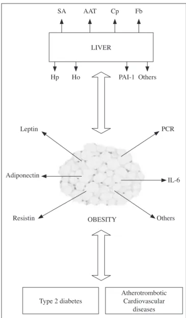
Amongst the hepatic biomarkes of inflammation related to the atherosclerose process are: sialic acid,
α1-antitrypsin and the C-terminal fragment of
al-pha1-antitrypsin, ceruloplasmin, fibrinogen, hapto-globin, homocystein and plasminogen activator inhi-bitor-1 (Fig. 1).
Siliac acid
The siliac acid or N-acetylneuraminic is compo-nent of some acute phase protein terminal part, as alpha1- antichemotrypisin, alpha1-antitrypsin, hapto-globin and orosomucoid. These glucoproteins together explain 70% of sialic acid plasmatic concentration16.
Its production in hepatocytes is stimulated in inflam-mation and metabolic/oxidative stress situations. It could be considered a biomarker of serum concentra-tion of many acute phase protein, beyound, could be considered a systemic inflammatory biomarker since predict the risk for type 2 diabetes and cardiovascular diseases7,16.
Non-diabetic normotensive obese individuals pre-sent the sialic acid concentration significantly increa-sed as compared to non-obese controls. The sialic acid correlates positively with Homeostatic Model Assessment- Insulin Resistance formula (HOMA-IR), body mass index (BMI), waist and hip circumferen-ce and, negatively with Quantitative Insulin Check index (QUICKI) and insulinic sensitivity index (SI)17.
Yet, the siliac acid correlate to the following
measure-ments of adiposity and /or metabolic syndrome com-ponents: glycemia, triacylglycerol, HDL-cholesterol, systolic and diastolic arterial pressure, besides it corre-lates with body weight, insulin, total cholesterol and LDL-cholesterol. In this same study, how much big-ger the number of metabolic syndrome components, bigger was the sialic acid concentration. For each 0.34 mmol/L of increase in sialic acid concentration, a OR for metabolic syndrome was 2.5 (1.8 to 3.4), and per-sisted significant after the adjustment to BMI, with OR of 1.9 (1.3 ato 2. 6)18,19. In ARIC Study, individuals
with the sialic acid and orosomucoid concentration bigger than average, showed odds ratio (OR) of 3.7 (1.4 to 9.8) e 7.9 (2.6 to 23.7), respectively, to develop type 2 diabetes. When adjusted for BMI and waist-hip index, the association of sialic acid and orosomucoid kept significants, with OR of 2.8 (1.01 to 8.1) and
SA
Hp
Leptin
Adiponectin
Resistin OBESITY Others LIVER
Type 2 diabetes
Atherotrombotic Cardiovascular
diseases IL-6 PCR Others PAI-1 Ho
Fb Cp AAT
7.1 (2.1 to 23.7), respectively7. In other study,
indivi-duals with sialic acid concentration on major quartile showed relative risk (RR) for cardiovascular mortality of 2.4 (2.0 to 2.8) and of 2.6 (1.9 to 3.6) in men and women, respectivelly16.
Moreover, serum concentrations of sialic acid co-rrelated to leptin concentrations, suggesting that high sialic acid concentration are related to biomarkers of obesity and adipose tissue metabolism, which could justify the fact of high concentrations of sialic acid pre-cede the development of type 2 diabets and could also explain its function as cardiovascular risk indicator20.
Then, the sialic acid correlate to adiposity measure-ment and/or metabolic syndrome components, besides predict the increase of the risk to develop metabolic syndrome and type2 diabetes, independently on BMI; as well as the increase of the risk to cardiovascular mortality. So, in individuals with elevation of its serum concentration, the sialic acid could add prognostic in-formation that could be useful about obesity and chro-nic diseases.
Alpha1-Antitripsin and the C-terminal fragment of alpha1-antitrypsin
The α1-antitripsinis the main inhibitory endogenous human plasm proteases with serin which present a big variety of anti-inflammatory propriety, besides to exe-cute an important function in reducing the proteolytic damage in the tissues. It is an acute phase protein and its concentration increases quickly in response to me-tabolic/oxidative stress due to infection and inflam-mation21.
Even its production be mainly hepatic, some cells of immune system (neutrophils, monocytes, macro-phages) also express it in response to one variety of inflammatory mediators. Some studies in vitro showed that alpha1- antitrypisin inhibit the TNF-β e da IL-1 syntesis and release, however it helps the inflammatory cytokines release, as IL-10 in human monocytes21.
Ad-dtionally, alpa1-antitrypsin is related to atherogenic process due to its union to LDL-cholesterol receptors and carriers receptors as the CD-36, which recognize the oxidate LDL-cholesterol and mediate the accumu-lation of lipid and foam cells formation. The result of this interaction is the inflammatory molecules pro-duction by activated monocytes22.
The C-36 carboxiterminal fragment, a product of al-pha1-antitrypsin degradation, is found in atheroma and related to inflammatory transcription factors, as acti-vation of NF-kβ, PPAR-α e PPAR-γ in primary culti-vation of human monocytes23. Moreover, the C-36
mo-dulates the human monocytes activation, actuating the TNF-α, IL-1β, IL-8 and the NF-kβ nuclear factor24.
Then, alpha1-antitrypsin, as well as, the C-36 executes a very important function on expression of regulation and on modulation of pro-inflammatory and anti-in-flammatory mediator21.
In ARIC Study, individual with alpha1-antitrypsin concentration bigger than average, showed an OR of 1.8 (1.6 to 4.9) to develope type2 diabetes in the first 3years of following, after adjustments including smo-king, glycemia in fasting, BMI and waist-hip index7.
In other study, men with alpha1-antitrypsin concen-tration in major quartile when compared to smallers quartiles, the OR for type2 diabetes increased (OR= 3.9; 6.2; 19.2), in accordance to the increase of BMI (BMI = <25.0; 25 a 29.9; ≥30), respectively6.
In relation to cardiovascular diseases, individual with alpha1-antitrypsin concentration in major quar-tile showed a RR of 2.3 (1.8 to 2.9), 1.3 (0.89 to 1.8) e 2.2 (1.7 to 3.0) to cardiacs events, acute myocar-dial infarction and cardiovascular mortality, respec-tively9.
Nowadays, it is necessary more information about alpha1-antitrypsin and C-36 in relation to obesity. Nevertheless, it was published that elevated concen-trations of sensitive proteins to inflammation, as al-pha1-antitrypsin, could predict future gain of weight5.
Then, alpha1-antitrypsin executes a relevant func-tion on expression regulafunc-tion, as well as, on modula-tion of pro-inflammatory and anti-inflammatory me-diators21. Additionally, it seems to predict the increased
chance to develop type 2 diabetes, irrespective of some conventional risk factors and dependently of BMI; as well as the increased risk to cardiovascular diseases. At last, individuals with elevation of alpha1-antitryp-sin concentration could be come along with the in-flammatory process, and high risk to develop chronic diseases, which could be aggravated by obesity.
Ceruloplasmin
The ceruloplasmin is a glucoprotein which is a fa-mily member of inflammatory sensitive protein, that include the alpha1-antitrypsin, haptoglobin, orosomu-coid and fibrinogen5,6. It is considered the main copper
transporter plasmatic protein (95%) for containing 6 copper atoms per molecule. It is syntetized, mainly, by liver, but other cells (monocytes, astrocytes, Sertoli cells) can also express it25. Its syntesis is increased in
infection, inflammation and associated diseases situa-tions26 and its concentrations could be associated to
cardiovascular risk factors, as hypertension, dyslipde-mias, diabetes and increase in body weight5.
Besides its transport function, ceruloplasmin exerts ferroxidase activity, modulation function of coagula-tion, angiogenesis, inactivity of biogenetic amines and defense to oxidative stress27. Due to its ferroxidase
Otherwise, many researches have proposed a pro-oxidant effect28. The abdominal/visceral obesity,
also relates to high ceruloplasmin concentration, pos-tulating that the determination of this protein in patient with central obesity could be useful in order to identify patients with high risk to acute myocardial infarction (AMI)29. In other study, individuals with
ceruloplas-min concentrations in bigger quartile showed RR of 2.1 (1.6 to 2.6), 2.0 (1.4 to 3.0) e 2.2 (1.6 to 3.1) for cardiac events, heart failure and cardiovascular morta-lity, respectively9.
So that, high ceruloplasmin and copper concentra-tions associate with glucose tolerance and diabetes30,
as well as it is an important factor of cardiovascular risk when associated with homocistein concentra-tions31. In a study, men with ceruloplasmin
concentra-tions in bigger quartile when compared with smallers quartiles, the OR for type 2 diabetes increases (OR= 4.2; 6.7; 18.4), in accordance to BMI increase (IMC= <25.0; 25 a 29.9; ≥30), respectively6.
The suggested mechanism that ceruloplasmin could contribute to development of these diseases is related to situations which disfavour the oxidative stress pro-moting the copper release of the ceruloplasmine mo-lecule and then, allowing the reation of free copper with pro-oxidants factors, which generate the free radical. Moreover, the enzymes activity in which the copper is a good cofactor (example: with the superoxi-de dismutase), will be prejudiced and the same way, the ferroxidase activity that depend on the molecule integrity, modifing the iron metabolism and favoring its accumulation3,27.
So, high ceruloplasmin concentrations predict the increased of the risk to develop cardiovascular disea-ses, as well as, the increase of the chance to present type 2 diabetes, irrespective of BMI. But, it is impor-tant to stand out that high concentrations could not be pathologics, necessarily.
In clinical practice, the best way to know it, is un-derstand the oxidative degree, and then, determine IF this elevation is pathologic27. Anyway, the
ceruloplas-min could be pro-oxidant or antioxidant effect, depen-ding on the integrity of its structure. The ceruloplasmin functions in stress oxidative situations and as biomar-ker of inflammatory state request new investigations3.
Fibrinogen
The fibrionogen (Factor I) is a glucoprotein syntheti-zed in the liver and is involved in final stage of coagu-lation, which consists on its conversion in fibrin under trombin action15. It is an acute phase protein, similar to
C-reactive protein (CRP), its production is apparent-ly controlled by IL-632. The fibrinogen promotes the
arterial and venous thrombosis through of increase in fibrin formation, plaquetary aggregation and plasma viscosity, promoting the atherosclerosis by prolifera-tions of endothelial and smooth vascular muscle cells15.
Since the obesity is associated with atherosclero-sis, many researches have been conducted in order to know possible associations between the fibrinogen and obesity and its comorbities. In individual without diabetes, the fibrinogen concentration correlated to fo-llowing adiposity measurement and/ or metabolic syn-drome components: fasting glycemia, waist circumfe-rence, HDL-cholesterol, systolic and diastolic arterial pressure, besides BMI, insulin, pro-insulin values and SI33. Additionally, overweight individuals have higher
fibrinogen concentration when compared to normal34;
and individuals with metabolic syndrome have fibrino-gen concentrations significantly bigger than individual without metabolic syndrome35. Otherwise, fibrinogen
concentrations could decrease in weight loss36.
A study showed positive correlation between con-centrations of insulin and fibrinogen, during many periods of glucose tolerance (normal tolerance, pre-judiced tolerance to glucose and type 2 diabetes, res-pectively. The decrease of insulin sensitivity was an independent factor associated with high fibrinogen concentrations37. These results suggest that fibrinogen
is a metabolic syndorme biomarker, in an insulin resis-tance state. In ARIC Study, individuals with fibrinogen concentrations in the bigger quartile, showed OR of 1.2 (1.0 to 5.0) to develop type 2 diabetes in a period of 7 years7. In other study, men with fibrinogen
concen-trations in the major quartile when compared with the smaller quartiles, the OR to type 2 diabetes increases (OR= 4.2; 7.8; 21.6), in accordance to the BMI increa-se (BMI = <25.0; 25 a 29.9; ≥30), respectively6.
In relation to cardiovascular diseases, individuals with fibrinogen concentration in major quartile showed a RR of 2.3 (1.8 to 2.9), 1.9 (1.3 to 2.7) e 2.5 (1.8 to 3.4) to cardiac events, heart failure and cardiovascular mortality, respectively9. In a prospective cohort study,
each increase of 100md/dL of fibrinogen level yielded a hazard ratio (HR) of 1.49 (1.11 to 2.22) for cardio-vascular mortality, after adjusting for sex, age, hyper-tension, diabetes mellitus, obesity, total cholesterol, HDL-cholesterol/triacylglycerols ratio, smoking habit, and history of previous cardiovascular disease. Within the population of this study, fibrinogen is an indepen-dent predictor of cardiovascular mortality38.
Fibrinogen concentrations also predict weight gain. It was demosntrated by a study in which individuals wich serum concentrations of fibrinogen in higher quartile, acquired approximately 0.23 kg/year when compared to them of smallers quartiles. The OR ad-justmented for the biggest weigth gain (bigger than percentil 90) for that in the bigger quartile of fibri-nogen concentration was de 1.65 (1.38 – 1.97) times when compared with them in the smaller quartile, for a period of 3 years. These OR values were also different for distinct degrees of obesity. Individuals with BMI < 25, 25 to 30 e > 30 kg/m2 have their OR of 1.43, 1.59
e 2.02, respectively39. Then, high fibrinogen
increa-se on oxidative and inflammatory state, favoured by increase in body adiposity.
The fibrinogen correlate to adiposity measument and/ or metabolic syndrome components and its con-centrations increase proporcionally to insulin resistan-ce state, increasing the chanresistan-ce to occur type 2 diabetes, which happen in dependent way of BMI. So, in cli-nical practice, the same could add usefull prognostic information to individuals with metabolic syndorme, an aggravated process by obesity, which predispose individuals to tromboembolic diseases. At last, there is an addicional advantage associated with the deter-mination of fibrinoogen concentration, since it is con-sidered an independent very important risk factor to cardiovascular diseases2.
Haptoglobulin
The haptoglobulin (α2-globulin) is a glucoprotein produced, mainly, in the hepatocytes, whose function principal is to fix the free hemoglobin and remove it of circulation by the reticuloendothelial system. As an acute phase protein, its synthesis is increased in inflam-matory process3. Besides its hepatic synthesis, it has
been demonstrated its presence in adipose tissue, as well as its release by primary cultivation. Its quantity is major in visceral than subcutaneous tissue, but in both cases are much inferior than circulant concentrations41.
Amongst the cytokines, TNF-α, IL-6 and others, regu-late its secretion in the liver and adipose tissue42.
In healthy individuals, the haptoglobulin concen-tration correlate to following adipose measuriments and/or metabolic syndrome: insulin, total cholesterol, percentual of body fat, lipid oxidation43, leptin, CRP44
e BMI43, 44. The haptoglobulin, together the other
plas-matic proteins sensitive to inflammation, relate to wei-ght gain in long-term and, consequently, to the increa-se in cardiovascular risk in obeincrea-se individuals12.
In ARIC Study, individuals with haptoglobulin con-centration bigger than average, showed an OR of 2.1 (0.7 to 6.0) to develop type 2 diabetes in 3 first years of following, after the adjustment including smoking, fasting glycemia, BMI and waist-hit index7. In other
study, men with Hp concentration in the major quar-tile when compared to smallers quarquar-tiles, the OR to type 2 increase (OR= 3.2; 8.4; 21.6), accordant to BMI increase (BMI= <25.0; 25 a 29.9; ≥30), respectively6.
In relation to cardiovascular diseases, individuals with haptoglobuin concentration in bigger quartile showed an RR of 2.0 (1.6 to 2.5), 1.9 (1.3 to 2.7) and 2.0 (1.5 to 2.7) for cardiac events, acute myocardial infarction and cardiovascular mortality, respectively9.
Despite the found association between haptoglo-bulin concentration and adipose measurement and/or components of metabolic syndrome42-44, as well as its
capacity to predict the risk to type 2 diabetes6,7,
depen-dently of BMI, and to cardiovascular diseases9, its use
as marker of inflammatory state in clinical and
epi-demiologic researches should be done and elucidated with caution, due to different behaviour showed by its 3 main phenotypes (haptoglobulin1-1; haptoglobu-lin1-2 e haptoglobulin 2-2)42.
Homocystein
The homocistein is an aminoacid which contain a thiol (sulphidril or SH-) that exerts an important func-tion in folate and methionine metabolism. The homo-cistein is metabolized by two pathways: (a) when the metionin stock is sufficient, the homocistein enter on transulfuration pathway and is converted in cistein in one reaction catalyzed by β-sintase cistationin depen-dent on B6 vitamin; (b) when the metionin conserva-tion is necessary, the homocistein receive a methyl group of N5 methyl-tetrahydrofolate (catalyzed by N5, N10 methylenetetrahydrofolate reductase enzime) and is converted to metionin by methionine synthase (demethylation), whose cofactor in this last reaction is the B12 vitamin45,46.
The hyperhomocysteinemia was, recently, recogni-zed as a factor of cardiovascular risk, independent on others risk factors as diabets, hypertension, hypercho-lesterolemia and smoking47,48. The estimated
prevalen-ce to hyperhomocysteinemia (Ho>14 µmol/L) vary between 5% and 30% in population49,50, and occur in
approximately 5 to 7% of population in generaly, and in 5 to 40% in patients with cardiovascular diseases51.
In a populational study, HOOGEVEN et al., (2000) de-tected a prevalence of 25.8%50.
The relation power between hyperhomocysteinemia and death seems to be stronger between individuals with diabetes than that one’s without this disturb50.
In fact, an interaction of hyperhocysteinemia with diabetes is biologically plausible. High homocystein concentrations could exert atherothrombotic effects by oxidative stress increase, which could induce endo-thelial disfunction46. The homocystein can also affect
the propriety of extracellular matrix and increase the smooth muscle cells proliferation46. In diabetes, the
oxidative stress is enhanced, and extracellular matrix alterations are proeminent features of diabetes. The both could become individuals with diabetes more susceptible to adverse effects of hyperhomocysteine-mia50. Additionally, the oxidative stress caused by the
increased concentration of tiacilglycerols and free fat acids is known in to cause the hyperinsulinemia and insulin resistance. This process could be aggravated in obesity in decurrency of the stock increase of body fat. The process is bidirectional and the insulin resistance could enhance the homocystein concentration52.
However, the specific mechanism through the ho-mocystein promote atherotrombose continuous unk-nowed, there are strong epidemiolgical evidences to association between hyperhomocysteinemia and athe-rotrombotic cardiovascular diseases46,47. Otherwise, as
treat-ment with pholic acid could reduce the homocystein concentration in 15 up to 40% into 6 weeks47. Then,
the hyperhomocysteinemia is an important risk factor but could be modified by diet habit.
In a prospective study, after the adjustment to the biggest cardiovascular risk factors, the serum albumin ( marker of healthy general state) and glycated hemog-lobin (HbA1c), the OR to mortality into 5 years was 1.56 (1.07 to 2.30) to hyperhomocysteinemia and 1.26 (1.02 to 1.55) for each 5-µmol/L of homocystein in-crease. Morover, the OR pto mortality in 5 years to hyperhomocysteinemia was 1.34 (0.87 to 2.06) in in-dividual without diabetes and 2.51 (1.07 to 5.91) in diabetics (p<0,08). For each 5-µmol/L of serum ho-mocystein increment, the risk to general mortality in 5 years increase in 17% patients without diabetes and 60% in diabetics50. In other study, how much big was
the quartile of homocystein concentration (Q1: <10.3; Q2: 10.3-12.49; Q3: 12.5-15.39; Q4: >15.4), bigger was the OR adjustment to heart failure, wich were 1.0, 1.2 (0.3 to 4.2), 2.6 (0.7 to 9.3) and 4.7 (1.1 to 20.0), respectively45.
Then, the hyperhomocysteinemia is related to mor-bimortality of independent form of biggest cardio-vascular risk factors45,50 and seems to be a strong risk
factor (about 2 times) to death in diabetics50, which
process could be aggravated in hyperinsulinemics52.
So, in individuals with its high concentrations, the ho-mocystein per si could be usefull in clinical practice to supply prognostic in relation to chronic diseases. Still, is important to stand out that the risk to chronic disea-ses, followed by the increase in homocystein concen-tration, could be modified by diet habit.
Plasminogen activator inhibitor-1
The PAI-1 is a protein that inhibits the tecidual plas-minogen activator (tPA), which cleaves the plamine to plasminogen, thus is the first physiological inhibitor of fibrinolysis in vivo. It occurs while present the capaci-ty to inhibit the plamine forerunner, whose function is the rupture of fibrine network, avoidding the thrombus formation53.
The PAI-1 is produced in many tissues, including the liver and adipocytes. Many factores contribute to in-crease of the expression and release of PAI-1 in adipose tissue (specially, the visceral), amongst them there are insulin, TGF-β, PCR, IL-6, FNT-α and IL-115,54. These
factors associated with the stock increase of body fat can explain theirs enhanced concentration, generally, verified in obese individuals and/or insulin resistants54.
These high concentration compromise the normal fibrine clearance, causing the fibrinolisys system de-terioration, and consequently promote the thrombose, which is part of cardiovascular complication of obe-sity15,53.
The PAI-1 concentration correlates to following adi-posity measurements and/or metabolic syndrome
com-ponents: waist circumference, insulin resistance and triacylglycerols concentrations. High concentration of PAI-1 predicts cardiac and coronary diseases and acu-te myocardial infarction. Then, high concentrations of PAI-1 found in obesity and metabolic syndrome could predispose to many micro and macrovasculars, arte-rials and venous, including the thrombose15.
A study demonstrated positive correlation between insulin and PAI-1 concentration, during many periods of glucose tolerance (normal, prejudiced tolerance to glucose and type 2 diabetes), respectively. The decrea-se decrea-sensitivity to insulin was an independent factor as-sociated with high PAI-1 concentrations37.
The IRAS Study evaluated the link between PAI-1 and the incidence of type2 diabetes during 5 years and observed that PAI-1, which has a knowed relation to metabolic syndrome components, it seems to be a precocious inflammatory biomarker to type2 diabe-tes. The PAI-1 concentrations are enhanced in insulin resistant individuals, who become diabetics indepen-dently on insulin sensitivity and BMI8. These results
suggest that PAI-1 is a metabolic syndrome marker, in insulin resistance state, due to reflect subclinical in-flammation.
The PAI-1 activity coud be stimulated by others in-flammatory mediators, as the PCR which stimulate the expression and PAI activity in endothelial cells. This effect is additional in hyperglicemia situation. So, the increase of PAI-1 concentration on diabetes and meta-bolic syndrome, is also due to a stimulation of mono-cytes and endothelial cells by PCR, in which situations are also increased55,56.
A study with elders without diabetic, after to ad-just the age, the PAI-1 concentrations correlated to following adiposity measurements and/or metabolic syndrome components: waist circumference, glyce-mia, HDL-cholesterol, body weight and insulin. In this study, the authors also related the PAI-1 concentration to body mass factors (body weight, waist circumferen-ce, insulin and glycemia). Then, individuals with high concentrations of PAI-1 together with body mass fac-tor support the relation between obesity and prejudiced fibrinolysis57.
Obeses with metablic syndrome have higher PAI-1 concentrations than individuls with normal weight wi-thout metabolic syndrome35. Additionally, in the same
Then, the determination of PAI-1 concentration has the advantage to be related to adiposity measurement and/or metabolic syndrome components and the fibri-nolysis system. This parameter is also considered an important risk factor to type2 diabetes and to athero-trombotic cardiovascular diseases, aggravated by insu-lin resistance degree.
Inflammatory biomarkers set (fibrinogen, haptoglobuline, ceruloplasmine, orosomucoid,
α1-antitripsin)
In a study, the OR to type2 diabetes in men with high concetrations of biggest quartile for no biomarker (OR= 3.3; 3.8; 13.9), presence of one (OR= 4.0; 6.3; 12.8), for two (OR= 3.3; 6.7; 20.8), for three (OR= 5.4; 6.9; 16.9) e for four or five (OR= 2.5; 8.7; 28.0), when compared to smallest quartiles, increased with BMI (BMI= <25.0; 25 to 29.9; ≥30), respectively. The pre-valence of diabetes was significantly associated with studied biomarkers concentrations among overweight and obese individuals, but not in individuals with BMI <25 kg/m2. This association was similar to insulin
re-sistance in accordance to HOMA-IR6.
In this same study, individuals with and without diabetes, as well as the presence of two up to five bio-markers on fourth quartile, when conpared to lowest quartiles, had different OR to atherothrombotic car-diovascular diseases. After adjusted to age, smoking, cholesterol, triacilglycerols, sedentary life, systolic and diastolic arterial pressure and hypertension me-dicaments; men without diabetes, with two up to five biomarkers had OR of 1.6 (1.4 to 1.8); 1.7 (1.4 to 2.2); 1.6 (1.3 to 1.9); 1.4 (1.1 to 1.9) and 1.4 (1.0 to 1.9) for every cardiovascular mortality causes (cardiovascu-lar diseases, cardiac events, infarct, ischemic infarct, respectively). And men with diabetes and presence of two up to five biomarkes had possuíam OR of 1.5 (0.93 to 2.4); 2.2 (1.2 to 4.2); 2.2 (1.2 to 3.8); 2.4 (0.98 to 5.8) and 2.0 (0.8 to 5.0) for all cardiovascular mor-tality causes (cardiovascular diseases, cardiac events, infarct, ischemic infarct, respectively). The authors concluded that on this study population (6.050 men), the diabetes was associated with the increase of in-flammatory biomarkers concentrations (fibrinogen, haptoglobulin, ceruloplasmin, orosomucoid, α1 -anti-tripsin) between individuals with overweight and obe-sity, but not between individuals with normal weight. Additionally, high concentrations of inflammatory biomarkers increase the cardiovascular risk similary in diabetics when compared to individuals without diabetes6.
Final Considerations
This review allows to verify the relation between some biomarkers (sialic acid, fibrinogen,
haptoglobu-lin and PAI-1) with obesity and chronic diseases, since them presented correlation to adiposity measurements and/or metabolic syndorme components. The sialic acid predicts the chance increase to become type2 dia-betic independently of BMI. Moreover, the α1 -antitrip-sin, ceruloplasmine, fibrinogen and haptoglobulin bio-markers, seem predict the chance increase to become type2 diabetic, dependently, of BMI. So, this process could be aggravated by obesity. The concentrations of fibrinogen, homocystein and PAI-1 increase propor-tionally to insulin resistance, showing its relation to metabolic syndrome (insulin resistance state) and with type2 diabetes.
In relation to cardiovascular diseases, every biomar-kers reported in this review seem to increase the risk, becoming usefull in add important prognostic. It seems that fibrinogen and homocystein, in specially, have an additional advantage in its use in clinical practice, be-cause they are considered independent risk factors to cardiovascular diseases.
The high concentration of ceruloplamin, should be interpretated with caution, because could be not ne-cessarily pathologic. In the clinical practice, the best way to confirm this situation is to evaluating the oxi-dative state. In any way, the ceruloplasmin function of inflammatory biomarker in oxidative stress situation request new investigations.
It is important to stand out that the strong relation between inflamation biomarkers and chronic disea-ses could be equal as much in healthy as in sick in-dividuals (after have been developed type 2 diabetes or had myocardial infarction), as well as in eutrofic and overweight individuals. In the same way, the car-diovascular risk varies widly between eutrofic and overweight individuals, with low or high concentra-tions of inflammatory biomarkers. Their relation to sensitive protein to inflammation contributes, but do not explain completly in increase of cardiovascular risk in obeses. It suggets that the contribution of in-sulin resistance on inflammatory process in not only a phenomenon restricted to individual with diseases or overweigth. All factors which modulate the liver and adipose tissue to product inflammatory biomarkers should be more explored; however, there is an inability in find differences on inflammatory biomarkers con-centrations between healty and sick individuals, and between eutrofic and overweight individuals.
An context, specially on food patterns58 and
physi-cal activity, should be considered into determinants of chronic diseases and not only in biochemistry and an-thropometric values, and body composition.
The inflammatory process is a very complex reac-tion. All of these reported biomarkers, alone or toge-ther, seem execute many functions and following va-rious biochemistry routs in human body metabolism59.
References
1. Willerson JT, Ridker PM. Inflammation as a cardiovascular risk factor. Circulation. 2004; 109: 2-10.
2. Wu JT, Wu LL. Linking inflammation and atherogenesis: So-luble markers identified for the detection of risk factors and for early risk assessment. Clin Chim Acta. 2006; 366: 74-80. 3. Zulet Mª A., Puchau B, Navarro C, Martí A, Martínez JA.
Biomarcadores del estado inflamatorio: nexo de unión con la obesidad y complicaciones asociadas. Nutr Hosp. 2007; 22(5): 511-27.
4. Volp ACP, Alfenas RCG, Costa NMB, Minim VPR, Stringueta PC, Bressan J. Capacidade dos biomarcadores inflamatórios em predizer a síndrome metabólica. Arq Bras Endocrinol Me-tabol. 2008; 52(3): 537-49.
5. Engstrom G, Stavenow L, Hedblad B, Lind P, Tydén P, Janzon L, et al. Inflammation sensitive plasma proteins and incidence of myocardial infarction in men with low cardiovascular risk.
Arterioscler Thromb Vasc Biol. 2003; 23: 2247-51.
6. Engstrom G, Stavenow L, Hedblad B, Lind P, Eriksson K-F, Janzon L, et al. Inflammation sensitive plasma proteins, diabe-tes, and incidence of myocardial infarction and stroke. Diabe-tes. 2003; 52: 442-7.
7. Schmidt MI, Duncan BB, Sharrett AR, Lindberg G, Savage PJ, Offenbacher S, et al. Markers of inflammation and prediction of diabetes mellitus in adults (Atherosclerosis Risk in Com-munities Study): A cohort study. Lancet. 1999; 353: 1649-52. 8. Festa A, D’Agostino R Jr, Tracy RP, Haffner S. Insulin
re-sistance atherosclerosis study. Elevated levels of acute-phase proteins and plasminogen activator inhibitor-1 predict the de-velopment of type 2 diabetes: The Insulin Resistance Atheros-clerosis Study (IRAS). Diabetes. 2002; 51: 1131-7.
9. Engstrom G, Lind P, Hedblad B, Stavenow L, Janzon L, Lind-garde F. Effects of cholesterol and inflammation-sensitive plas-ma proteins on incidence of myocardial infarction and stroke in men. Circulation. 2002; 105: 2632-7.
10. Kim F, Tysseling KA, Rice J, Pham M, Haji L, Gallis BM, et al. Free fatty acid impairment of nitric oxide production in endothelial cells is mediated by IKKbeta. Arterioscler Thromb Vasc Biol. 2005; 25: 989-94.
11. Luca C, Olefsky JM. Inflammation and insulin resistance.
FEBS Letters 2008; 582: 97-105.
12. Engstrom G, Hedblad B, Stavenow L, Jonsson S, Lind P, Jan-zon L, et al. Incidence of obesity- associated cardiovascular disease is related to inflammation- sensitive plasma proteins: A population based cohort study. Arterioscler Thromb Vasc Biol. 2004; 24:1498-502.
13. Kannel WB, Wilson PW, Nam BH, D`Agostino RB. Risk stra-tification of obesity as a coronary risk factor. Am J Cardiol. 2002; 90: 697-701.
14. Jonsson S, Hedblad B, Engstrom G, Nilsson P, Berglund G, Janzon L. Influence of obesity on cardiovascular risk. Twenty-three-year follow-up of 22,025 men from an urban Swedish population. Int J Obes Relat Metab Disord. 2002; 26: 1046-53. 15. Darvall KAL, Sam RC, Silverman SH, Bradbury AW, Adam DJ. Obesity and thrombosis. Eur J Vasc Endovasc Surg. 2007; 33: 223-33.
16. Lindberg G, Rastam L, Gullberg B, Lundblad A, Nilsson-Ehle P, Hanson BS. Serum concentration of total sialic acid and sia-loglycoproteins in relation to coronary heart disease risk mar-kers. Atherosclerosis. 1993; 103: 123-9.
17. Rajappa M, Ikkruthi S, Nandeesha H, Satheesh S,Sundar I, Ananthanarayanan PH., et al. Relationship of raised serum total and protein bound sialic acid levels with hyperinsuline-mis and indices of insulin sensitivity and insulin resistance in non-diabetic normotensive obese subjects. Diabetes & Meta-bolic Syndrome: Clinical Research & Reviews. 2013;7:17-9. 18. Browning L, Krebs JD, Jebb SA. Discrimination ratio analysis
of inflammatory markers: implications for the study of inflam-mation in chronic disease. Metabolism. 2004; 53(7): 899-903. 19. Browning LM, Jebb SA, Mishra GD, Cooke JH, O`Connell
MA, Crook MA, et al. Elevated sialic acid, but not CRP,
pre-dicts features of the metabolic syndrome independently of BMI in women. Int J Obes. 2004; 28: 1004-10.
20. Crook MA, Miell J, Ameerally P, Lumb, P, Singh N, Russe-ll-Jones D, et al. Serum sialic acid, a reputed cardiovascular risk factor, is related to serum leptin concentrations in Fijians.
Clin Chim Acta. 2003; 331:1-5.
21. Janciauskiene S, Larsson S, Larsson P, Virtala R, Jansson L, Stevens T. Inhibition of lipopolysaccharide-mediated human monocyte activation, in vitro, by alpha1-antitrypsin. Biochem Biophys Res Commun. 2004; 321: 592-600.
22. Janciauskiene S, Moraga F, Lindgren S. C-terminal fragment of alpha1-antitrypsin activates human monocytes to a proin-flammatory state through interactions with the CD36 scaven-ger receptor and LDL receptor. Atherosclerosis. 2001; 158: 41-51.
23. Moraga F, Janciauskiene S. Activation of primary human monocytes by the oxidized form of alpha1-antitrypsin. J Biol Chem. 2000; 275:7693-700.
24. Subramaniyam D, Glader P, Von Wachenfeldt K, Burneckiene J, Stevens T, Janciauskiene S. C-36 peptide, a degradation pro-duct of alpha1-antitrypsin, modulates human monocyte activa-tion through LPS signaling pathways. Int J Biochem Cell Biol.
2006; 38: 563-75.
25. Fox PL, Mazumder B, Ehrenwald E, Mukhopadhyay CK. Ce-ruloplasmin and cardiovascular disease. Free Radic Biol Med.
2000; 28: 1735-44.
26. Uriu-Adams JY, Keen CL. Copper, oxidative stress, and human health. Mol Aspects Med. 2005; 26: 268-98.
27. Shukla N, Maher J, Masters J, Angelini GD, Jeremy JY. Does oxidative stress change ceruloplasmin from a protective to a vasculopathic factor? Atherosclerosis. 2006;187: 238-50. 28. Giurgea N, Constantinescu MI, Stanciu R, Suciu S, Muresan
A. Ceruloplasmin: Acute-phase reactant or endogenous an-tioxidant? The case of cardiovascular disease. Med Sci Monit.
2005; 11: 48-51.
29. Cignarelli M, Depergola G, Picca G, Sciaraffia M, Pannacciu-lli N, Tarallo M, et al. Relationship of obesity and body fat distribution with ceruloplasmin serum levels. Int J Obes Relat Metab Disord. 1996; 20:809-13.
30. Kim CH, Park JY, Kim JY, Choi CS, Kim YII, Chung YE, et al. Elevated serum ceruloplasmin levels in subjects with meta-bolic syndrome: A population based study. Metabolism. 2002; 51: 838-42.
31. Exner M, Hermann M, Hofbauer R, Hartmann B, Kapiotis S, Gmeiner B. Homocysteine promotes the LDL oxidase activity of ceruloplasmin. FEBS Lett. 2002; 531: 402-6.
32. Kerr R, Stirling D, Ludlam CA. Interleukin 6 and haemostasis.
Br J Haematol. 2001; 115:3-12.
33. Festa A, D’Agostino R, Howard G, Mykkanen L, Tracy RP, Haffner SM. Chronic subclinical inflammation as part of the insulin resistance syndrome: The Insulin Resistance Atheros-clerosis Study (IRAS). Circulation. 2000; 102: 42-7. 34. Celik A, Saricicek E, Saricicek V, Sahin E, Ozdemir G,
Kili-ne M., et al. Relation between the Kili-new anthropometric obesity parameters amd inflammatory markers in healthy adult men.
SCIRJ. 2014; 11(3): 6-10.
35. Bahia L, Aguiar LG, Villela N, Bottino D, Godoy-Matos AF, Geloneze B, et al. Relationship between adipokines, inflam-mation, and vascular reactivity in lean controls and obese sub-jects with metabolic syndrome. Clinical Sciences. 2006; 61(5): 433-40.
36. Ditschuneit HH, Flechtner-Mors M, Adler G. Fibrinogen in obesity before and after weight reduction. Obes Res. 1995; 3:43-8.
37. Festa A, D`Agostino R, Mykkanen L, Tracy RP, Zaccaro DJ, Hales CN, et al. Relative contribution of insulin and its pre-cursors to fibrinogen and PAI-1 in a large population with different states of glucose tolerance: The Insulin Resistance Atherosclerosis Study (IRAS). Arterioscler Thromb Vasc Biol. 1999; 19: 562-68.
39. Duncan BB, Schmidt MI, Chambless LE, Folsom AR, Car-penter M, Heiss G. Fibrinogen, other putative markers of in-flammation, and weight gain in middle-aged adults: The ARIC Study. Obesity Research. 2000; 8(4): 279-86.
40. Mertens I, Van Gaal LF. Obesity, haemostasis and the fibri-nolytic system. Obes Rev. 2002; 3: 85-101.
41. Fain JN, Bahouth SW, Madan AK. Haptoglobin release by hu-man adipose tissue in primary culture. J Lipid Res. 2004; 45: 536-42.
42. Taes YE, De Bacquer D, De Backer G, Delanghe JR. Hapto-globin and body mass index. J Clin Endocrinol Metab. 2005; 90: 594.
43. Heliovaara MK, Teppo AM, Karonen SL, Tuominen JA, Ebe-ling P. Plasma IL-6 concentration is inversely related to insulin sensitivity, and acute-phase proteins associate with glucose and lipid metabolism in healthy subjects. Diabetes Obes Metab.
2005; 7: 729-36.
44. Chiellini C, Santini F, Marsili A, Berti P, Bertacca A, Pelosini C, et al. Serum haptoglobin: A novel marker of adiposity in humans. J Clin Endocrinol Metab. 2004; 89:2678-83. 45. Perry IJ, Refsum H, Morris RW, Ebrahim SB, Ueland PM,
Shaper AG. Prospective study of serum total homocysteine concentration and risk of stroke in middle-aged British men.
Lancet. 1995; 346:1395-8.
46. Welch GN, Loscalzo J. Mechanisms of diseases: Homocys-teine and atherothrombosis. N Engl J Med. 1998; 338: 1042-50.
47. Boushey CJ, Beresford SAA, Omenn GS, Motulsky AG. A quantitative assessment of plasma homocysteine as a risk fac-tor for vascular disease. JAMA. 1995; 274: 1049-57. 48. Mayer EL, Jacobsen DW, Robinson K. Homocysteine and
co-ronary atherosclerosis. J Am Coll Cardiol. 1996; 27:517-27. 49. Hoogeveen EK, Kostense PJ, Beks PJ, Mackaay AJC, Jakobs
C, Bouter LM, et al. Hyperhomocysteinemia is associated with an increased risk of cardiovascular disease, especially in non-insulin-dependent diabetes mellitus: a population-based study. Arterioscler Thromb Vasc Biol. 1998; 18: 133-8.
50. Hoogeveen EK, Kostense PJ, Jakobs C, Dekker JM, Nijpels G, Heine RJ et al. Hyperhomocysteinemia increases risk of death, especially in type 2 diabetes: 5-year follow-up of the Hoorn Study. Circulation. 2000; 101: 1506-11.
51. Eikelboom JW, Lonn E, Genest J Jr, Hankey G, Yusuf S. Ho-mocysteine and cardiovascular disease: A critical review of the epidemiologic evidence. Ann Intern Med. 1999; 131: 363-75. 52. Sánchez-Margalet V, Valle M, Ruz FJ, Gascón F, Mateo J. Ele-vated plasma total homocysteine levels in hyperinsulinemic obese subjects. J Nutr Biochem. 2002; 13(2): 75-9.
53. Juhan-Vague I, Alessi MC. PAI-1, obesity, insulin resistance and risk of cardiovascular events. Thromb Haemost. 1997; 78(1): 656-60.
54. Samad F, Loskutoff DJ. Tissue distribution and regulation of plasminogen activator inhibitor-1 in obese mice. Mol Med. 1996; 2(5): 568-82.
55. Yudkin JS, Stehouwer CDA, Emeis JJ, Coppack SW. C-reac-tive protein in healthy subjects: associations with obesity, in-sulin resistance, and endothelial dysfunction: A potential role for cytokines originating from adipose tissue? Arterioscler Thromb Vasc Biol. 1999; 19: 972-8.
56. Devaraj S, Xu DY, Jialal I. C-reactive protein increases plas-minogen activator inhibitor-1 expression and activity in human aortic endothelial cells: Implications for the metabolic syndro-me and atherothrombosis. Circulation. 2003; 107:398-404. 57. Sakkinen PA, Wahl P, Cushman M, Lewis MR, Tracy RP.
Clus-tering of procoagulation, inflammation, and fibrinolisis varia-bles with metabolic factors in insulin resistance syndrome. Am J Epidemiol. 2000; 152: 897-907.
58. Volp ACP, Hermsdorff HHM, Bressan J. Glycemia and in-sulinemia evaluation after high-sucrose and high-fat diets in lean and overweight/obese women. J Physiol Biochem. 2008; 64(2):103-114.